Disappearing Butterflies

HOW TO SOLVE A BIOLOGICAL MYSTERY USING MUSEUM COLLECTIONS AND DNA TECHNOLOGY


By Rebecca Whitla, PhD student at Oxford Brookes University


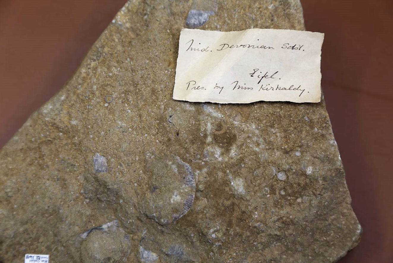
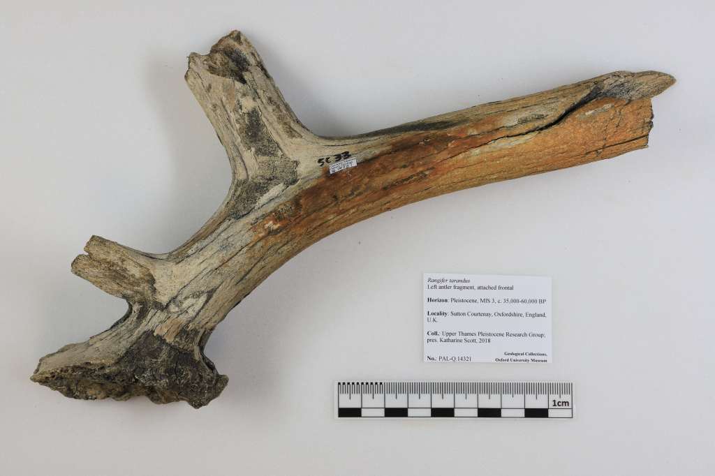
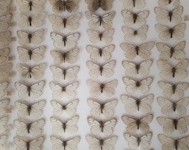
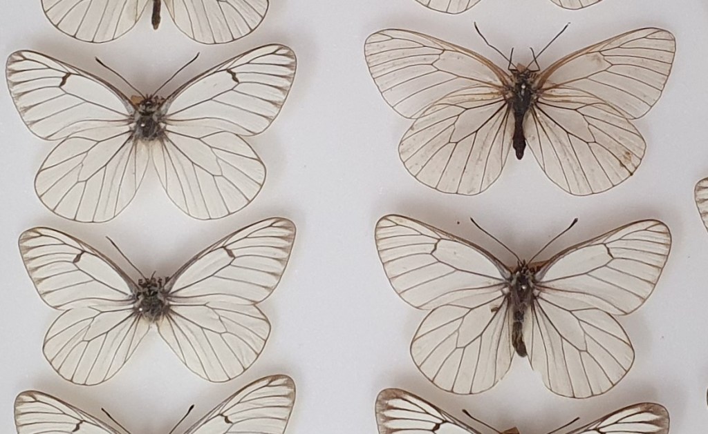
The Black-veined white butterfly (Aporia crataegi) was a large, charismatic butterfly with distinctive black venation on its wings. Once commonly found in the UK, the species unfortunately went extinct here in around 1925, with the last British specimens collected from Herne Bay in Kent. It isn’t fully understood why the species disappeared from the UK, but climate change, predation, parasites, and disease have all been suggested to have caused its disappearance — perhaps with several of these factors contributing to its decline. Central to solving the mystery of the disappearance of the Black-veined white will be the collections of butterflies that are stored in museums like OUMNH.

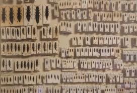
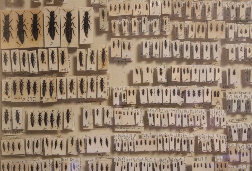
Butterflies tend to be well-represented in museum collections, and the Black-veined white is no exception. While the species has now been extinct in the UK for around 100 years, Lepidoptera enthusiasts from previous centuries often captured wild Black-veined white specimens for their personal collections. The abundance of Black-veined white butterflies in museum collections, like the collections at OUMNH, serve as a valuable repository for scientific research — including my own!

Black-veined white butterflies in the collections at OUMNH

Between June and December 2021, I undertook a research project using OUMNH’s Black-veined white butterflies. My task was to extract enough DNA from the butterflies to use for ‘whole genome sequencing’ — in other words, I was attempting to extract DNA from butterfly specimens to decode their complete DNA sequence. Getting DNA sequences from the historical specimens that are kept in Museums is no easy task, as DNA degrades over time. Nonetheless, animal specimens from natural history museums have successfully been used for whole genome sequencing and genetic analysis in the past, including species as diverse as longhorn beetles and least Weasels.

In order to work out how to extract DNA from the specimens, I had to try a variety of methods. This included experimenting to find out whether butterfly legs or abdomen fragments yielded more DNA, and whether non-destructive methods of DNA extraction were as effective as destructive methods. An example of a non-destructive method of DNA extraction would be a process like soaking a sample overnight and using the leftover liquid for DNA extraction, whereas a destructive method might involve mashing a whole leg or abdomen segment to use as a DNA source.

Preparing a DNA sample

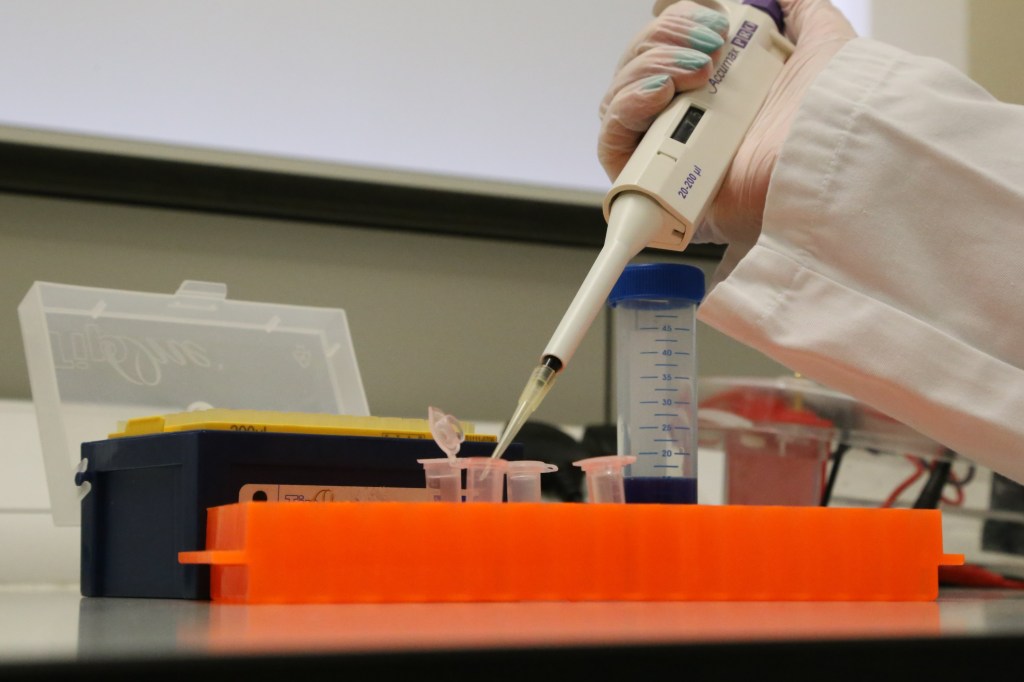
Overall, I found that destructively sampling the legs of the butterflies gave the most reliable results, and also had the added benefit of not destroying the wings or abdomen of the specimens. Keeping the wings and abdomens of the butterflies intact will likely prove useful for conducting morphological studies in future.

Now that I have a reliable DNA extraction method, the next step in my research will be to analyse more Black-veined white specimens from a span of different time periods leading up to the species’ disappearance. I will then compare samples collected from each time period to calculate the genetic diversity of the species at each point in time, leading up to its disappearance. If I find a steady decline in the species’ genetic diversity over time, this may indicate a gradual extinction of the species. This is because we expect that, as numbers of a species decrease, inbreeding will become common, resulting in less diversity in the species’ DNA. However, if the populations of Black-veined white butterflies went extinct very suddenly, the decline in genetic diversity will probably be less pronounced. Learning more about the fate of the Black-veined White could not only help us unlock the historical mystery of the species’ decline in Britain, but will also help us understand more about the species’ decline in other parts of the world.


British Insect Collections: HOPE for the Future is an ambitious project to protect and share the Museum of Natural History’s unique and irreplaceable British insect collection. Containing over one million specimens – including dozens of iconic species now considered extinct in the UK – it offers us an extraordinary window into the natural world and the ways it has changed over the last 200 years. The HOPE for the Future project is funded by the National Lottery Heritage Fund, thanks to National Lottery players.

Re-Collections: Jane Willis Kirkaldy

By Evie Granat, Project Officer Trainee with the Freshwater Habitats Trust and Museum volunteer


The Museum is lucky enough to house several specimens presented by Jane Willis Kirkaldy (1867/9 – 1932). They serve as a reminder of a passionate and dedicated tutor, and of a key figure behind the development of women’s education at Oxford University.


Jane Willis Kirkaldy was born somewhere between 1867 and 1869, and spent her youth in London with her parents and five siblings. After completing her secondary education at Wimbledon High School, Kirkaldy gained entry to Somerville College (Oxford) on an exhibition scholarship in 1887. She finished her degree in 1891, becoming one of the first women to achieve a First Class Hons in Natural Sciences (Zoology). However, since the University didn’t award women degrees in the nineteenth century, it wasn’t until 1920 that Kirkaldy received her MA.

Upon completing her undergraduate studies, Kirkaldy worked for a short period as a private tutor in Castle Howard before returning to Oxford in 1894. Whilst researching at the University, she produced two papers for the Quarterly Journal of Microscopical Science, including an article entitled “On the Head Kidney of Myxine”. This study of the renal systems of hagfish was written with the aid of experimental work carried out by renowned zoologist Walter Weldon at his UCL laboratory. She also studied lancelets under the Oxford Linacre Professor of Zoology, publishing “A Revision of the Genera and Species of Branchiostomdae” in 1895.

Kirkaldy’s achievements are especially noteworthy given how few women studied Natural Sciences at Oxford during the nineteenth century. In addition to her contributions to the scientific field, she also helped advance women’s education at Oxford University. In 1894, The Association of the Education of Women named Kirkaldy a tutor to female students in the School of Natural Sciences. The following year she ceased all research to concentrate fully on teaching, co-authoring ‘Text Book of Zoology’ with Miss E.C. Pollard in 1896, and Introduction to the Study of Biology with I. M. Drummond in 1907. She eventually became a tutor or lecturer at all of Oxford’s Women’s Societies, and a Director of Studies at all five of the women’s colleges. Amongst the many female scientists that came under her care was the Nobel Prize-winning chemist Dorothy Crowfoot Hodgkin.

Left: Page from one of our donations books listing Jane Willis Kirkaldy as the donor of a series of Middle Devonian fossils (from the Eifel) to the Museum in October 1901. Right: Chromite from East Africa, also donated to the Museum by Kirkaldy.

Beyond the Department of Natural Sciences, Kirkaldy was an important figure at Oxford — she served as a member of the Council of St. Hugh’s College for 14 years, and was made an honorary fellow of Somerville College in 1929. At the Museum of Natural History, she presented beetles from New Guinea (1890), Devonian Fossils from the Eiffel (1901), and Chromite from near Beira, Mozambique (1924).

Kirkaldy retired from the University in 1930 due to ill health, before passing away in a London care home in 1932. Oxford University subsequently dedicated the junior and senior ‘Jane Willis Kirkakdy Prizes’ in her memory, which still exist to this day.


References

https://www.firstwomenatoxford.ox.ac.uk/article/principals-and-tutors

https://archive.org/details/internationalwom00hain/page/160/mode/2up

https://www.ias.ac.in/article/fulltext/reso/022/06/0517-0524

http://wimbledonhighschool.daisy.websds.net/Filename.ashx?tableName=ta_publications&columnName=filename&recordId=72

http://wimbledonhighschool.daisy.websds.net/Filename.ashx?tableName=ta_publications&columnName=filename&recordId=71

https://archive.org/details/internationalwom00hain/page/160/mode/2up

Dorothy Crowfoot Hodgkin: Patterns, Proteins and Peace: A Life in Science, by Georgina Ferry

Quarterly Journal of Microscopical Science

Reindeer are not just for Christmas

WHAT WE CAN LEARN FROM BRITAIN’S ICE AGE RANGIFER


By Emily Wiesendanger, Volunteer


If you’ve ever visited the Skeleton Parade in the Main Court of the Museum, you may have noticed that nestled between the Malayan tapir and the rhinoceros is the skeleton of a reindeer, or caribou if you are from North America.

Today, reindeer are found throughout the Arctic and Subarctic in places like Canada, Alaska, Russia, and Lapland (Norway, Sweden, and Finland). However, their range was not always so limited. During the Late Pleistocene – around 126,000 to 11,700 years ago – it would not have been unusual to see herds of reindeer roaming freely across most of Britain and western Europe. In fact, reindeer sub-fossils in the form of bones, teeth, and antlers have been found at a number of Oxfordshire sites including the excavations at Cassington and Sutton Courtenay, which are kept behind the scenes in the Museum’s extensive Paleontological Collections.

Studying these Ice Age reindeer can teach us as much about the future as they can about the past. Pleistocene reindeer were likely similar to their modern counterparts, which undertake large, bi-annual migrations between summer and winter grazing pastures. Looking at the movements of Ice Age populations of reindeer can therefore help us to understand how modern reindeer may respond to climactic and environmental changes in the future. This is possible because reindeer only come together in large herds at certain times of the year. During these seasonal aggregations, the herd is characterised by different combinations of ages and sexes. Therefore, by looking at the age and sex of the remains of reindeer present at a site, we can tell the time of year that they were left there — in particular, we can infer the sex of reindeer from their bones, their age from their teeth, and their age and sex from their antlers.

Modern reindeer are highly adapted to cold environments (-45 to +15°C) with two layers of fur (the tips of which turn white in the winter), short and furry ears and tails, and large feet to make walking on snow and digging for food much easier. Reindeer even make a clicking noise with their feet, produced by a tendon slipping over a bone, to help keep track of each other in blizzards or fog.

Unfortunately, it is extremely rare to find anything so complete as the reindeer in the skeleton parade. Instead, you are much more likely to find remains like the antler below, which was excavated from Sutton Courtenay. Despite being only a fragment, it is exactly this kind of sub-fossil that can help us to understand more about the movements of reindeer during the Late Pleistocene.

This left antler base and skull from a male reindeer found at Sutton Courtenay can be used to determine which season reindeer were present at the site.

Reindeer grow and shed a new pair of antlers every year, and this happens at different times of the year for males and females. If you can identify whether an antler is male or female, shed or unshed, you can also tell the season of death. The Sutton Courtenay antler featured above would have belonged to a male reindeer. At its base, we can see it is still clearly attached to some skull bone, and so is unshed. Because males only have their fully grown antlers between September and November, this particular reindeer must have been in the area around Sutton Courtenay during the autumn. It is by using similar deductions that we can also tell that Rudolph and his antlered friends would have actually all been females — by the 24th December, males have already shed their antlers, but females will keep them until the spring!

After studying thousands of these kinds of remains from all over Britain, we can start to build a picture of where reindeer were at different times of the year. It’s amazing to think that we can learn so much from simple skeletons. So, the next time you visit the skeleton parade, take a moment to think about the secrets they may be hiding.

Conservation in the Genomic Era

HAVE DNA TECHNOLOGIES REPLACED THE NEED FOR MUSEUMS?


By Sotiria Boutsi, Intern

I am PhD student at Harper Adams University with MSc in Conservation Biology, currently doing a professional internship at the Museum of Natural History in the Public Engagement office. My PhD uses genomic data to study speciation in figs and fig wasps.


The year 1995 marked the first whole-genome sequencing for a free-living organism, the infectious bacterium Haemophilus influenza. Almost three decades later, biotechnological advances have made whole-genome sequencing possible for thousands of species across the tree of life, from ferns and roses, to insects, and – of course – humans. Ambitious projects, like the Earth BioGenome Project, aim to sequence the genomes of even more species, eventually building the complete genomic library of life. But do these advancements help us with conservation efforts? Or are the benefits of biotechnology limited to industrial and biomedical settings?

The value of genetic information is becoming increasingly apparent: from paternity tests and DNA traces in forensic investigations, to the characterization of genes related to common diseases, like cancer, we are becoming familiar with the idea that DNA can reveal more than meets the eye. This is especially the case for environmental DNA, or eDNA — DNA molecules found outside living organisms. Such DNA is often left behind in organic traces like tissue fragments and secretions. Practically, this means that water or air can host DNA from organisms that might be really hard to observe in nature for a variety of reasons — like being too small, too rare, or just too shy.

So, how do we determine which species left behind a sample of eDNA? The method of identifying a species based on its genomic sequence is called barcoding. A barcode is a short genomic sequence unique to a species of organism. Therefore, every time we encounter a barcode sequence, whether it is taken from a living animal or eDNA, we can associate it to the species which it belongs to.

When we have a mix of different species to identify, things become a bit more complicated. Sometimes we will pick up samples which represent an entire ecological community, and must sort through these using a process called meta-barcoding.

How does meta-barcoding work? Well, we want to be able to identify species based on the shortest possible species-specific sequence. Traditional laboratory methods for DNA amplification (PCR) are combined with DNA sequencing to read the DNA sequences found in any given water or air sample. Then, having a database of reference genomes for different species can serve as the identification key to link the sample sequences to the species they originated from.

Pinned insects can be found in the Upper Gallery of the Museum. There are currently 5 million insect specimens at the Museum, serving as a record of biodiversity at the time and space of collection. Museum collections are invaluable ways of monitoring biodiversity but rely on capturing live animals.

So, what does this mean for the future of ecology and conservation? Traditional monitoring of biodiversity can involve capturing and killing live animals. This is the case with insect specimens found in museums across the world. Although museum collections are irreplaceable as a record of the history of wild populations, regular monitoring of endangered species should rely on non-invasive methods, such as meta-barcoding of eDNA. Indeed, eDNA has been used to monitor biodiversity in aquatic systems for almost a decade. Monitoring terrestrial ecosystems through air samples is now also becoming possible, opening new possibilities for the future of conservation.

During March, the Museum delivered practical molecular workshops in our laboratory, reaching more than 200 Key Stage 5 students. Students have had the opportunity to learn about the use of eDNA in ecology, as well as get some hands-on experience in other molecular techniques. These include DNA extraction, PCR, the use of restriction enzymes, and gel electrophoresis.  The workshops were delivered by early-career researchers with practical experience in working in the laboratory, as well as Museum staff with a lot of experience in delivering teaching. Through the Museum’s workshops, which run regularly, the next generation of scientists is introduced not only to both human genetics, but also molecular tools used in ecological research, which without a doubt will become increasingly relevant for future conservationists.

Since 2009, the Museum runs practical workshops for Key Stage 5 students in the molecular laboratory at the Museum’s main facilities. Workshops started again this March, after the mandatory 2-year covid-19 break. Students can learn about and discuss the use of molecular techniques in biology by extracting their own DNA.  

We cannot conserve what we do not know. Monitoring biodiversity is the cornerstone of any conservation practice. Doing it efficiently, by making use of both traditional as well as molecular tools, can allow more accurate predictions for the future of biodiversity under the lens of anthropogenic change.


More Information:


British Insect Collections: HOPE for the Future is an ambitious project to protect and share the Museum of Natural History’s unique and irreplaceable British insect collection. Containing over one million specimens – including dozens of iconic species now considered extinct in the UK – it offers us an extraordinary window into the natural world and the ways it has changed over the last 200 years. The HOPE for the Future project is funded by the National Lottery Heritage Fund, thanks to National Lottery players.

Sisters of Science

THE PIONEERING LEGACIES OF KATHLEEN LONSDALE AND DOROTHY CROWFOOT HODGKIN


By Leonie Biggenden, Volunteer


As Women’s History Month comes to a close, this blog post looks at two ‘sisters of science’, friends and contemporaries Dorothy Crowfoot Hodgkin (1910 – 1994) and Kathleen Lonsdale (1903-1971), and considers some links between these remarkable women.

Dorothy Crowfoot Hodgkin is the only female bust in the Oxford Museum of Natural History and is the only British woman to have been awarded the Nobel Prize for science.  When she was awarded the Prize in 1964 – for her ground-breaking discovery of the structures of vitamin B12 and penicillin – there was much scepticism about whether women belonged in the field of science. One newspaper commemorated her achievement with the headline “Nobel prize for a wife from Oxford”.

Hodgkin was assisted and supported in her endeavours by fellow scientist Kathleen Lonsdale, who worked in London while Hodgkin was based in Oxford.  Both women were pioneers who advanced the x-ray crystallography technique, in which x-rays are fired at crystals of molecules to determine their chemical structure. Lonsdale applied the technique to diamonds, benzene, and later kidney stones.  For her efforts, she had a type of diamond named after her: Lonsdaleite.  It was not just any diamond, but one formed in meteorites, as a result of the heat and pressure of impact into the Earth’s atmosphere.

Both had similar difficulties as girls wanting to study science.  Hodgkin was initially not allowed to take chemistry at her grammar school as it was considered a ‘boy’s subject’, but she thankfully managed to reverse the school’s decision, allowing her to pursue her scientific career.  Lonsdale had to transfer to a boys’ school to be able to study maths and science, as these were subjects not offered at her girls’ school. She later described how her love of maths was inspired by learning to count at school using yellow balls.

Both women were supported by strong male advocates and mentors, such as the scientist William Bragg. Bragg first met Lonsdale when he was assigned as one of her examiners, and subsequently asked her to join his research school at University College London (UCL).  Lonsdale would later follow Bragg when he moved his laboratory to the Royal Institution.  Bragg was also responsible for inspiring Hodgkin’s interest in the properties of atoms, giving her a copy of ‘Concerning the Nature of Things’ when she was 15 years old.

Lonsdale and Hodgkin worked hard to show that science was a viable option for girls.  Lonsdale’s essay, ‘Women in Science – why so few?’, argued that social expectations placed on women discouraged them from pursuing science [1]. In fact, she was so determined to encourage girls’ interest in the subject that, while ill in hospital, she received special permission to be able to leave to award prizes for science at a local girls’ school. Hodgkin advocated for female scientists and directly mentored several who went on to become important crystallographers in their own right.

Both women eventually became professors, and Lonsdale was one of the first two women elected as Fellows of the Royal Society (FRS) in 1945. In 1947, Hodgkin was one of the youngest people to be elected FRS. 

Both Hodgkin and Lonsdale were extremely concerned about the threat of nuclear war, and in 1976 Hodgkin became president of the Pugwash Conference which advocated for nuclear disarmament.  Lonsdale was also involved with Pugwash and was president of the Women’s International League of Peace and Freedom.  A lifelong pacifist, she went to Holloway prison in London for a month for failing to register for war service and not paying her £2 fine.  She became a dedicated advocate for prison reform after seeing the conditions of the women first-hand.

My favourite facts about both Lonsdale and Hodgkin are those that give us a glimpse of their ingenuity.  Lonsdale made her own hat to meet the Queen and have her Damehood conferred upon her.  It was constructed with lace, cardboard and 9d worth of ribbon.  Similarly, when awarded her first honorary degree, Lonsdale pinned a strip of beautiful material inside her gown as a substitute for buying a whole new dress.  Hodgkin was also very creative. As a child, she created her own personal laboratory in the attic and acquired acids from the local chemist to experiment with.

The two women held each other in great respect, as testified to by the fact that Hodgkin wrote a biographical memoir of Lonsdale.  She said of her friend: “There is a sense in which she appeared to own the whole of crystallography in her time.”  Let’s agree that both women can claim that crown. Looking back, we can remember these women for their remarkable stories, featuring precious gems, prisons, penicillin and peace. But, most importantly, we should remember Hodgkin and Lonsdale as pioneers who paved the way for future women scientists.


References

[1] Hodgkin D (1975) Kathleen Lonsdale 28 January–1 April 1971. Biogr Mems Fell R Soc 21:447–484

Reading Archival Silences

MAUD HEALEY AND HER GEOLOGICAL LEGACY


By Chloe Williams, History Finalist at Oxford University and Museum Volunteer

Email: chloegrace1000@gmail.com


“The professor regrets to have to record the loss of the invaluable services of Miss Healey, who as a result of overwork has been recommended to rest for an indefinite period. This will prove a serious check to the rate of progress which has for some time been maintained in the work of rearrangement, and it is hoped that her retirement may be only temporary.” So ends the Oxford University Museum of Natural History’s 1906 Annual Report, marking the near-complete departure of Maud Healey from the archival record.

Despite how little of her history has been preserved, it is clear that Maud Healey made significant contributions to the field of geology. After studying Natural Sciences at Lady Margaret Hall in 1900, Healey worked at the Museum as an assistant to Professor William Sollas from 1902–1906. Here, she catalogued thousands of specimens and produced three publications. These publications were at the centre of debates about standardising the geological nomenclature, and turning geology into a practical academic discipline that could sustain links across continents. However, Healey was continually marginalized on the basis of her gender. Closing the Geological Society of London’s discussion of one of her papers, “Prof. Sollas remarked that he had listened with great pleasure to the complimentary remarks on the work of the Authoress, and regretted that she was not present to defend before the Society her own position in the disputed matter of nomenclature.”[1] Predating the Society’s 1904 decision to admit women to meetings if introduced by fellows, Healey had been unable to attend the reading of her own paper.

Photo of the Geological Society of London centenary dinner in 1907, at which Maud Healey was present. Healey can be seen seated in the fourth row from the front, three chairs to the left. Of the 263 guests, 34 were women, 20 of whom were the wives or daughters of academics, and only 9, including Healey, were present ‘in their own right’. [2] Source: Burek, Cynthia V. “The first female Fellows and the status of women in the Geological Society of London.” Geological Society, London, Special Publications 317, no. 1 (2009): 373-407.

Healey later worked with specimens collected by Henry Digges La Touche in colonial Burma (now Myanmar). While Healey worked with the identification of species, acknowledged by La Touche himself as ‘a more difficult lot to work at’ than similar specimens assigned to her male contemporaries, the physical collection and therefore its name and record is attributed to a male geologist. [3] She continued her work identifying La Touche’s collection of Burmese fossils after retiring from the Museum in 1906 and published a report about them in 1908. What happened to her afterwards is unclear. Tantalizing snippets like a 1910 marriage record might suggest that she turned to a life of domesticity, but whether Healey continued to engage with geology as a hobby remains uncertain.

It is almost unbelievable that a professional of Healey’s calibre could abandon the work in which she excelled. However, Healey lacked any familial connections to geology, and apparently did not marry into money, which would have made it difficult for her to retain access to organizations like the Geological Society of London. The diagnosis of ‘overwork’ mentioned in the Annual Report makes it possible that a medical professional could have discouraged her from engaging further in academia. Unfortunately, any diaries or letters which might have provided us with further clues were not deemed worthy of preservation.

Maud Healey on a dig site (location unknown). Image from the Archives at Oxford University Museum of Natural History.

Tracing Maud Healey’s history to 1910, it might seem as though we hit a depressing dead end. Healey is one of many nineteenth-century female geologists who participated in an international community in a range of roles including collecting, preserving samples, and actively producing knowledge. However, like many of her colleagues, her contributions are largely absent from the historical record. My research doesn’t aim to simply ‘rediscover’ these exemplary women after previously being ‘hidden’ from history, but instead considers how history itself is constructed from a material archive created along lines of gender and class. A subjectivity which surfaces only rarely in appended discussions to academic papers, and in spidery cursive on ancient fossils, Maud Healey ultimately suggests the need for women’s history to read archival silences as their own stories.


Works cited

[1] Healey, M. ‘Notes on Upper Jurassic Ammonites, with Special Reference to Specimens in the University Museum, Oxford: No. I’, Quarterly Journal of the Geological Society of London 60, (1904), p.1-4.

[2] Burek, Cynthia V. “The first female Fellows and the status of women in the Geological Society of London.” Geological Society, London, Special Publications 317, no. 1 (2009): 373-407.

[3] La Touche, H.D. Letter to Anna La Touche, 1 August 1907. La Touche Collection. MSS.Eur.C.258/77. Asian and African Studies Archive, The British Library, London, UK.


Read more