The Beginning of the End: Do locusts still spell danger for humanity?


By Ella McKelvey, Web Content and Communications Officer


A few days ago, I was working from home when a delivery driver arrived with a strange parcel – a cardboard box stamped with the letters FRAGILE that seemed to be producing a peculiar, scratching sound. Tentatively, I opened the cardboard box and pulled out a plastic punnet filled with newspaper, old egg cartons, and… wait! Was that an antenna? 

The parcel turned out to be a box of locusts, ordered by my housemate who uses them to feed her pet reptiles. I set the punnet down beside me and tried to continue with my morning’s work. But over the next few hours, the locusts grew increasingly restless, bouncing against the walls of their punnet like hot, microwaved popcorn. The sight and sound of the insects began to return memories of the infamous locust swarms of 2020 — one in a series of near-apocalyptic events that befell us that fateful year. Worryingly, climate change is set to make locust swarms increasingly common, with Sardinia currently facing its worst locust swarm in thirty years.1 

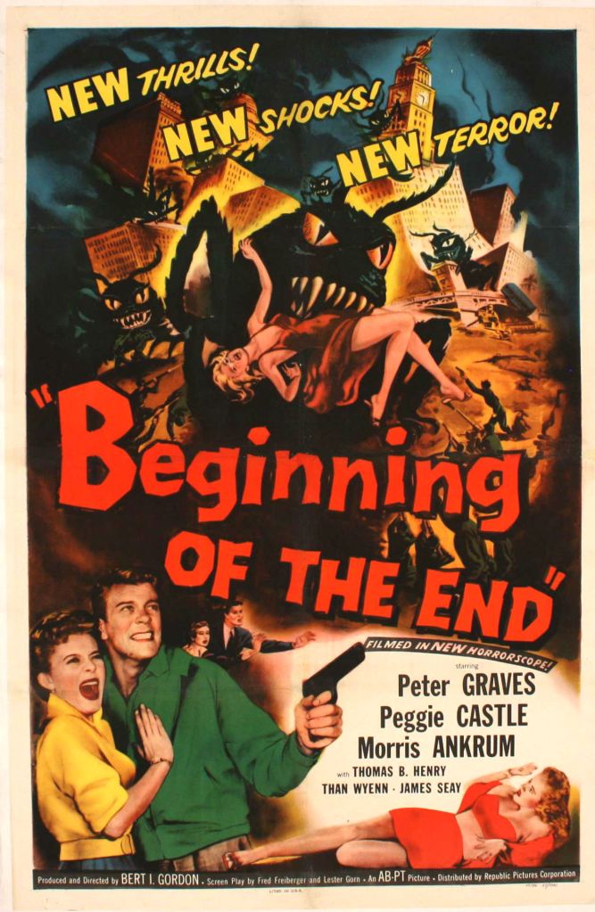
Left: A poster for The Beginning of the End (1957) about a fictional invasion of giant, mutant locusts in Illinois. Right: A real-life locust swarm near Satrokala, Madagascar (2014).

Throughout history, locusts have been widely understood as symbols of maleficence and misfortune. One of the oldest written references to locusts is, of course, the Biblical story of the ten plagues of Egypt, in which locusts were sent as a punishment from God. Since then, these infamous insects have been featured in art, books, music, and films as harbingers of destruction. Americans of the mid-twentieth century were somewhat obsessed with giant locusts and grasshoppers which were featured everywhere from cartoons to postcards. 1957 saw the release of the movie The Beginning of the End – a schlocky Hollywood sci-fi tale about a swarm of giant, mutant locusts invading Illinois. The film’s principal Entomologist describes locusts as “deadly killer[s]”, both “intelligent and strong”. Real-life locusts are, indeed, very strong for their size, with back legs that can catapult them up to a metre from standing. This means that it would be feasible for the human-sized locusts in The Beginning of the End to jump as far as forty metres — a terrifying thought!2  

While The Beginning of the End is ridiculous both in premise and execution, I can’t deny that I find the concept of giant locusts pretty nightmarish. Earlier in the week, I sent an email to the Life Collections team to enquire about the possibility of looking through our pinned locusts and snapping a few photos of the biggest and grisliest specimens. As I walked upstairs to entomology, I braced myself for an encounter with some fearsome insects. But what I found were a few drawers of modest-sized locusts that looked about as benign as garden grasshoppers. Many of them were even stuffed with wool; more like teddy bears than agents of Armageddon. 

Left: Anacridium aegyptium or Egyptian Locust from the Collections at OUMNH. Right: Underside of a locust specimen showing cotton wool stuffing.

According to Collections Assistant Rob Douglas, stuffing large insect specimens with cotton wool used to be a common entomological practice. Insects with fatty insides, like locusts, must be gutted to ensure good preservation. Following the removal of the insects’ insides, cotton was often used to return their abdomens to their usual size and shape. Locusts’ ample fat stores contribute as much to their physical prowess as their powerful hind legs; sustaining them through migrations of up to 310 miles a day.3 Such migrations occur when locusts are exposed to a dry spell followed by wet weather, allowing for the sudden regrowth of vegetation. These conditions will cause locusts to switch their solitary lifestyles for gregariousness, coming together to chomp their way through crops and vegetation at a density of 80-160 million insects per square mile. A large migrating swarm of locusts has been estimated to need as many calories in a day as 1.5 million human males, explaining why even ordinary-sized locusts are capable of causing agricultural annihilation.

If it weren’t for government and international interventions, the 2020 locust swarms in East Africa could have caused up to $8.5 billion in economic damages by the year-end.5 But locusts can do much worse. One of the most notorious locust swarms on record was that of the Rocky Mountain locust in the USA between 1874 and 1877. According to some accounts, the swarm caused damages to agriculture equivalent to $116 billion in today’s money, leaving behind piles of locust carcases up to six feet high.6 

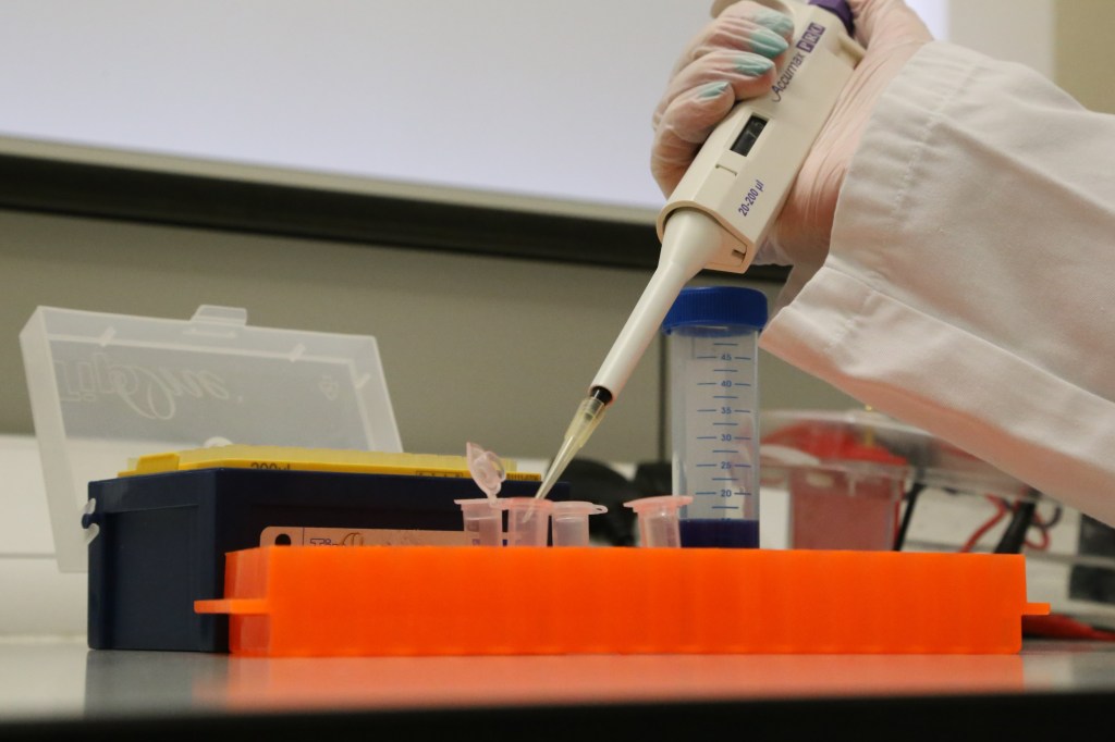
When it comes to protecting crops from locusts, prevention is better than cure. Likely locust outbreaks can be pre-empted by studying weather patterns and using satellite imagery to keep an eye on vegetation growth.7 Once a (potential) locust swarm has been identified, traditional methods of locust management involve the use of pesticides to wipe out the insects as soon as possible. Back in the 1950s, this meant dowsing locusts with DDT. But as the drawbacks of synthetic pesticides become increasingly apparent, chemical interventions are being replaced with the application of naturally occurring ‘pesticides’ like the fungus Metarhizium acridum.  

Our understanding of locusts has come a long way since the release of The Beginning of the End. One of my favourite news stories of the past month was the announcement by a laboratory at Michigan State University that locusts have been successfully used to ‘sniff out’ mouth cancer.8 It turns out that locusts no longer just spell danger for humanity — they can smell danger for humanity too! These cancer-detecting locusts are, in my opinion, far more ‘sci-fi’ than the giant bugs imagined by scriptwriters of the 1950s, reminding us that, when it comes to science, the truth is often stranger than fiction. Reports like these demonstrate that scientific research has the power to transform our relationship with the pests that have tormented us for thousands of years.


[1] Sardinian farmers suffer worst locust invasion in over 30 years | Reuters 

[2] https://www.st-andrews.ac.uk/~wjh/jumping/perform.html

[3] https://www.nationalgeographic.com/animals/invertebrates/facts/locusts

[4] Weis-Fogh T. 1952 Fat combustion and metabolic rate of flying locusts (Schistocerca gregaria Forskål)Phil. Trans. R. Soc. Lond. B2371–36http://doi.org/10.1098/rstb.1952.0011

[5] Dominy, Nathaniel J., and Luke D. Fannin. “The sluggard has no locusts: From persistent pest to irresistible icon.” People and Nature 3, no. 3 (2021): 542-549.

[6] Lockwood, Jeffrey A. Locust: The Devastating Rise and Mysterious Disappearance of the Insect that Shaped the American Frontier. London: Hachette (2004).

[7] Zhang, Long, Michel Lecoq, Alexandre Latchininsky, and David Hunter. “Locust and grasshopper management.” Annu. Rev. Entomol 64, no. 1 (2019): 15-34.

[8] https://www.technologyreview.com/2022/06/21/1054532/cyborg-locust-brain-hacked-sniff-out-cancer/

A Fashion Flea-esta


By Danielle Czerkaszyn, Librarian and Archivist


In September 2021, the Museum initiated its first “Specimen Showdown” on Twitter and Instagram, where followers could vote on their favourite specimens from our collections. Over the course of the month, followers narrowed down their favourite among 32 specimens from four collections: The Library Legends, The Bygone Beasts, The Rock Stars, and The Birds and The Beetles. The final showdown was between the Connemara Column (found in the Main Court of the Museum) and the Pulgas Vestidas from the Library and Archives. In a nail-biting race, the Pulgas Vestidas narrowly beat the column with 53.9% of the vote.

But what are Pulgas Vestidas? And why are they so popular?

Dressed fleas, you say?

The delicate art of dressing fleas in tiny costumes, known as ‘Pulgas Vestidas’ in Spanish, flourished in Mexico for over two centuries. It is believed that the craft began in Mexican convents where nuns would fashion tiny pieces of clothing onto dead fleas. An important point to note is that the fleas themselves were not actually dressed — instead, they formed the heads of the figures. The individual fleas were set in matchboxes and decorated with elaborate human costumes, hats, shoes, and accessories. Sometimes the fleas were set in whole scenes, often as married couples in miniature dioramas of everyday life. The bride and groom sets were the most popular, with the bride sporting a long veil and the groom in his best suit. The nuns would then sell the fleas for a small amount of money to passing tourists. The trade was later picked up by the local villagers and Pulgas Vestidas were widely sold to tourists visiting Mexico in the early twentieth century.

Dressed fleas were popular with tourists until the 1930s when the art declined in popularity. An increasing awareness of hygiene meant that fleas were rapidly regarded as unhealthy. Many dressed fleas were consigned to the bin, and Pulgas Vestidas became a lost art as tourists’ tastes for memorabilia changed. Examples of these tiny curiosities are now rare collectors’ items.

Pulgas Vestidas at Oxford University Museum of Natural History

The Museum’s dressed fleas were collected in 1911 by American archaeologist and anthropologist Zeila M. M. Nuttall who specialised in Mexican history and culture. She sent the dressed fleas to her brother, bacteriologist George H.F. Nuttall. George formally donated a collection of 50 Ixodidae (ticks) to the Museum, and it is likely he also gifted the dressed fleas at the same time. The dressed fleas would have been considered more of a Victorian novelty, and so were not formally recorded or accessioned into our collections.

Although most of OUMNH’s dressed fleas reside behind-the-scenes, one example is on public display in the Upper Gallery of the Main Court. Sporting tiny clothes and a backpack, the flea is just visible with the help of a magnifying glass. Clearly, this one was born to flea wild.

OUMNH’s Pulgas Vestidas are definitely among the more unusual items in the Museum’s collections, and they were clearly head and shoulders above other specimens in the September Specimen Showdown competition, despite being no more than 5mm tall! Pulgas Vestidas may be small, but they certainly are mighty.

I’ll Flea There

The dressed fleas will be on display, with a flea-tastic craft, for the Museum’s free evening event Late Night: A Buzz in the Air‘ on 27 May from 7-10pm.

Disappearing Butterflies

HOW TO SOLVE A BIOLOGICAL MYSTERY USING MUSEUM COLLECTIONS AND DNA TECHNOLOGY


By Rebecca Whitla, PhD student at Oxford Brookes University


The Black-veined white butterfly (Aporia crataegi) was a large, charismatic butterfly with distinctive black venation on its wings. Once commonly found in the UK, the species unfortunately went extinct here in around 1925, with the last British specimens collected from Herne Bay in Kent. It isn’t fully understood why the species disappeared from the UK, but climate change, predation, parasites, and disease have all been suggested to have caused its disappearance — perhaps with several of these factors contributing to its decline. Central to solving the mystery of the disappearance of the Black-veined white will be the collections of butterflies that are stored in museums like OUMNH.

Butterflies tend to be well-represented in museum collections, and the Black-veined white is no exception. While the species has now been extinct in the UK for around 100 years, Lepidoptera enthusiasts from previous centuries often captured wild Black-veined white specimens for their personal collections. The abundance of Black-veined white butterflies in museum collections, like the collections at OUMNH, serve as a valuable repository for scientific research — including my own!

Black-veined white butterflies in the collections at OUMNH

Between June and December 2021, I undertook a research project using OUMNH’s Black-veined white butterflies. My task was to extract enough DNA from the butterflies to use for ‘whole genome sequencing’ — in other words, I was attempting to extract DNA from butterfly specimens to decode their complete DNA sequence. Getting DNA sequences from the historical specimens that are kept in Museums is no easy task, as DNA degrades over time. Nonetheless, animal specimens from natural history museums have successfully been used for whole genome sequencing and genetic analysis in the past, including species as diverse as longhorn beetles and least Weasels.

In order to work out how to extract DNA from the specimens, I had to try a variety of methods. This included experimenting to find out whether butterfly legs or abdomen fragments yielded more DNA, and whether non-destructive methods of DNA extraction were as effective as destructive methods. An example of a non-destructive method of DNA extraction would be a process like soaking a sample overnight and using the leftover liquid for DNA extraction, whereas a destructive method might involve mashing a whole leg or abdomen segment to use as a DNA source.

Preparing a DNA sample

Overall, I found that destructively sampling the legs of the butterflies gave the most reliable results, and also had the added benefit of not destroying the wings or abdomen of the specimens. Keeping the wings and abdomens of the butterflies intact will likely prove useful for conducting morphological studies in future.

Now that I have a reliable DNA extraction method, the next step in my research will be to analyse more Black-veined white specimens from a span of different time periods leading up to the species’ disappearance. I will then compare samples collected from each time period to calculate the genetic diversity of the species at each point in time, leading up to its disappearance. If I find a steady decline in the species’ genetic diversity over time, this may indicate a gradual extinction of the species. This is because we expect that, as numbers of a species decrease, inbreeding will become common, resulting in less diversity in the species’ DNA. However, if the populations of Black-veined white butterflies went extinct very suddenly, the decline in genetic diversity will probably be less pronounced. Learning more about the fate of the Black-veined White could not only help us unlock the historical mystery of the species’ decline in Britain, but will also help us understand more about the species’ decline in other parts of the world.


British Insect Collections: HOPE for the Future is an ambitious project to protect and share the Museum of Natural History’s unique and irreplaceable British insect collection. Containing over one million specimens – including dozens of iconic species now considered extinct in the UK – it offers us an extraordinary window into the natural world and the ways it has changed over the last 200 years. The HOPE for the Future project is funded by the National Lottery Heritage Fund, thanks to National Lottery players.

Re-Collections: Jane Willis Kirkaldy

By Evie Granat, Project Officer Trainee with the Freshwater Habitats Trust and Museum volunteer


The Museum is lucky enough to house several specimens presented by Jane Willis Kirkaldy (1867/9 – 1932). They serve as a reminder of a passionate and dedicated tutor, and of a key figure behind the development of women’s education at Oxford University.


Jane Willis Kirkaldy was born somewhere between 1867 and 1869, and spent her youth in London with her parents and five siblings. After completing her secondary education at Wimbledon High School, Kirkaldy gained entry to Somerville College (Oxford) on an exhibition scholarship in 1887. She finished her degree in 1891, becoming one of the first women to achieve a First Class Hons in Natural Sciences (Zoology). However, since the University didn’t award women degrees in the nineteenth century, it wasn’t until 1920 that Kirkaldy received her MA.

Upon completing her undergraduate studies, Kirkaldy worked for a short period as a private tutor in Castle Howard before returning to Oxford in 1894. Whilst researching at the University, she produced two papers for the Quarterly Journal of Microscopical Science, including an article entitled “On the Head Kidney of Myxine”. This study of the renal systems of hagfish was written with the aid of experimental work carried out by renowned zoologist Walter Weldon at his UCL laboratory. She also studied lancelets under the Oxford Linacre Professor of Zoology, publishing “A Revision of the Genera and Species of Branchiostomdae” in 1895.

Kirkaldy’s achievements are especially noteworthy given how few women studied Natural Sciences at Oxford during the nineteenth century. In addition to her contributions to the scientific field, she also helped advance women’s education at Oxford University. In 1894, The Association of the Education of Women named Kirkaldy a tutor to female students in the School of Natural Sciences. The following year she ceased all research to concentrate fully on teaching, co-authoring ‘Text Book of Zoology’ with Miss E.C. Pollard in 1896, and Introduction to the Study of Biology with I. M. Drummond in 1907. She eventually became a tutor or lecturer at all of Oxford’s Women’s Societies, and a Director of Studies at all five of the women’s colleges. Amongst the many female scientists that came under her care was the Nobel Prize-winning chemist Dorothy Crowfoot Hodgkin.

Left: Page from one of our donations books listing Jane Willis Kirkaldy as the donor of a series of Middle Devonian fossils (from the Eifel) to the Museum in October 1901. Right: Chromite from East Africa, also donated to the Museum by Kirkaldy.

Beyond the Department of Natural Sciences, Kirkaldy was an important figure at Oxford — she served as a member of the Council of St. Hugh’s College for 14 years, and was made an honorary fellow of Somerville College in 1929. At the Museum of Natural History, she presented beetles from New Guinea (1890), Devonian Fossils from the Eiffel (1901), and Chromite from near Beira, Mozambique (1924).

Kirkaldy retired from the University in 1930 due to ill health, before passing away in a London care home in 1932. Oxford University subsequently dedicated the junior and senior ‘Jane Willis Kirkakdy Prizes’ in her memory, which still exist to this day.


References

https://www.firstwomenatoxford.ox.ac.uk/article/principals-and-tutors

https://archive.org/details/internationalwom00hain/page/160/mode/2up

https://www.ias.ac.in/article/fulltext/reso/022/06/0517-0524

http://wimbledonhighschool.daisy.websds.net/Filename.ashx?tableName=ta_publications&columnName=filename&recordId=72

http://wimbledonhighschool.daisy.websds.net/Filename.ashx?tableName=ta_publications&columnName=filename&recordId=71

https://archive.org/details/internationalwom00hain/page/160/mode/2up

Dorothy Crowfoot Hodgkin: Patterns, Proteins and Peace: A Life in Science, by Georgina Ferry

Quarterly Journal of Microscopical Science

Thanks for the Myrmories

AMAZING ANTS AND THE LEGACY OF E.O. WILSON


By Jordan Wernyj – Deputy Visitor Services Manager


If you happen to encounter one of the 50+ ant types in Britain, observe their hurried activities and interactions with each other. One cannot help but compare the complex functioning of an ant society to our own, and consider its advanced societal structures in relation to humans. The way an ant colony organises itself is highly industrial and commanding, subdivided into castes including queens, males, and worker ants, the latter of which contribute to their colony through roles as diverse as tending to larvae, foraging, or attacking rival threats.

Having worked at the Museum of Natural History for a few months, my interactions with specimens and discussions with the entomology department have reignited an intrigue in myrmecology, the study of ants. This began with locating the ant case on the Upper Gallery on the south side of the Museum. You can find fantastic British insects on display, selected from our ginormous British Insect Collection. Specimens include Lasius fuliginosus (Jet Black Ant) and Formica saunguinea (Slave-Making Ant) —the latter aptly named given its tendency to attack ants from other colonies and force its victims to work for them.

Slave-making Ant and Jet Black Ant on display in the Museum

Outside of the Museum, a viral video of a group of ants following each other in a circle led me to the even more surprising discovery that ants can mistakenly cause their own demise. The name of this circular march is an ‘ant mill’ which, rather morbidly, is a circle of death. Ants use pheromones to communicate with and organise each other during normal behaviour. However, these chemical trails can be lost, which for worker or army ants that leave the colony to forage or attack, it is a prominent risk. Ants follow one another, and if the leading ant loses the trail and begins to follow an ant behind, a rotational spiral motion occurs. Sadly, an ant mill can cause tragic consequences, with either the ants picking up the trail back to the colony, or continuing in the rotation until they die of exhaustion.

Having expressed curiosity in myrmecology, an entomologist at the Museum provided me with a fascinating book Tales of the Ant World by Edward O. Wilson. Wilson’s enlightening work within myrmecology and ecology gave him the nickname ‘Dr. Ant’. Wilson, highlighting his scholarship on the ant species Camponotus femoratus – one of the most aggressive in the world.

These intriguing invertebrates are located within the depths of the Amazon rainforest and are largely arboreal, territorial, and scary! Nonetheless, the intrepid Wilson decided to test out the ants’ offensive tactics. A mere brush up against an inhabited tree would provoke swarming formations, snapping mandibles and, if the pain wasn’t already discomforting enough, a release of formic acid. Edward Osbourne Wilson sadly passed away on Boxing Day 2021, while I was halfway through reading this book. It is a fascinating work that not only informs the reader of ant facts, but tells the most interesting story of a myrmecologist’s life and his discovery of ant species.

Lungfish, lithographs and libel


By Mark Carnall, Collections Manager


In addition to the many thousands of biological specimens that can be found at Oxford University Museum of Natural History, we also possess a variety of objects that originate from historical versions of the Museum’s displays. These include models, casts, and illustrations of various kinds, used to represent organisms that were otherwise difficult to preserve and display.

That any of these exhibition materials survive at all is down to pure happenstance and luck. At the time when they were removed from display, these artefacts would have just been seen as outdated ‘display furniture’ and all but destined to have been thrown away. One surviving piece of ex-display material, which catches my eye almost daily as it sits in my office, is a rather large pair of illustrations showing a South American and a West African lungfish mounted on a black backing board.

Mounted illustrations of West African lungfish, Protopterus annectens (top) and South American lungfish, Lepidosiren paradox (bottom). The board they are mounted on measures 93cm across.

By pure coincidence, I recently came across lithograph reproductions of these illustrations in an 1895 publication by E. Ray Lankester. Had these fish not have been my office-mates, I might not have paid the lithographs in the paper much attention, nor recognised their significance. 

E. Ray Lankester was a noted Zoologist who studied at Oxford University and was the holder of the Linacre Chair. He was also heavily involved in adding to the collections and displays here at OUMNH. His 1895 paper – a smash hit I’m sure we all remember – was titled On the Lepidosiren of Paraguay, and on the external characters of Lepidosiren and Protopterus, and sought to add more reliable evidence on the appearances of lungfishes. 

Lungfishes were of particular interest to scientists at the end of the nineteenth century. Though seemingly related, the different species of lungfish caused no small amount of head-scratching, given that they were found in freshwater ecosystems as far apart as Australia, Africa, and South America. As their name suggests, they are fish but also air-breathing, and the fact that they possess lungs also marked them for scientific interest at the time.

Comparison of Bayzand’s original drawing of Protopterus annectens (top) and screen-capture of the published figure (bottom). You’ll no doubt agree with Lankester that the changes to the scales are egregious and vexing. 

Interestingly (well, interesting to me!) is that Lankester adds an extensive note in the paper about the illustration of the specimens, explaining that he is unhappy with how Bayzand’s original drawings have been modified in the process of transforming them into lithographs for publication. According to Lankester, these modifications introduced inaccuracies. In particular, he complained that the lithographer had made it look like the lungfishes were covered in scales, and stresses that “[a]s a matter of fact, no scales at all[,] or parts of scales[,] are visible on the surface” of the lungfish. Instead, he makes clear that in real life (or, in this case, in preserved life) the scales of the fish are overlaid with soft tissue. Comparing the figure in the paper with the illustrations in my office confirms that the lithographer had, indeed, inaccurately reproduced the original drawings.

The happy coincidence of me finding Lankester’s paper led me to several important revelations. Firstly, we now know that Bayzand’s original drawings of the lungfish can still be found here at OUMNH. Secondly, we can surmise that, at some point in the past, these drawings were displayed in the Museum’s galleries. We can also corroborate that the original illustrations are different to the published versions, meaning that, if we are to believe Lancaster, they are also more accurate than those in the publication. Finally, we now know that two of the Museum’s specimens were cited with extra biographical information in Lankester’s paper.

Sadly, these exciting findings mean that my office mates will probably have to be relocated and take up residence in the Museum’s archives alongside their subject matter…