A cemetery may seem like an unusual location for a geology fieldtrip, but for rock hounds from beginner to professor there’s a treasure trove of different rock types in gravestones. Whether it’s shells of oysters from the time of the dinosaurs, or beautiful feldspar crystals formed deep within the Earth’s crust, rocks are uniquely placed to tell the story of the history of our planet.
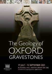
This incredible resource is elegantly celebrated in a new temporary exhibition in the Weston Library in Oxford. Compiled by two of the Museum’s Honorary Associates, Nina Morgan and Philip Powell, The Geology of Oxford Gravestones brings together the geological and human history of Oxford’s cemeteries.
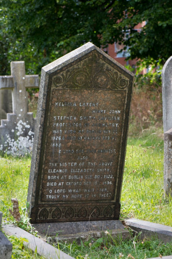
The gravestone for Henry John Stephen Smith, second keeper of the Museum, in St Sepulchre’s cemetery. It is a wonderful example of Shap Granite from quarries in Cumbria. Image: Mike Tomlinson.
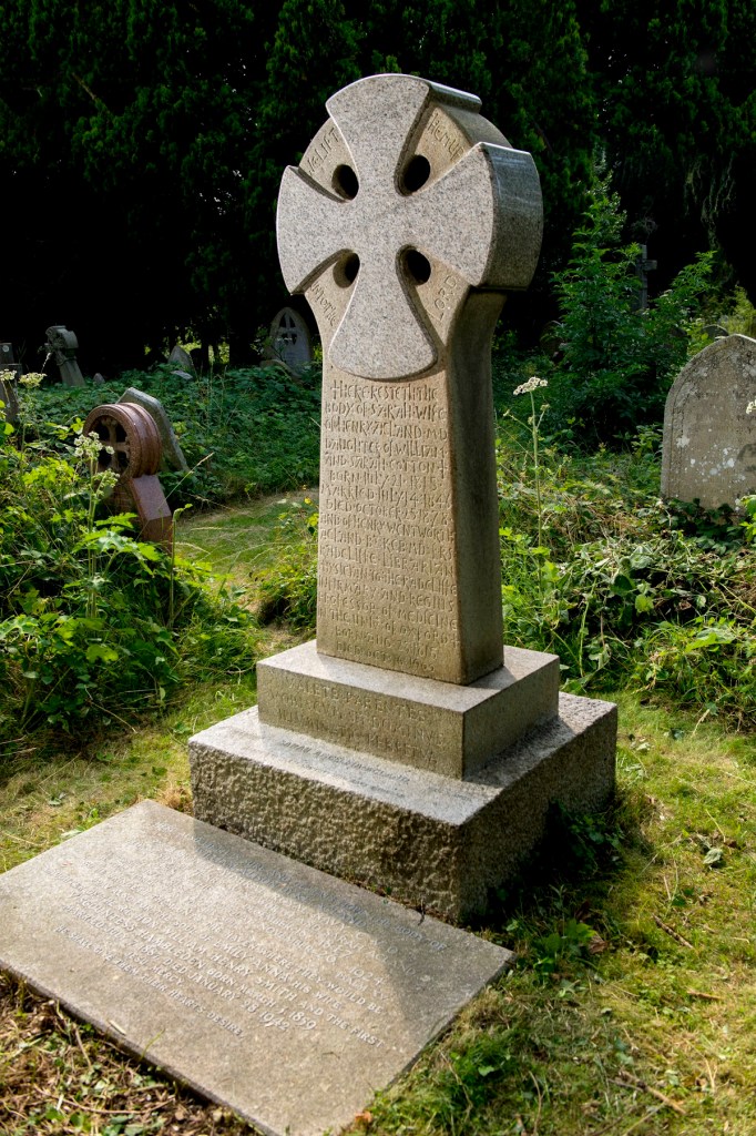
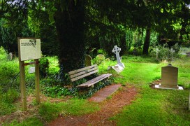
This imposing monument in Holywell cemetery, made of granite from Cornwall, marks the graves of the Acland family, including that of Henry Acland, one of the founders of Oxford University Museum of Natural History. Image: Mike Tomlinson.
The exhibition is illustrated with artefacts including undertakers’ trade cards and ‘rules of burial’, rock samples from the Museum’s collections, and photographs of headstones from Museum luminaries such as Henry John Stephen Smith, our second Keeper, and Henry Acland, one of our founders.
Although compact, the exhibition is full of fascinating snippets for fans of geology and social history alike, even bringing the science right up to date with a study using lichen on gravestones to understand our changing environment. The text and objects on display are enhanced by rolling digital displays that give more insight and colour to the story.
As the exhibition says, “visit a cemetery with a hand lens and you’ll be amazed at what you can see, you’ll never look at cemeteries in the same way again”. Just make sure you visit the display in the Weston Library first!
The Geology of Oxford Gravestones, is in the Blackwell Hall foyer of the Weston Library in Broad Street, Oxford and runs from 17 July to 12 September 2021. You can also find out more about Gravestone Geology here and in our previous post Celebrate science in a cemetery.
We like to think we know a lot about our collections, but with millions of items to care for some inevitably remain mysterious, with little record of their history. Luckily, every now and then someone gets in touch with a story about an object or specimen we know very little about. We were delighted when this happened recently for one of the most overlooked items on display: a delicate scale model of the Sun, Earth and Moon.
The model is a long-standing feature of the upper gallery: an astronomical moment hidden amongst the zoological and the geological. Yet we knew very little about it. Who made it, when was it installed, and what was its intention?
Meet the maker: Ted Bowen (1898-1980)
Edmund ‘Ted’ John Bowen. Image courtesy of Dr Will Bowen.
Thanks to a chance remark by Dr Will Bowen we can reveal that the model was created by his grandfather, Edmund ‘Ted’ John Bowen, lifelong fellow in Chemistry at University College. Ted Bowen was passionate about communicating science effectively, and the model was intended as a simple yet powerful representation of the true scale of our Solar System.
Born in Worcester in 1898, Ted Bowen won the Brackenbury Scholarship in 1915 to the University of Oxford, where he studied chemistry in the Balliol/Trinity labs. It was here that, from necessity, he started to create his own scientific apparatus and models, all made from whatever was to hand.
In 1935, Bowen was elected a Fellow of the Royal Society for his research into fluorescence and in 1963 was awarded the society’s Davy Medal in recognition of his distinguished work explaining photochemical reactions. While Bowen devoted his working life to the field of chemistry, he had many other scientific interests, especially palaeontology, but also our planetary system.
Creation of the Sun, Earth, Moon model
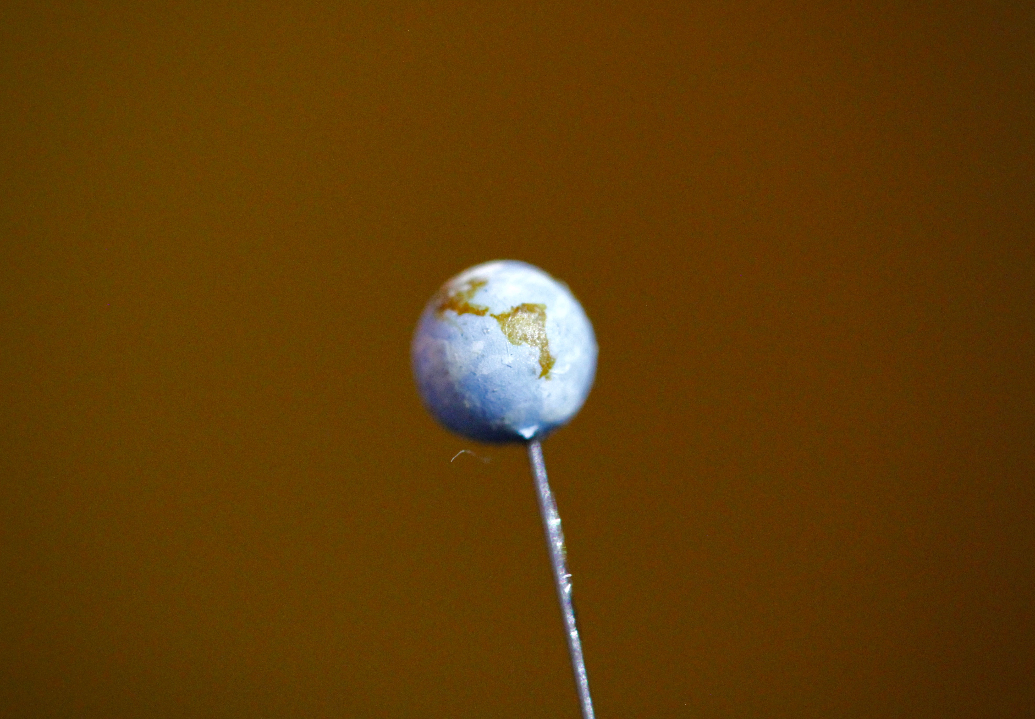
The Earth model is no larger than a pea, but still beautifully detailed.
Although we don’t know for sure, it is likely that the model was made between 1965 and 1971, and donated while Bowen was a member (and later chairman) of the Committee for the Scientific Collections in the University Museum, as the Museum was then known.
The distance across the Museum’s main court, around 37 metres, represents the distance between the Earth and the Sun – one Astronomical Unit, or 150 million kilometres. This makes the model scale to roughly 1:4,000,000,000!
The Sun itself is the size of a small beach ball, while the Earth and the Moon become tiny objects: the Earth the size of a small pea, and the Moon little more than a dot. Yet Bowen’s attention to detail is striking: the Earth is decorated with continents and even the miniscule Moon has texture to its surface.
If you haven’t seen it before, be sure to look out for the model on the upper gallery of the Museum: the Earth and Moon are on one side, where the Museum Café is currently located, and the Sun glistens on the far side, nestled in our temporary exhibition gallery.
Many thanks to Dr Will Bowen for his reminiscences, which have illuminated an object that was hidden in plain sight.
Around 120 years ago, William Sollas, Professor of Geology at the University of Oxford, developed a special technique for grinding down and imaging certain kinds of fossils. Sollas was based at the Museum at the time, and the process he pioneered is still used here today, as our PalaeobiologyTechnician Carolyn Lewis explains to mark the anniversary of Sollas’ birthday on 30 May.
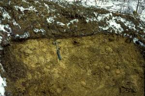
Site of the Herefordshire Lagerstätte, showing the nodules embedded in soft volcanic ash.
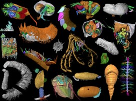
Here at the Museum, I work on a collection of exceptionally well-preserved fossils from the Silurian Herefordshire Lagerstätte. They were deposited on the seabed 430 million years ago when the animals were buried by a volcanic ash flow. The fossils range in size from less than a millimetre up to a few centimetres, and represent a diverse collection of marine invertebrates that includes sponges, echinoderms, brachiopods, worms, molluscs and a wide variety of arthropods.
These Herefordshire Lagerstätte fossils are unusual in that many of them have preserved soft tissues in remarkable detail, including eyes, legs, gill filaments, and even spines and antennae only a few microns in diameter. The key to this extraordinary preservation is that as the fossils developed, calcium carbonate nodules formed around them, protecting and preserving the fossils since the Silurian Period.
Usually, only the hard parts of fossil invertebrates are preserved – the carapace of trilobites or the shells of brachiopods, for example – so the Herefordshire material provides us with a great opportunity to work out the detailed anatomy of these early sea creatures.
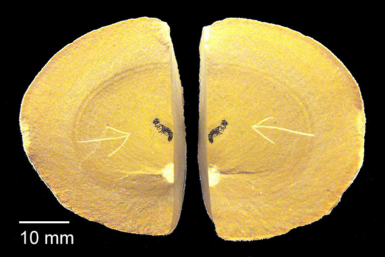
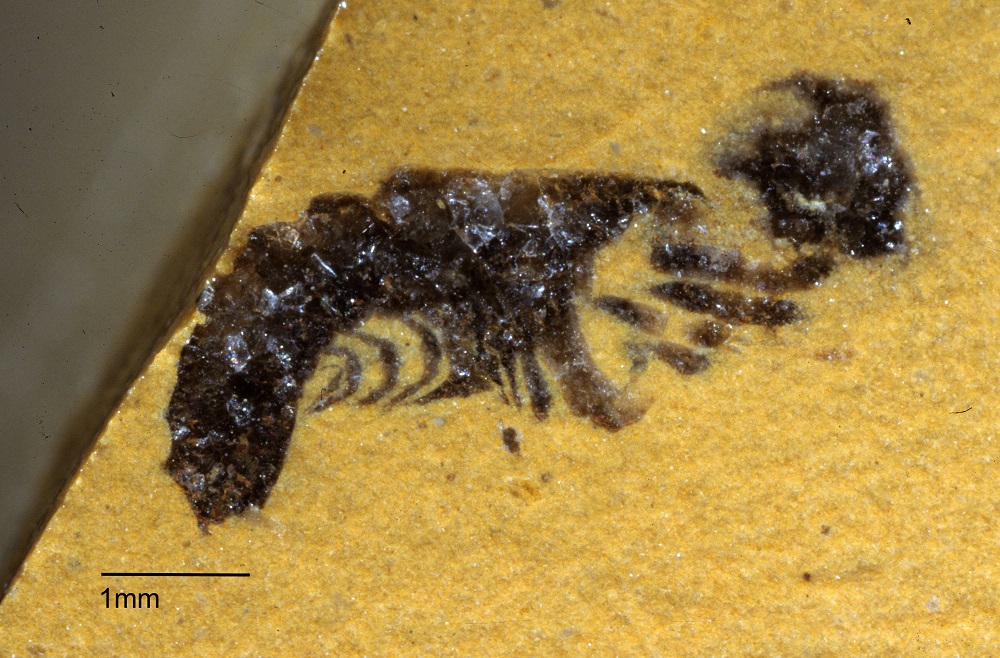
Split rock nodule showing fossil of Offacolus kingi inside.Close-up of the fossil of Offacolus kingi
But the problem we face is how to extract the specimen from the rock nodule without losing the information it contains. The fossils cannot be separated from the surrounding rock by dissolution, because both fossil and nodule are made mainly of calcium carbonate, so would dissolve together. And they are too delicate to be extracted mechanically by cutting and scraping away the surrounding nodule. Even high resolution CT scans cannot, at present, adequately distinguish between the fossils and the surrounding rock material.
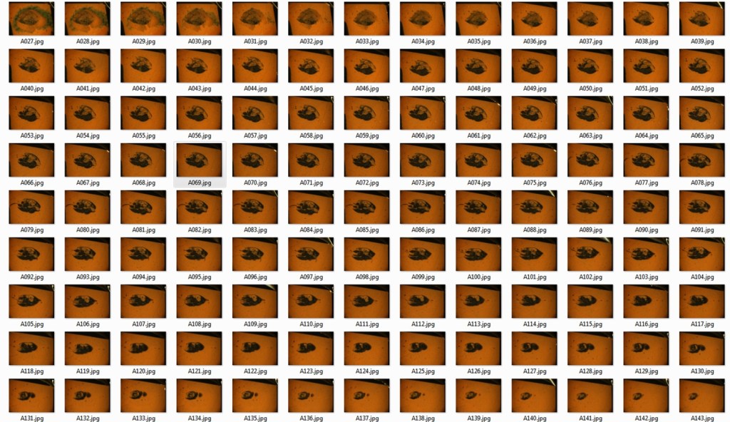
To get round this problem we use a method of serial grinding and photography based on the technique developed by William Sollas in the late 19th century. We grind the fossils in increments of 20 microns then photograph each newly ground surface using a camera mounted on top of a light microscope. This generates hundreds of digital images of cross sections through the specimen.
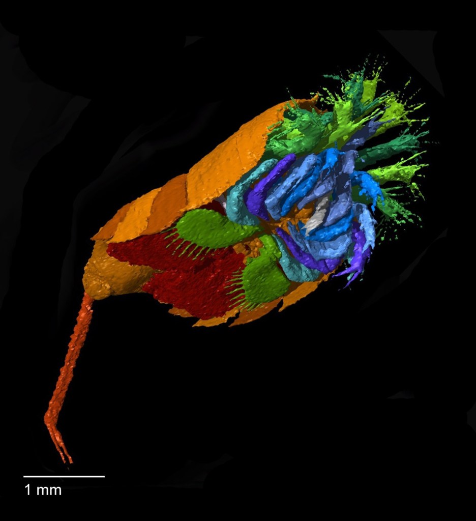
Then, using specially developed software we convert the stack of two-dimensional images into a 3D digital model that can be viewed and manipulated on screen to reveal the detailed form of the animal. These 3D models are artificially coloured to highlight different anatomical structures and can be rotated through 360o, virtually dissected on screen, and viewed stereoscopically or in anaglyph 3D.
3D virtual reconstruction of Offacolus kingi, a chelicerate arthropod related to horseshoe crabs.
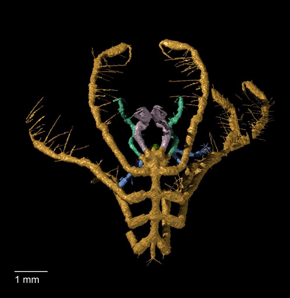
Haliestes dasos (sea spider)
Enalikter aphson (arthropod)
Although our method of serial grinding is still fairly labour intensive, it is far less laborious and time-consuming than the process used by William and his daughter Igerna Sollas. Compared to the photographic methods of the early 20th century, where each photographic plate required long exposure and development times, digital photography is almost instant, enabling us to grind several specimens simultaneously.
Sequential serial grinding images of an ostracod
Computer software also allows us to create 3D virtual models rather than building up physical models from layers of wax. Yet despite our modern adaptations, we are using essentially the same technique that William Sollas developed here at the Museum 120 years ago. And using this technique to study the fossils of the Silurian Herefordshire Lagerstätte has yielded a wealth of new information that opens up a unique window into the evolution and diversification of early life in our oceans.
With lockdown and the long winter nights shuffling the nation’s emotions like a ham-fisted magician with a damp deck of cards, we have no doubt all suffered from a case of the winter blues at some point recently. While the Museum and its inspiring specimens have been closed to visitors we have tried out some new approaches to bring you the solace and creative inspiration that nature can provide.
Events manager Laura hard at work on a lighting set-up for a Drawn to Nature live stream.
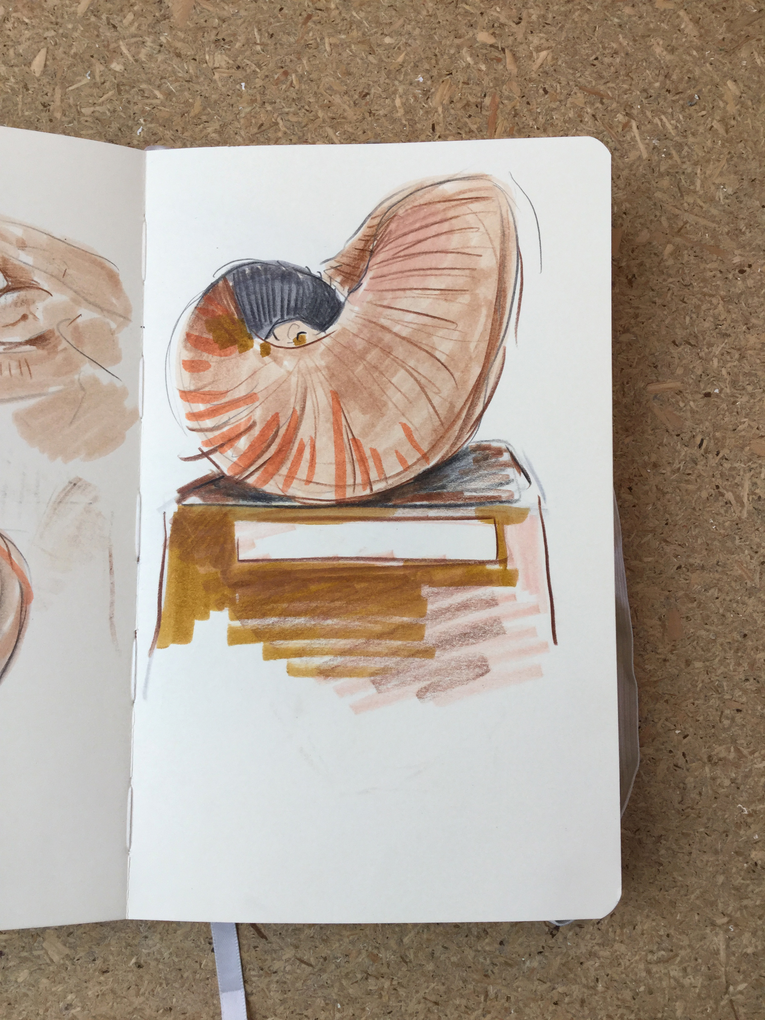
Drawn to Nature is a new series of online events designed to lift people’s spirits with a combined art and science activity. Originally planned as a wellbeing event to take place in the Museum, the online version was created in response to the last lockdown. We start each session with a short talk by a member of the Museum’s collections or research team, who share their passion for a selection of favourite specimens. The talk is followed by a chance for viewers to explore their creative sides by drawing the specimens, while learning more by about them through some Q&A.
It’s not an art lesson as such, but more a chance for people to find inspiration from some of the jewels of the natural world held in our collections. We hope it helps people to relax, find inspiration, immerse themselves in a creative activity, and learn a little natural history at the same time.
Click the gallery images to zoom and see credit information.
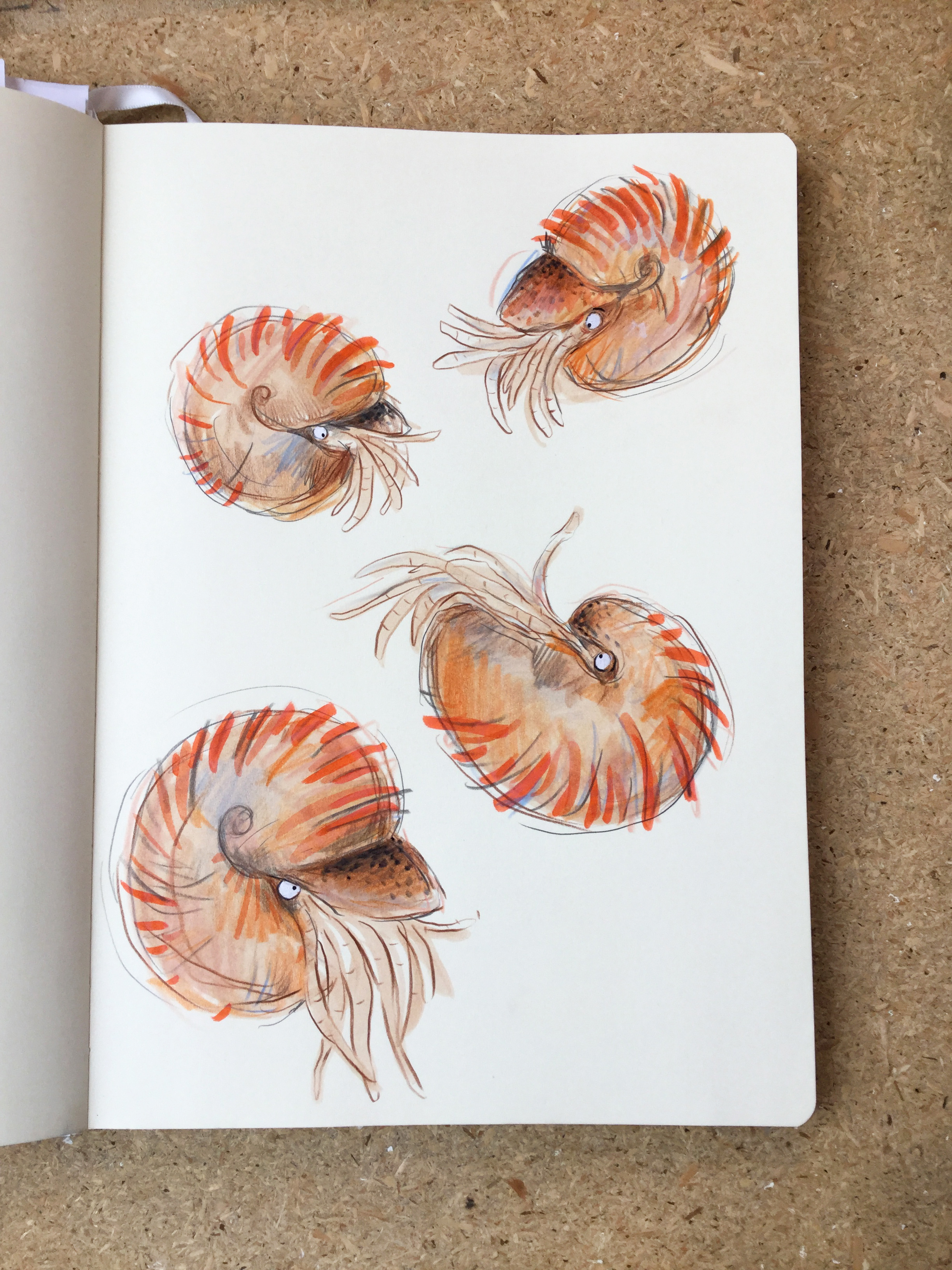
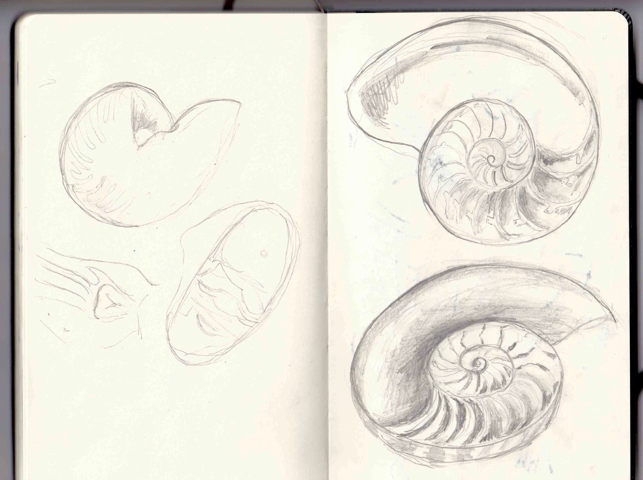
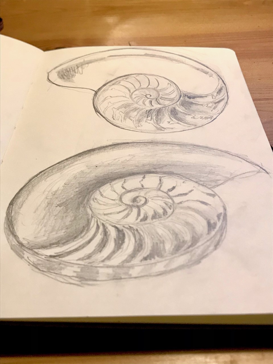
Our first session came from Life Collections manager Mark Carnall, who talked us through the natural history of Nautiloids, the fascinating shelled molluscs that are related to other cephalopods such as squid and octopus. Lit and arranged beautifully by our Events Manager Laura Ashby, their intricate chambered shells and 100 tentacles proved a challenging subject, but one that resulted in an array of wonderful artworks in a variety of styles and media shared on social media.
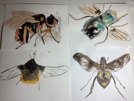
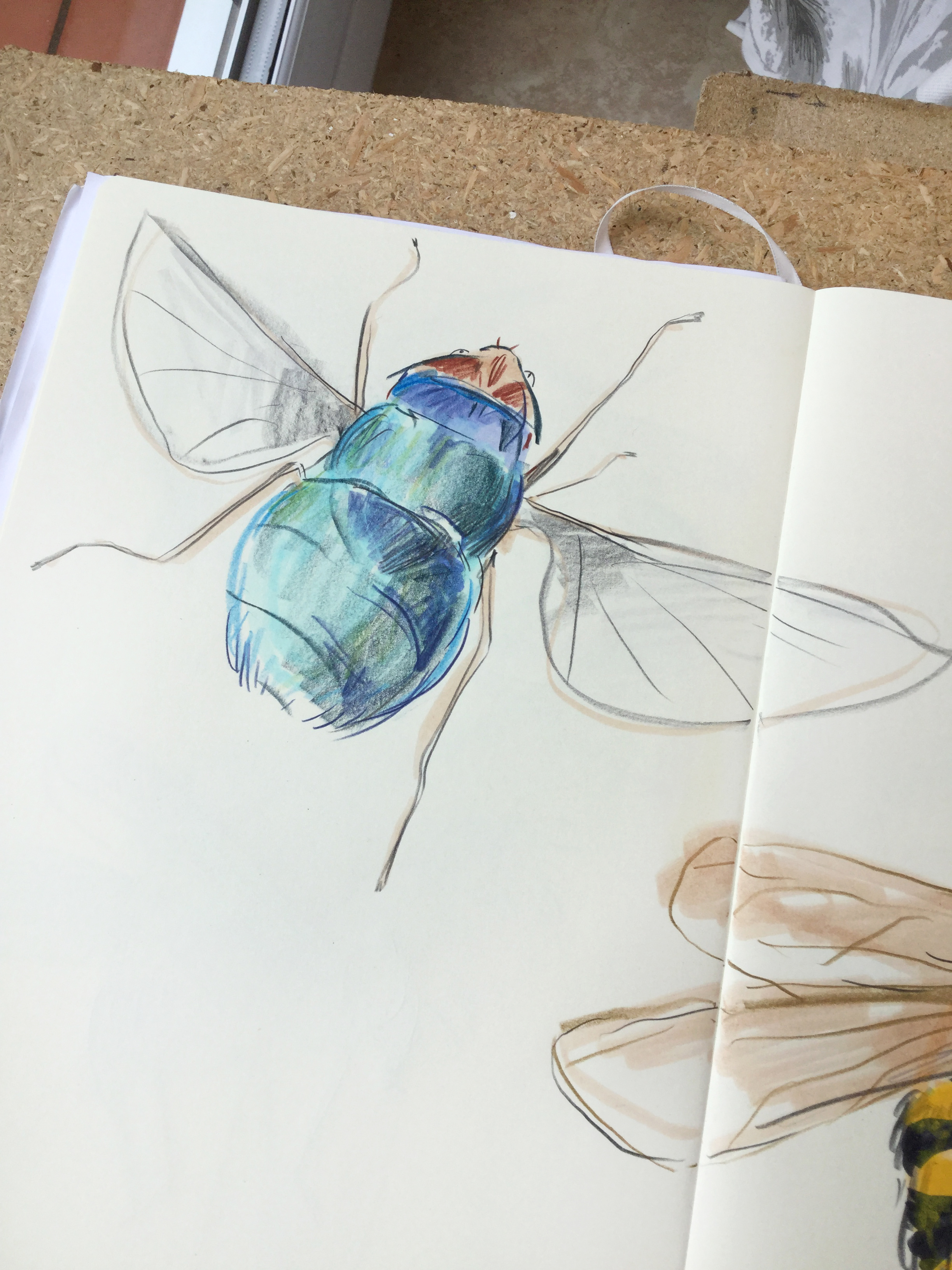
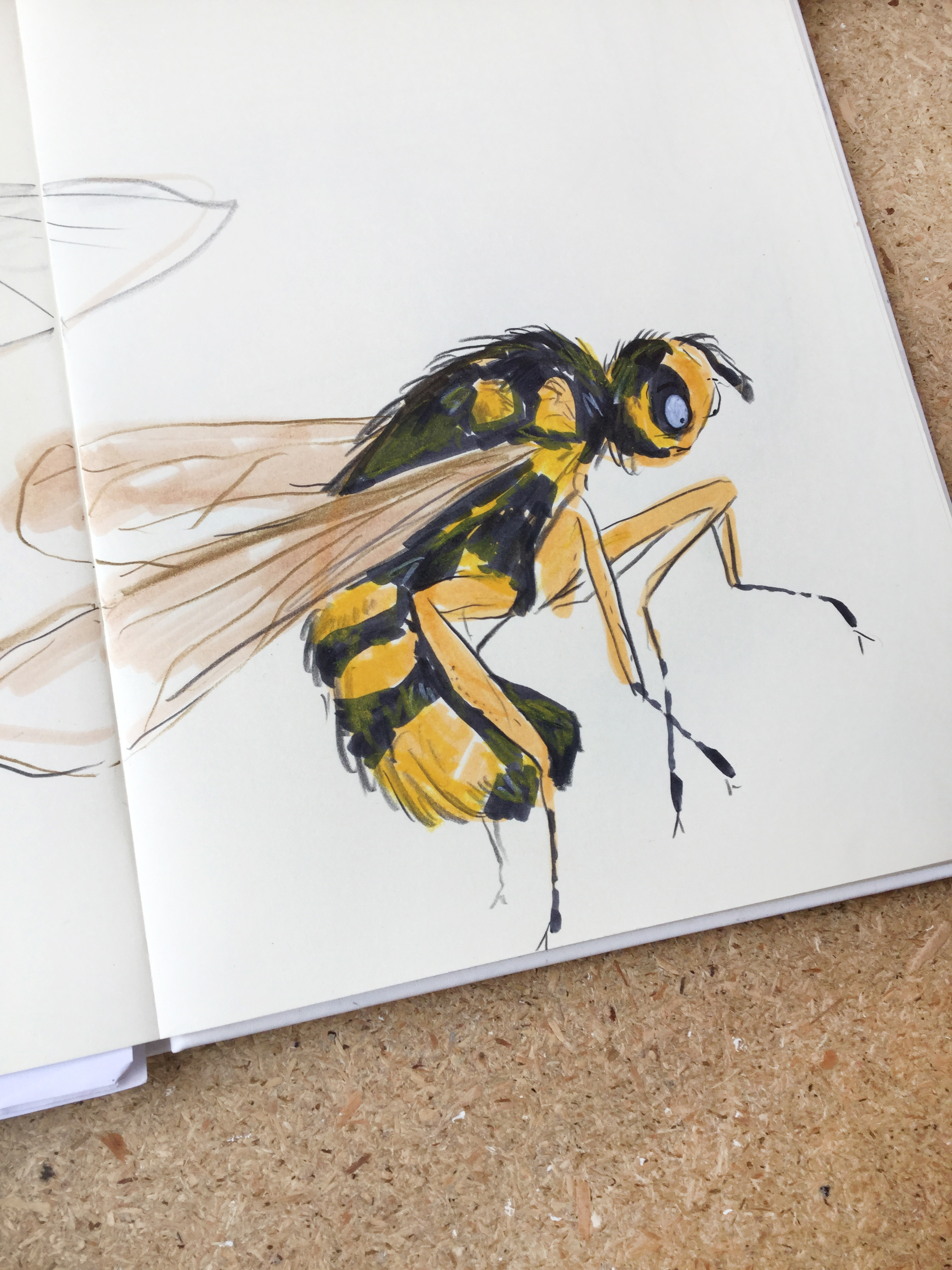
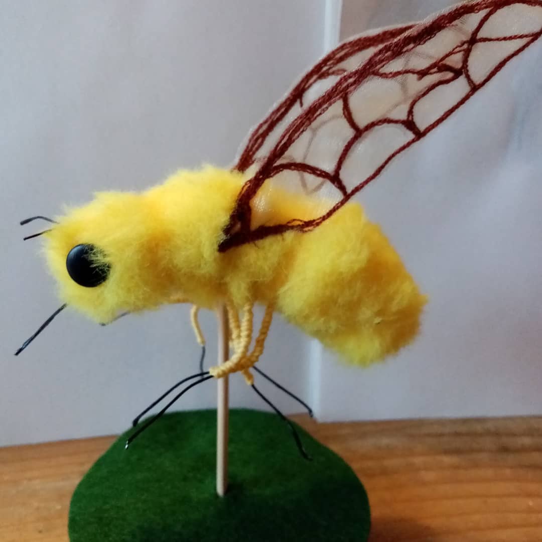
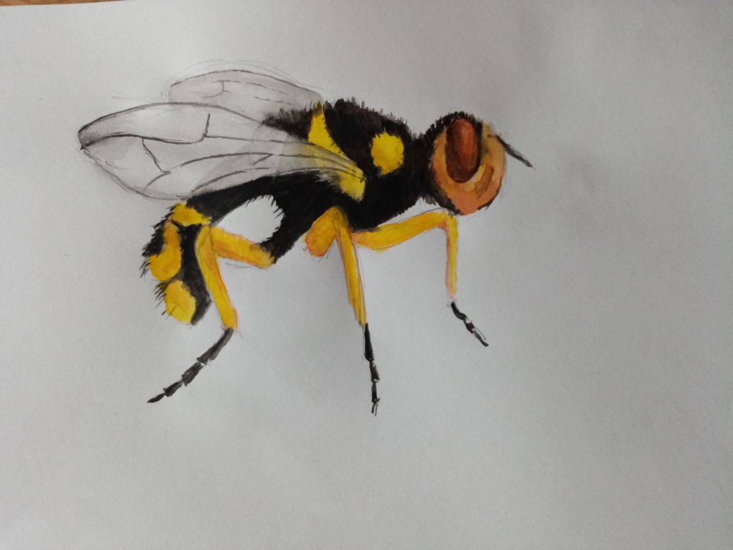
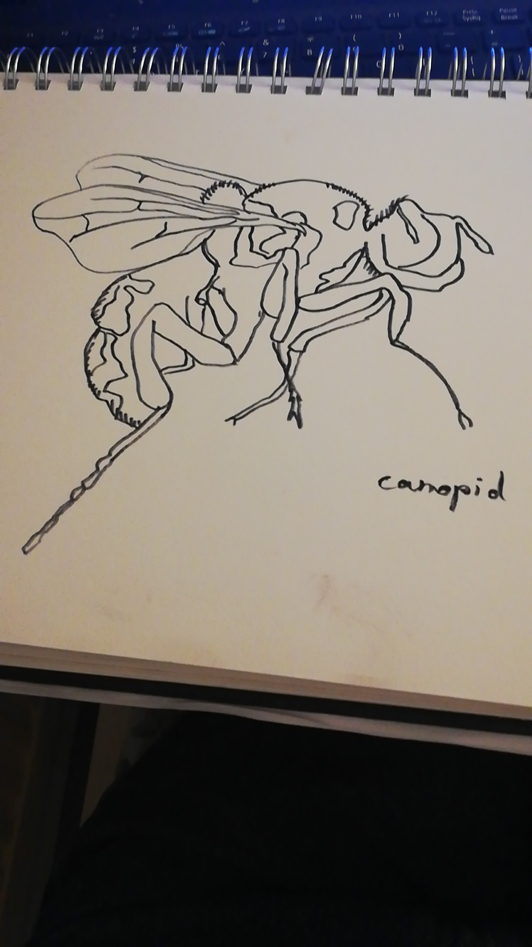
Following Mark’s talk, we explored some of the wonderful specimens from our entomology collections with our expert speaker Zoe Simmons, Head of Life Collections. Zoe picked out some of the most beautiful flies from our five million-plus insects. Using microscopes, specialist lighting, and careful placement, the specimens were a real hit and again followed by some inspired artworks posted online by attendees from across the world (‘best night of lockdown yet!’ enthused one attendee).
Click the gallery images to zoom and see credit information.
And there’s more to come. Tomorrow evening (Wednesday 10 March), Earth Collections Manager Dr Hilary Ketchum introduces the strange-looking carnivorous marine reptiles of the Jurassic – the plesiosaurs. We hope to bring you yet more of the inspirational well-being the natural world has to offer.
You can watch the Drawn to Nature streams online in the playlist on our YouTube channel:
This article is taken from European research magazine Horizon as part of our partnership to share natural environment science stories with readers of More than a Dodo.
In the summer of 2014 a strange building began to take shape just outside MoMA PS1, a contemporary art centre in New York City. It looked like someone had started building an igloo and then got carried away, so that the ice-white bricks rose into huge towers. It was a captivating sight, but the truly impressive thing about this building was not so much its looks but the fact that it had been grown.
The installation, called Hy-Fi, was designed and created by The Living, an architectural design studio in New York. Each of the 10,000 bricks had been made by packing agricultural waste and mycelium, the fungus that makes mushrooms, into a mould and letting them grow into a solid mass.
This mushroom monument gave architectural researcher Phil Ayres an idea. ‘It was impressive,’ said Ayres, who is based at the Centre for Information Technology and Architecture in Copenhagen, Denmark. But this project and others like it were using fungus as a component in buildings such as bricks without necessarily thinking about what new types of building we could make from fungi.
That’s why he and three colleagues have begun the FUNGAR project – to explore what kinds of new buildings we might construct out of mushrooms.
Mushrooms might sound like an outlandish building material. But there is certainly good reason to drastically rethink construction. Buildings and construction are responsible for 39% of anthropogenic carbon dioxide emissions – and a whopping 21% of those emissions come just from the making of steel and concrete. Construction also uses vast amounts of natural resources. Take sand, one of the principal ingredients in concrete. It takes a special sort, with just the right roughness, to make concrete. These days it is a lucrative commodity and controlled in some parts of the world by sand mafias and stolen by the boatload from islands.
Such troubles are set to worsen over the next decades as the world’s population grows faster and gets wealthier. We need a lot more homes and if you do the maths, the amount we need to build is staggering. ‘It’s like building a Manhattan every month for the next 40 years,’ said Ayres, borrowing a line from Bill Gates.
Fungi bricks
Can fungi really help? Absolutely, says mycologist Professor Han Wosten at Utrecht University in the Netherlands. Fungi are not consumers of CO2 like plants are. They need to digest food and so produce carbon dioxide, like animals do. However, the organic waste streams (such as straw or other low value agricultural waste) that the fungi digest would be degraded to CO2 anyway, either by composting or burning. Plus, fungi bricks permanently fix some of that waste inside them and so act as a store of carbon. All this makes fungi buildings a climate win – and certainly miles better than using concrete, steel and bricks.
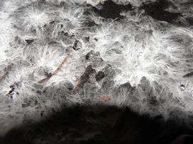
The mycelium composite can be grown over a woven scaffold for a period of 7-10 days, eventually encasing the structure. Image credit – FUNGAR/CITA, 2019-2020
The FUNGAR project began in late 2019 and so far Prof. Wosten has been experimenting with how to make building materials. At Prof. Wosten’s lab in Utrecht, the team have been combining mycelium, the ‘roots’ of fungi, with agricultural waste such as straw. Then they allow the fungi to grow for about two weeks, until the fungus has colonised the straw. This binds the straw together, producing a white-ish foam-like material. Then they heat-treat it to kill the organism. They can also process it, for example by applying coatings or by squashing it. ‘If we press it we can get a material like hardboard,’ said Prof. Wosten. By varying the type of fungi and agricultural waste, the growth conditions and the post-processing, Prof. Wosten says they are getting all sorts of candidate building materials with different mechanical properties.
‘It’s very early days to start saying your house will be made entirely of fungus,’ said Ayres. But parts of it already can be. Mogu, a company based near Milan in Italy, already produces and sells sound-dampening velvet-textured wall tiles and floor tiles based on mycelium foam. The company’s chief technology officer Antoni Gandia is another FUNGAR project partner. He said that Mogu is also developing mycelium-based insulation material for buildings.
Ayres is hoping that the FUNGAR project will go way beyond just using fungi-based products as components in existing building designs. He wants to think about what entirely new kinds of building might be made from fungi. Foremost in his mind is building with living fungus.
‘It’s very early days to start saying your house will be made entirely of fungus.’
Phil Ayres, Centre for Information Technology and Architecture, Copenhagen, Denmark
Living fungus
There are two principal advantages to this. First, living fungus might behave as a self-healing material, simply re-growing if it becomes damaged. Second, mycelium networks are capable of information processing. Electrical signals run through them and change over time in a manner almost akin to a brain. ‘We’ve discovered that fungal materials respond to tactile stimulation and illumination by changing their patterns of electrical activity,’ said Prof. Andrew Adamatzky at the University of the West of England in Bristol, UK, who is coordinating the project with Ayres.
The idea is that perhaps the very structure of a mushroom building might sense and respond to its environment independently. It might for instance sense when CO2 levels from the mycelium are building up and open the windows to release the gas, according to Gandia.
Building with living mycelium will be a big challenge. This is because the longer it grows, the more of the substrate material – the straw, or whatever waste – it decomposes. Since the straw gives the materials their structural integrity, allowing the fungi to grow for too long isn’t desirable. There may be ways around this though. Depriving the fungi of water puts it into a dormant state: alive but not growing. And so one of Ayres’ ideas is to construct walls with two layers of dead fungus enclosing a layer of living fungus inside. This set up would shut out water from the inner layer, keeping the fungus there dormant.
Mycolite panels are made by pouring the composite into a mould. Image credit – FUNGAR/CITA, 2019-2020
One of the few other people who have explored working with fungi in construction is Jonathan Dessi Olive at Kansas State University in the US. He says that working with living mycelium is a very interesting new idea because it offers the possibility of the building being able to heal itself. But for him the real attraction of what he calls ‘myco-materials’ is that they ‘give us a way of reshaping how we think about the permanence of architecture.
‘What if some – not all – of our buildings were meant to only last a couple of years and could thereafter be recycled into shelter, food, or energy?’ he said.
The next major goal for the FUNGAR project is to build a small, freestanding building. They plan to pull that off within a year and then spend time monitoring it as it ages. It is crucial, says Ayres, to be able to monitor the living structure and see how it changes. It isn’t yet clear exactly what sorts of structures might end up being made from fungi, but they will probably start small. ‘I wouldn’t be crossing a bridge made of fungi, would you?’ joked Prof. Wosten.
You might be wondering what happened to Hy-Fi, that igloo-like structure in New York. The answer points to one of the most beautiful things about mycelium buildings. No wrecking ball or slow decay for them. It was taken down and composted.
The research in this article was funded by the EU. If you liked this article, please consider sharing it on social media.
Cemeteries not only provide a peaceful place to commemorate the dead, and observe and enjoy nature; they are also wonderful repositories for the study of local history and art. But that’s not all. Cemeteries also offer an easy introduction to science that anyone can enjoy.
A visit to a cemetery presents a wonderful way to learn about geology and the other sciences, such as chemistry, physics and engineering, that underpin it. For geologists – whether amateur, student or professional – almost any urban cemetery provides a valuable opportunity to carry out scientific fieldwork at leisure, right on the doorstep, and at no cost.
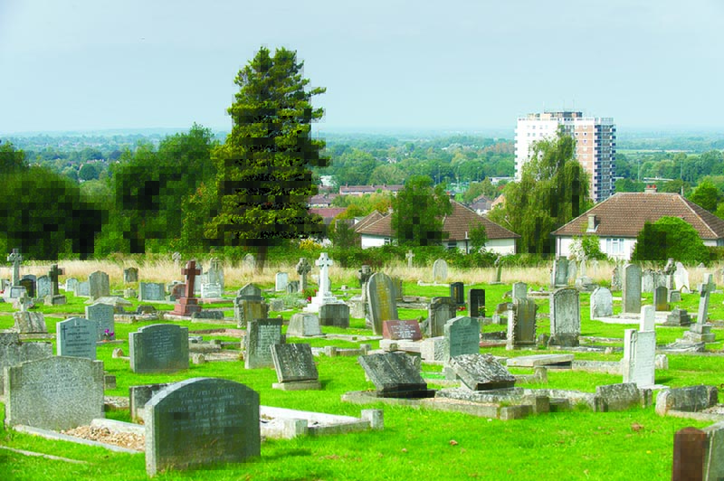
Headington Municipal Cemetery, Oxford
Geology on show
Because gravestones are made from a wide variety of rock types formed in a range of geological settings, cemeteries can be geological treasure-troves. Many headstones are made of polished stone, so reveal details – such as minerals and crystal features – that are not easy to see elsewhere. Some demonstrate the textures and mineral composition of igneous rocks – rocks formed when molten magma cooled and solidified. Others are happy hunting grounds for lovers of fossils. Some gravestones reveal sedimentary structures that show how the rock was originally deposited. Others provide clues to earth movements and environments that occurred hundreds of millions of years ago.
For those interested in engineering, examination of gravestones can also provide useful information about topics ranging from weathering of stone to atmospheric chemistry, effects of pollution, stability and settling in soils and land drainage.
St Andrews Church in Headington, Oxford
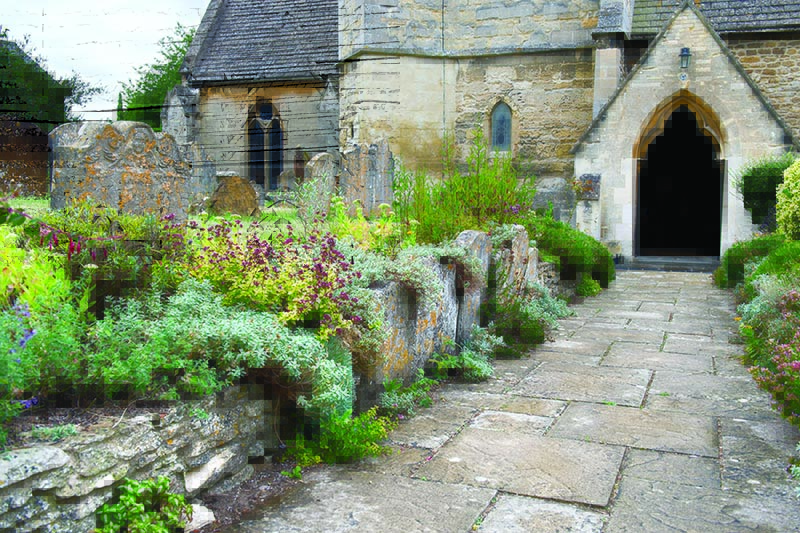
Cemeteries in Oxford include ancient churchyards, such as St Andrews Headington, as well as Victorian cemeteries like Holywell (pictured top) and St Sepulchres, and more modern burial grounds, such as Headington Municipal cemetery. Together they exhibit the main features and stone types that can be seen in cemeteries all around Britain.
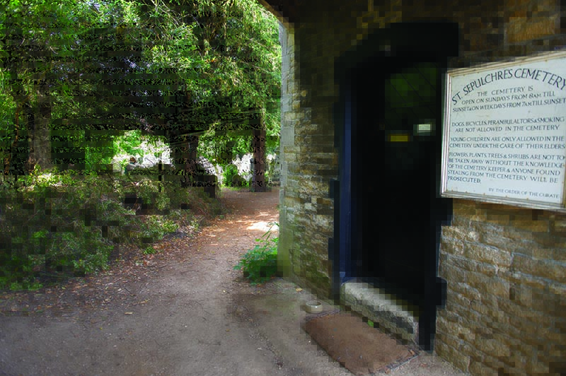
St Sepulchres Cemetery, Oxford
In the short video below, filmed in the churchyard of St Mary and John in Oxford’s Cowley Road, Philip Powell and I introduce the basics and show you how to get started in exploring these geological gems. If you want to learn more, visit www.gravestonegeology.uk. But be warned – gravestone geology can be addictive. Once you’ve got your eye in, you’ll never look at cemeteries in the same way again!