
Around 120 years ago, William Sollas, Professor of Geology at the University of Oxford, developed a special technique for grinding down and imaging certain kinds of fossils. Sollas was based at the Museum at the time, and the process he pioneered is still used here today, as our Palaeobiology Technician Carolyn Lewis explains to mark the anniversary of Sollas’ birthday on 30 May.
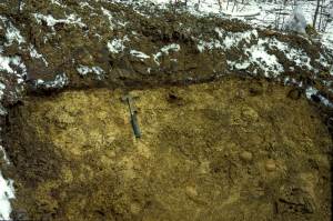
Here at the Museum, I work on a collection of exceptionally well-preserved fossils from the Silurian Herefordshire Lagerstätte. They were deposited on the seabed 430 million years ago when the animals were buried by a volcanic ash flow. The fossils range in size from less than a millimetre up to a few centimetres, and represent a diverse collection of marine invertebrates that includes sponges, echinoderms, brachiopods, worms, molluscs and a wide variety of arthropods.
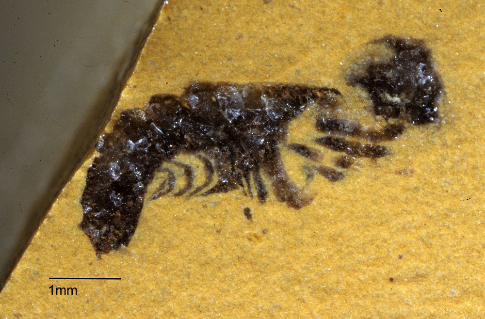
These Herefordshire Lagerstätte fossils are unusual in that many of them have preserved soft tissues in remarkable detail, including eyes, legs, gill filaments, and even spines and antennae only a few microns in diameter. The key to this extraordinary preservation is that as the fossils developed, calcium carbonate nodules formed around them, protecting and preserving the fossils since the Silurian Period.
Usually, only the hard parts of fossil invertebrates are preserved – the carapace of trilobites or the shells of brachiopods, for example – so the Herefordshire material provides us with a great opportunity to work out the detailed anatomy of these early sea creatures.
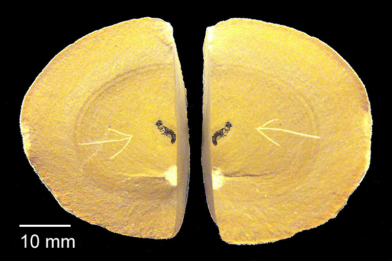
But the problem we face is how to extract the specimen from the rock nodule without losing the information it contains. The fossils cannot be separated from the surrounding rock by dissolution, because both fossil and nodule are made mainly of calcium carbonate, so would dissolve together. And they are too delicate to be extracted mechanically by cutting and scraping away the surrounding nodule. Even high resolution CT scans cannot, at present, adequately distinguish between the fossils and the surrounding rock material.
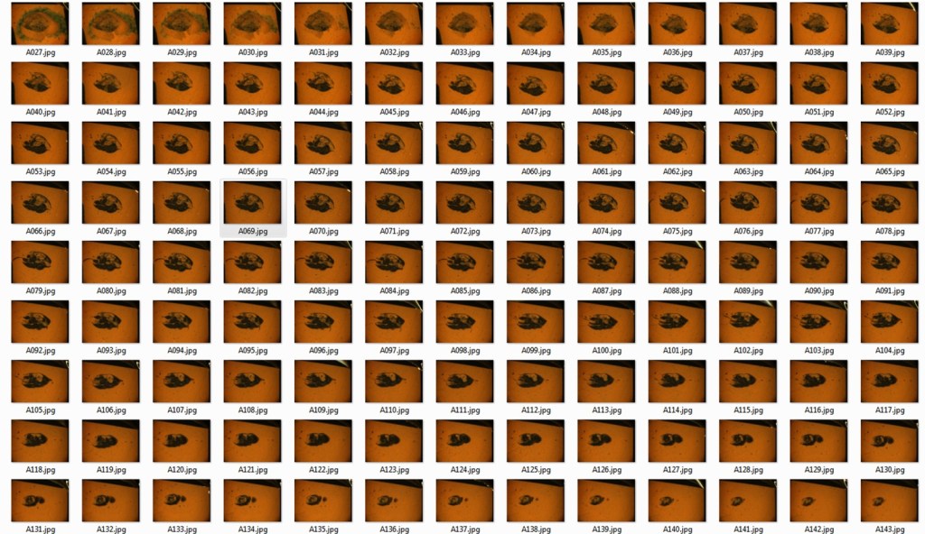
To get round this problem we use a method of serial grinding and photography based on the technique developed by William Sollas in the late 19th century. We grind the fossils in increments of 20 microns then photograph each newly ground surface using a camera mounted on top of a light microscope. This generates hundreds of digital images of cross sections through the specimen.
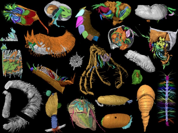
Then, using specially developed software we convert the stack of two-dimensional images into a 3D digital model that can be viewed and manipulated on screen to reveal the detailed form of the animal. These 3D models are artificially coloured to highlight different anatomical structures and can be rotated through 360o, virtually dissected on screen, and viewed stereoscopically or in anaglyph 3D.
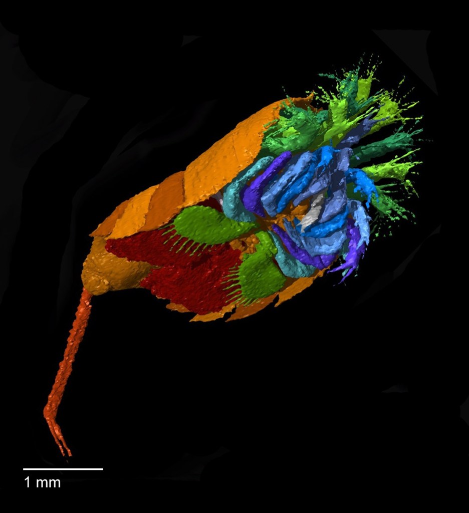
3D virtual reconstruction of Offacolus kingi, a chelicerate arthropod related to horseshoe crabs. 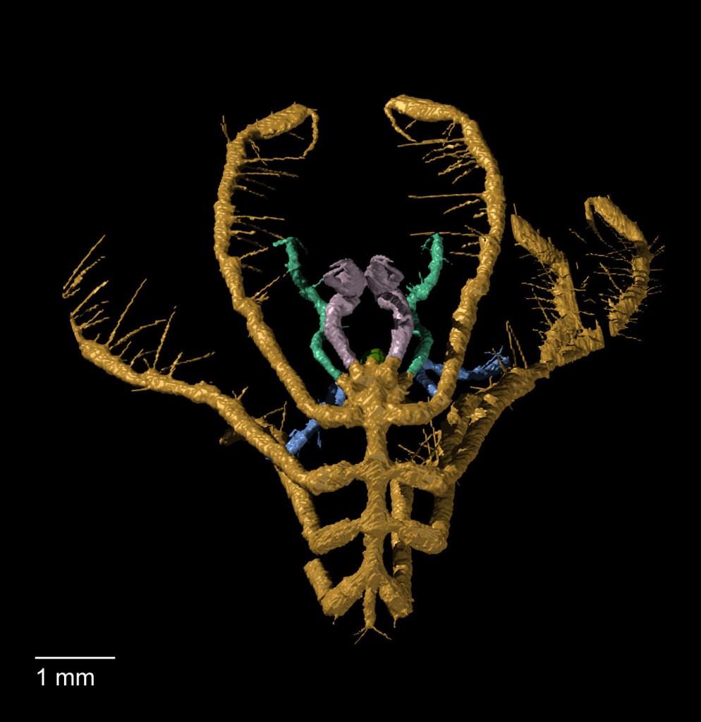
Haliestes dasos (sea spider) 
Enalikter aphson (arthropod)
Although our method of serial grinding is still fairly labour intensive, it is far less laborious and time-consuming than the process used by William and his daughter Igerna Sollas. Compared to the photographic methods of the early 20th century, where each photographic plate required long exposure and development times, digital photography is almost instant, enabling us to grind several specimens simultaneously.
Computer software also allows us to create 3D virtual models rather than building up physical models from layers of wax. Yet despite our modern adaptations, we are using essentially the same technique that William Sollas developed here at the Museum 120 years ago. And using this technique to study the fossils of the Silurian Herefordshire Lagerstätte has yielded a wealth of new information that opens up a unique window into the evolution and diversification of early life in our oceans.

Published by