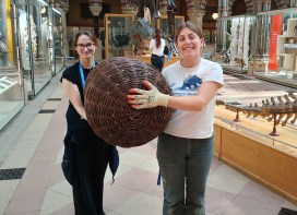
Bethany Milne from the Museum’s Visitor Experience team has spent some time working alongside the Exhibitions team to deinstall a temporary art exhibition. She has reflected on the day – what she enjoyed and what she learnt along the way.
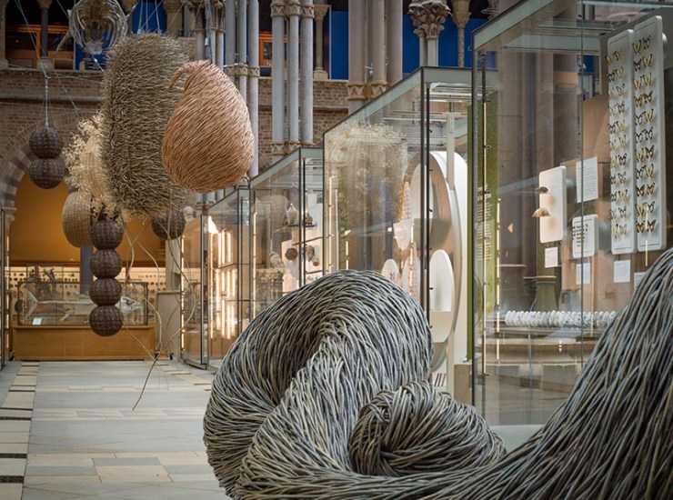
The deinstall of the Museum’s Deadly Six art installation is my first introduction into the world of exhibitions and an exciting one to start with. I have seen the display since it was first installed in September 2024 and I began working at the museum, watching it on my patrols of the court to make sure it stays intact and safe from all the little hands that pass through our doors every day. I didn’t imagine what it would be like to touch them myself until I found out I had the opportunity to join the team in taking them down.
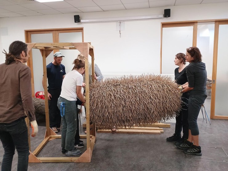
On September 8th I came into the museum at 8:30am to shadow the exhibitions team as a development day. Most of the staff and contractors were already there and so I started by talking to the Head of Exhibitions, Rachel, about what needed to be done. She showed me her schedule of the day, starting at 8am, going through to 5pm, in which we would derig all the sculptures and get them set up safely out of the public areas and into storage. After, I helped set up hoarding to keep the aisle where the work was being done secluded, as the museum would be open to the public as usual around us. This aspect was an interesting learning opportunity for me as I have no earlier experience with the manual labour aspect of the work, but I enjoyed being hands-on.
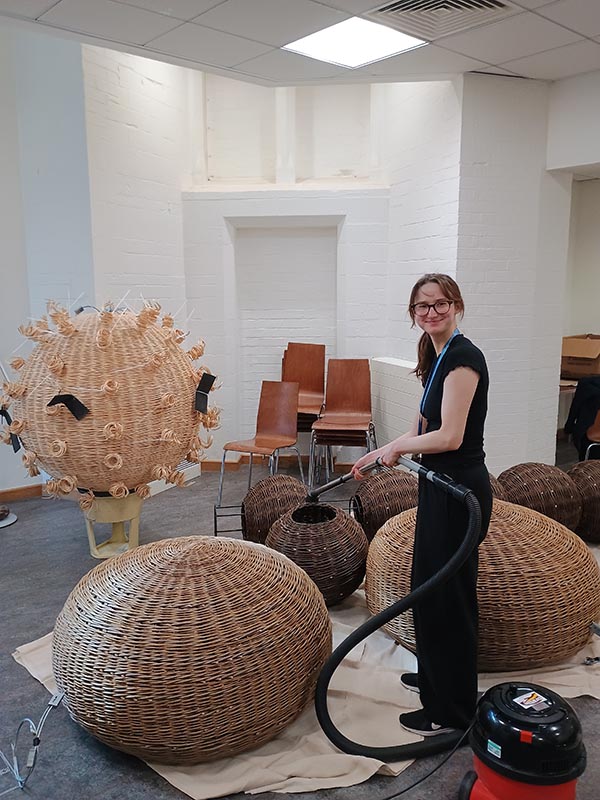
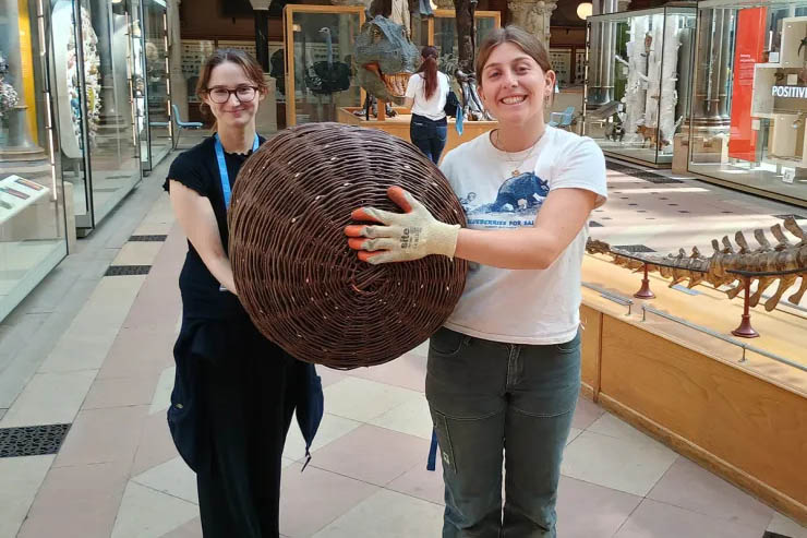
As I was doing this, the team from Outback Rigging worked on carefully lowering the suspended works to the ground and to our surprise; the take down was ahead of schedule. Most of them were removed by the middle of the day and these ones were simple to move. This was interesting to me as they seem so delicate, being large structures made of woven willow, but they proved sturdier than I thought. However, some were more difficult to take down than others, including the COVID sculpture. This was the most difficult to get down to the storage space, due to its intricate structure and its large size. Too wide to take down the stairs we usually use, Rachel organized with the Pitt Rivers to take it through their door, along the road and back into the Museum of Natural History through another door! What made this procedure more complicated was the sculpture itself, as its round construction meant there were no handholds. Luckily, the artist Issy Wilkes created some by looping some zip ties through the metal frame and, learning from the setup of the exhibition, used foam sheets to wrap around these painful to hold ties, to make the journey even easier. I felt it was an extremely rewarding process when we were finally able to put it down, as it required a lot of teamwork and shimmying about corridors to make it – I was very relieved we were able to keep it in one piece.
As the day went on and the sculptures were down and put away safely, I began to do smaller, but equally important tasks. The works themselves weren’t the only part of the exhibition, so I aided in removing the signage that was in the aisle that explained the meaning of each part. We also moved these down to the storage room, out of the way of the surrounding visitors and I began the task of removing the labels from inside. Another small step was vacuuming the sculptures. Despite regular conservation cleaning, hanging up in the court for so long had meant they had accumulated a lot of dust, which had to be cleaned out before they moved to their next home. These tasks were ones I wouldn‘t have thought of before the experience, but I realise that the smaller aspects are just as important as the larger ones when it comes to taking care of the exhibitions. It is our job to not only display the works as best we can, honouring their artistic intent and presenting to the audience in a way they understand, but this experience showed me how important the after care is, ensuring that they are well maintained to carry on their purpose and that the museum is returned to its original state.
LEARNING ABOUT ANCIENT FASHION FROM NATURAL HISTORY COLLECTIONS
By Ella McKelvey, Web Content and Communications Officer
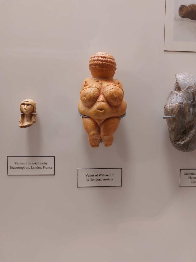
Tucked in a display case in the southwest corner of the Museum is a sculpture of an unidentified female figure, small enough to fit in your coat pocket. It is a replica of one of the most important examples of Palaeolithic artwork ever discovered; a 25,000-year-old carving known as the Venus of Willendorf. The Venus of Willendorf is one of several Palaeolithic statues found in Europe or Asia believed to depict female deities or fertility icons. Known collectively as the Venus Figurines, the carvings are similar in size and subject matter, but each has her own peculiarities. Many are naked, but some of the later examples are wearing distinctive garments, clothes we might describe today as ‘snoods’ or ‘bandeaux’. The Venus of Willendorf is easily distinguished by her statement headpiece; perhaps a spiralling hair-braid or ceremonial wig. But there is another, more exciting interpretation — this strange, thimble-like adornment might actually represent a woven fibre cap, making it the oldest ever depiction of human clothing.
The ‘Venus of Willendorf’ is known for the distinctive markings on her head. Are these the oldest representation of human clothing ever discovered?A cast of the ‘Venus of Willendorf’ is on display in the ‘Ancient Toolmakers’ case in OUMNH.
The Venus Figurines are incredibly important to the study of human fashion because they significantly predate any direct archaeological evidence of ancient clothing. The oldest surviving garment dates back an astonishing 5,000 years; an exceptionally-preserved linen shirt discovered in an Egyptian tomb. But our species, Homo sapiens, has a much longer history, perhaps up to a quarter of a million years. How much of this time have we spent wearing clothing? And why did we even begin to dress ourselves in the first place?
By comparing human genes to those of our furrier primate relatives, researchers have been able to estimate that modern humans lost their body hair around 240,000 years ago. A mutation in a gene called KRTHAP1 likely led to a decrease in our production of the protein keratin, the building block of hair. The exact reason why this mutation spread through the population is still up for speculation. One commonly held theoryis that, with less body hair, our ancestors could sweat and tolerate higher temperatures, allowing them to expand their habitats from sheltered forests into sun-drenched savannahs. But at some stage, our ancestors started covering their skin again — leaving us to wonder when nakedness became a nuisance.
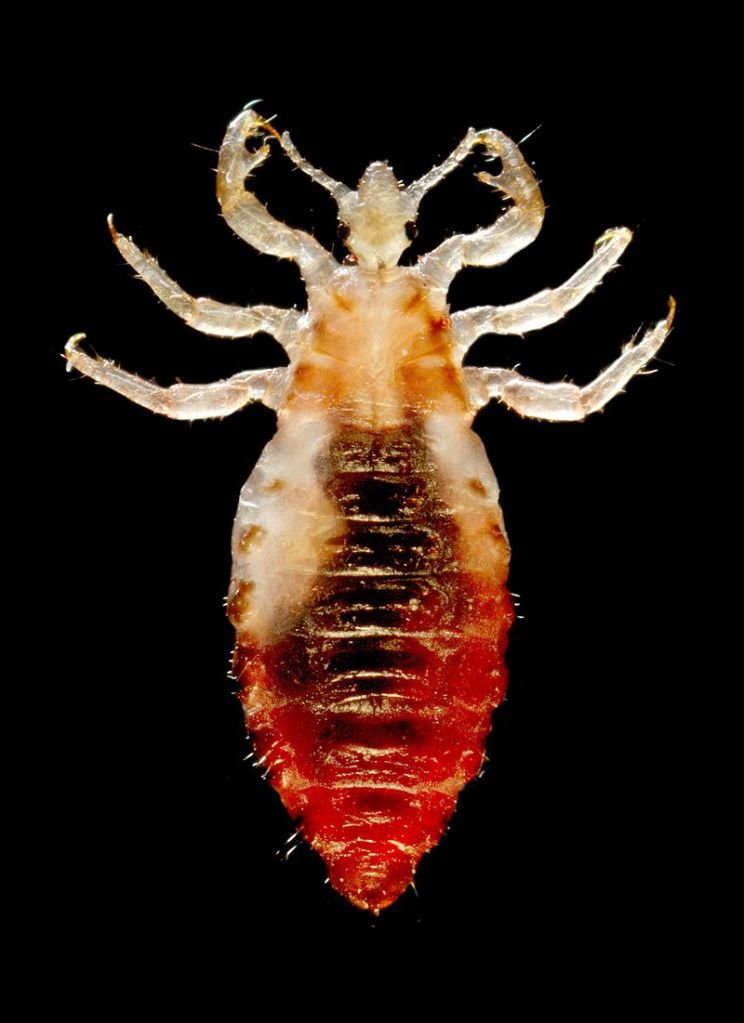
An intriguing clue about the circumstances that led to the adoption of clothing has come from studying the DNA of our parasites — namely, clothing lice. In 2010, researchers used genetic sequencing to determine that clothing lice split from their ancestral group, head lice, between 170,000 and 83,000 years ago. When compared with genetic data from our own species, we can begin to weave a story about the origins of clothing that ties in with human migration. Gene sequencing has helped us work out that Homo sapiens originated in Africa but must have begun migrating towards Europe between 100,000 and 50,000 years ago, a window which overlaps neatly with the evolution of clothing lice. Is it possible that clothing lice are a consequence of the widespread adoption of clothing; a result of humans migrating into more northerly latitudes with cooler temperatures?
Sharp-eyed visitors can spot body lice on display on the First Floor of the Museum.Studying the divergence of clothing lice and body lice allows us to estimate that humans have been wearing clothes for 170,000 years.
Curiously, there are indications in the archaeological record that human clothing could date to an even earlier stage in our species’ history than the expansion of humans into Europe. In 2021, researchers uncovered 120,000-year-old bones from a cave in Morocco believed to be used to process animal hides. There is a strong possibility that humans would have used these tools to make wearable items out of hunted animals, including blankets, cloaks, or perhaps more structured garments.
It seems likely that the first clothes humans made from hides were loose-fitting capes or shawls, which may have been more important for protection or camouflage than keeping warm. There are numerous reasons why other animals cover themselves with foreign objects besides thermoregulation. ‘Decorating’ behaviours occur in animals as diverse as crabs, birds, and insects, allowing them to disguise themselves from predators, or protect themselves from UV radiation. While early humans might have only needed simple clothing items to aid with disguise, as the climate began cooling 110,000 years ago, cloaks probably wouldn’t have cut it; our species must have learned how to make multi-layered and closer-fitting garments to maintain high enough body temperatures. Archaeology provides a similar estimate for the adoption of constructed garments, based on the discovery of 75,000-year-old stone awls — tools used for puncturing holes in hides to prepare them to be sewn together.
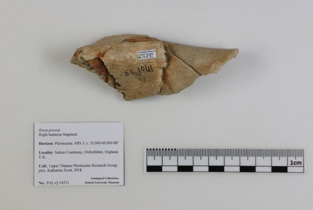
Homo sapiens‘ ability to make complex clothing items may have helped give our ancestors a competitive edge over the Neanderthals in Europe. Researchers have studied sub-fossil material in museum collections to learn about the changing distributions of European mammals throughout human history, allowing them to deduce that Neanderthals only had access to large animals like bison to make cape-like clothing from. But, in addition to bison, Homo sapiens lived alongside other, fluffier animals like wolverines during the last Ice Age, which could have been hunted to make warm trims for our clothing. Studies like these are highly speculative, but with such a threadbare archaeological record, they contribute valuable insight into the landscapes of ancient Europe.
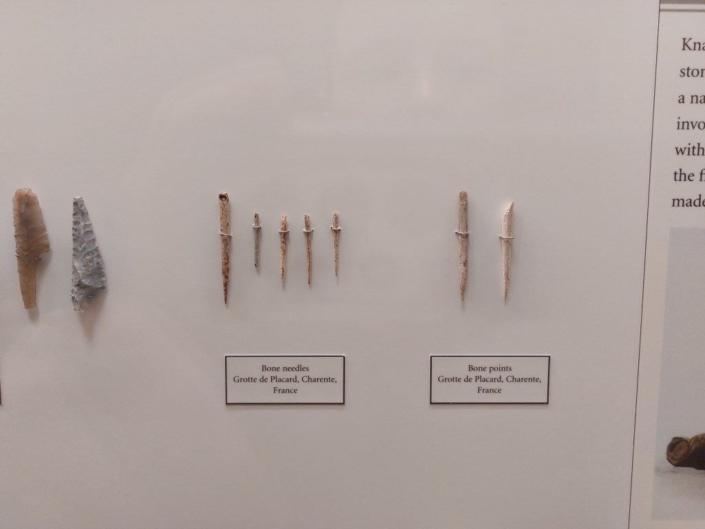
Museum collections can teach us about the species that lived alongside humans in ancient Europe. Homo sapiens and Homo neanderthalis might have used the hides of species like bison to make clothes.On display in the Ancient Toolmakers case are bone needles from the Placard Cave, around 17,000 years old. But huans may have been sewing clothes for much longer, perhaps 75,000 years.
The Neanderthals might have been less well-dressed than our Homo sapiens ancestors, but we can’t be certain that humans of our own species were the only prehistoric fashionistas. The oldest sewing needle to have ever been discovered dates to 50,000 years before present and was actually found in a cave associated with Denisovans — a group of extinct hominins we know little about. The Denisovans may be an extinct subspecies of Homo sapiens, but they might also have formed an entirely separate species altogether, perhaps learning how to sew independently of modern humans.
Following the invention of sewing was another crucial innovation in the history of human clothing — the ability to make textiles. In 2009, a group of researchers discovered 36,000-year-old evidence of textile-based clothing in the form of microscopic flax plant fibres that had been dyed and twisted together. There are many potential uses of twisted fibres such as these, but scientists have been able to study the organisms associated with the fibres, finding the remains of skin beetles, moth larvae, and fungal spores that are all commonly associated with modern clothing. Humans do not simply fashion clothes, we also fashion microhabitats, capable of supporting organisms as diverse as insects, fungi, and bacteria.
The discovery that humans have been making textiles into clothing for 36,000 years lends credence to the theory that the Venus of Willendorf is wearing a woven cap — but we might never be able to draw any certain conclusions about such an ancient artefact. Until just ninety years ago, humans could only make textiles from biodegradable materials, meaning that we have very little evidence about the clothing that our ancient ancestors wore. Thankfully, however, the story of human fashion is closely interwoven with the natural histories of hundreds of other species, allowing us to stitch together a patchwork history, utilising evidence from all corners of the kingdom of life.
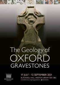
A cemetery may seem like an unusual location for a geology fieldtrip, but for rock hounds from beginner to professor there’s a treasure trove of different rock types in gravestones. Whether it’s shells of oysters from the time of the dinosaurs, or beautiful feldspar crystals formed deep within the Earth’s crust, rocks are uniquely placed to tell the story of the history of our planet.
This incredible resource is elegantly celebrated in a new temporary exhibition in the Weston Library in Oxford. Compiled by two of the Museum’s Honorary Associates, Nina Morgan and Philip Powell, The Geology of Oxford Gravestones brings together the geological and human history of Oxford’s cemeteries.
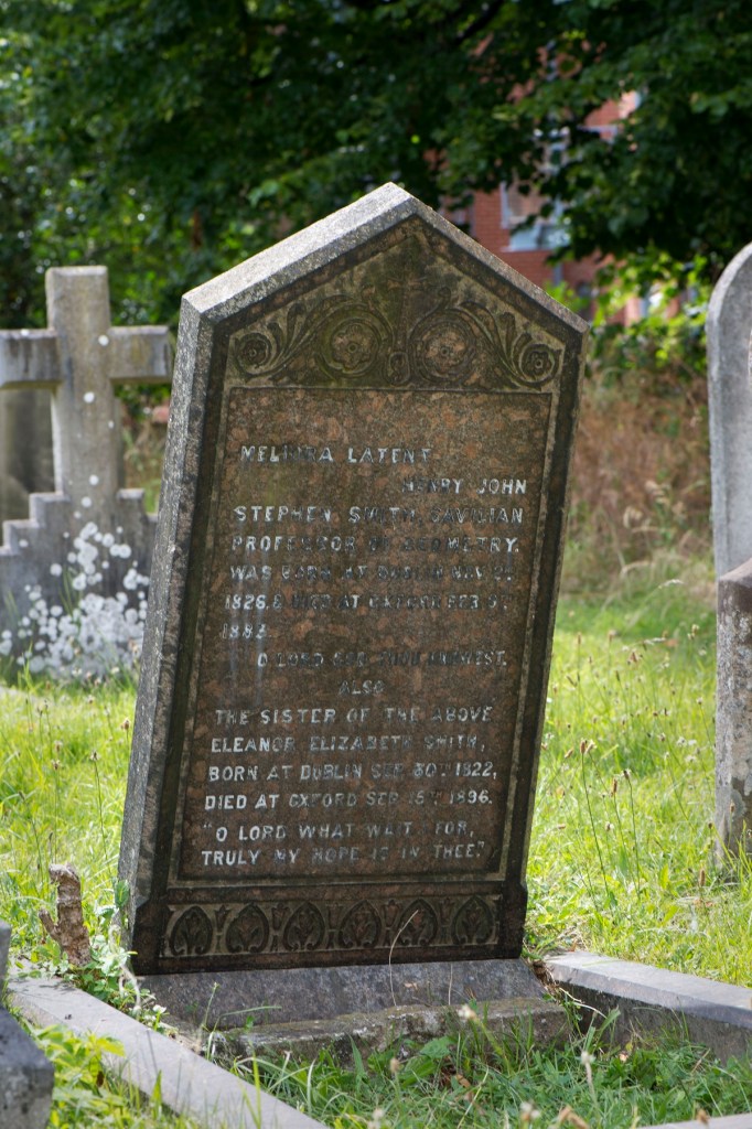
The gravestone for Henry John Stephen Smith, second keeper of the Museum, in St Sepulchre’s cemetery. It is a wonderful example of Shap Granite from quarries in Cumbria. Image: Mike Tomlinson.
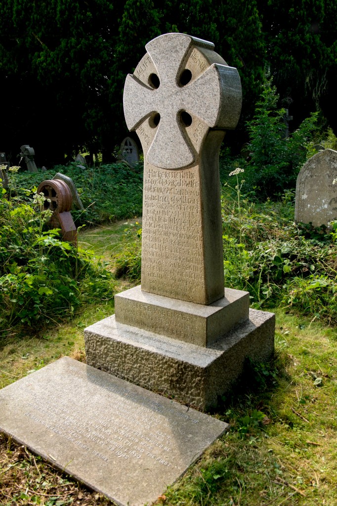
This imposing monument in Holywell cemetery, made of granite from Cornwall, marks the graves of the Acland family, including that of Henry Acland, one of the founders of Oxford University Museum of Natural History. Image: Mike Tomlinson.
The exhibition is illustrated with artefacts including undertakers’ trade cards and ‘rules of burial’, rock samples from the Museum’s collections, and photographs of headstones from Museum luminaries such as Henry John Stephen Smith, our second Keeper, and Henry Acland, one of our founders.
Although compact, the exhibition is full of fascinating snippets for fans of geology and social history alike, even bringing the science right up to date with a study using lichen on gravestones to understand our changing environment. The text and objects on display are enhanced by rolling digital displays that give more insight and colour to the story.
As the exhibition says, “visit a cemetery with a hand lens and you’ll be amazed at what you can see, you’ll never look at cemeteries in the same way again”. Just make sure you visit the display in the Weston Library first!
The Geology of Oxford Gravestones, is in the Blackwell Hall foyer of the Weston Library in Broad Street, Oxford and runs from 17 July to 12 September 2021. You can also find out more about Gravestone Geology here and in our previous post Celebrate science in a cemetery.
It goes without saying that 2020 has been a very unusual and troubled year, but it is also the 160th anniversary of the founding of the Museum, so we wanted to snatch a little breather from the difficulties of the pandemic, if possible, to take a positive look at the past and future of the Museum.
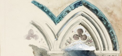
We have made a few special productions to mark this. Our new temporary exhibition – Truth to Nature – opens in the centre court on 18 October, and is accompanied by this online version for those who can’t make it to the Museum. The displays chart the philosophies and artistry underpinning the creation of the Museum in the mid-19th century and reflect on the role of natural history museums today, including the need for greater equity in science.
Taking a look at the unique and treasured building itself, this short film reveals some of the hidden secrets of the Museum’s architecture:
And finally, this week we have released a new five-part video podcast series looking in greater detail at the history of the Museum’s art and architecture, written and presented by John Holmes, Professor of Victorian Literature and Culture at Birmingham University, who is also an Honorary Associate of the Museum.
We’ll be sharing an episode a week here and on our social media channels, but you can dive into the series here or watch Episode 1, Oxford’s Pre-Raphaelite Natural History Museum, below.
Worms, fish and … Greenland? Hugely different topics which all have one thing in common – the Museum’s First Animals exhibition online lecture series. Running every other Wednesday from May until September 2020, this series provided a fantastic insight into a wide range of topics about how the first animals lived, died, and are studied. And illustrator Rachel Simpson tells us how she drew her way through them all…
I came across this lecture series just before the first talk and I knew I had to sign up. Drawing along to lectures is a hobby I seem to have developed in the past few months as we went into lockdown and didn’t have much to do. It’s the perfect combination for me – an opportunity to listen to interesting topics and brush up on my live drawing skills at the same time. There’s no pause button, there’s no asking the webinar speaker to just go back a few slides and hold on a minute whilst I draw; it’s fast paced, it’s inspiring and it’s a great way to just create art.
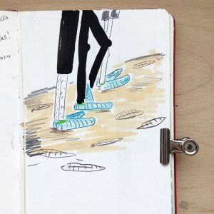
Barma Booties used on the rocks at Mistaken Point, and my first drawing of the series.
I’ve done some illustration work with the Museum before so I knew that it was going to be fun. In 2018, I worked with Dr Jack Matthews illustrating Ediacaran Fossils as part of a collaborative university project between the University of Plymouth and the Museum. I was also lucky enough to be able to go to Newfoundland and see some of the fossils myself, again with Jack. This was such an incredible opportunity and opened up a whole new world of science/art collaborative work which I didn’t know about before.
The First Animals series kicked off with Jack’s talk titled Don’t walk on the rocks! – an interesting insight into how protective “Barma Booties” (some rather funky socks worn to protect fossil sites such as Mistaken Point, Newfoundland) might actually be damaging to the fossils they’re meant to be protecting. Having been to Mistaken Point myself and worn these socks, it was interesting to hear about their possible impact and to learn about the experiments conducted to prove this fact.
Of course, at the same time as Jack was talking, I was scribbling away in my sketchbook trying to form some sort of visual response to the talk. At the end of the hour I’d managed a portrait of Jack and a family of Barma-Booted tourists trampling on the fossil site. It was a start. The beginning of my lecture drawings and a point at which I can retrospectively say started a new hobby.
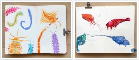
Annelid worms drawn with Tombow brush pens.
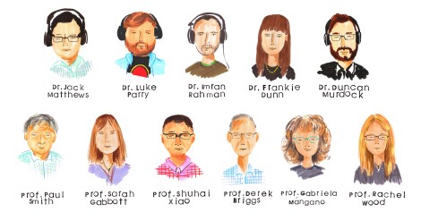
Over the following weeks we heard about worms from Dr Luke Parry; 3D reconstruction from Dr Imran Rahman; The Chronicles of Charnia by Dr Frankie Dunn; and the first animal skeletons from Dr Duncan Murdock. Luckily for me, all the speakers kindly included photos and descriptions of the topics they were discussing which meant that I was never short of visual inspiration for my drawings. After all, it’s hard to try and draw an annelid worm if you’ve never seen one before.
I love to look at the fossils being discussed and then try to draw a little character or creature inspired by them. They’re not scientifically accurate, nor are they always anatomically correct, but they have character and begin to bring to life the essence of something that’s been dead for many millennia. The fossils are obviously stone-coloured so I take as many liberties as possible when it comes to colour. I like to make them as vibrant and colourful as I can, so although they probably didn’t look like that, that’s how I like to think they looked.
Some fun little beasties from Dr. Imran Rahman’s talk.
Charnias galore! They come in all different shapes and sizes.
Small filaments which could have joined all those Charnia together.
Shells, bones and teeth from Dr. Duncan Murdock’s talk drawn in Tombow brush pen and Posca Pen.
Within my wider practice I like to use stamps as the basis of my illustrations. These however, are time consuming to make and therefore not very suitable for when I’m drawing along to lectures. As a result I’ve found myself using brush pens and pencils to make my lecture illustrations. If you’re interested in art, or thinking about getting into art, brush pens will be your best purchase. They create a wonderful quality of line and are quick and easy to use. Whereas a ballpoint pen will give you one line of a certain weight and thickness, brush pens are versatile and depending on the pressure applied, the line quality will change.
For the first few lectures I only used brush pens, but later on I decided to use coloured pencils as well, to add depth to the drawings. As I got more used to drawing in lectures I found that I was making more illustrations per talk. Early on, I managed to finish maybe a double page in my sketchbook but towards the end of the series I was filling four double pages! It’s amazing what a little bit of practice can do.
As the weeks went by the talks continued and we heard about the evolutionary origin of animals from Museum director Professor Paul Smith; an introduction to taphonomy, the study of fossilisation, by Professor Sarah Gabbott; and how the first animals moved by Professor Shuhai Xiao.
During this time I became a lot more confident drawing the specimens; looking back I can see that this was the period in which my work developed the most. My drawings began to have more character and life. The landscape drawings were slowly becoming more realistic and detailed. This was great news for me as this whole endeavour began as a way to practice my drawing skills in a timed environment.
Paul Smith’s lecture has to be my favourite of them all. He gave a wonderful talk all about the Evolutionary Origin of Animals and talked us through his fieldwork expedition to Greenland. How I would have loved to have been on that trip!
How I would have loved to have been on this trip! Drawings of Professor Paul Smith’s fieldwork to Greenland.
Some of the weird and wonderful fossils Professor Paul Smith found on his trip.
One of my favourite drawing from the lecture series! Drawn with Tombow brush pens and Polychromo pencils.
It was during Paul’s talk that I made one of my favourite drawings from the series – the plane –and coincidentally it was also at this point that I bought myself some new polychromo pencils. I started using these pencils in my illustrations on top of the Tombow brush pens. The pencils added a softer layer on top of the solid base colour from the brush pens and meant that I could add more details, shading and most importantly, the characterful eyes I love to add to my drawings.
Fish and animal studies from Professor Sarah Gabbott’s introduction to taphonomy, the study of the processes of fossilisation.
Imagine being the owner of this house and being told there were found fossils on your roof! Drawing from Professor Shuhai Xiao’s talk.
Buoyed by this development in my drawings, and some lovely responses to my work on Instagram and Twitter, I raced through the next few weeks of talks and made twelve pages of drawings over the next four talks. Professor Derek Briggs told us all about extraordinary soft-bodied fossils; Professor Gabriela Mángano told us about the trace fossil record; and Professor Rachel Wood gave us her thoughts about what triggered the Cambrian Explosion.
Another favourite drawings from the series, drawn from Professor Derek Briggs’ talk.
Close up of drawing from Professor Derek Briggs’ talk.
Trace fossil studies drawn in Tombow brush pens and Polychromo pencils.
The last drawings from the series from Professor Rachel Wood’s talk.
Another of my favourite drawings from the series was from Derek Briggs talk about extraordinary soft-bodied fossils. Here, I made a small series of drawings based on some of the animals mentioned in the talk and as soon as I’d finished drawing them I wished that they were real and that I could pop them in a fish tank and keep them as pets. These drawings got the best response on social media too and it’s wonderful now to look back and compare these drawings to the work I was creating at the beginning of the series.
Comparison between week 2, Luke Parry’s talk (left), and Week 9, Derek Briggs’ talk (right): What a difference 16 weeks of drawing practice makes!
The First Animals series may be over but keep your Wednesday evenings free because there are more talks to come! The next series, “Visions of Nature”, starts on 8 October so make sure you join us then! A huge thank you to all the speakers, to Jack for hosting and to the Museum for running the events.
Whether you’re a great white shark with a deadly conveyor belt of teeth, a deep sea snail with a coat of armour or a coral building the Great Barrier Reef one polyp at a time, mineralized skeletons are a crucial part of many animals’ way of life. These hard skeletons – shells, teeth, spines, plates and bones – are all around us.
The fossil record is full of the remains of the skeletons of long-extinct critters, so much so that entire layers of rocks can be composed almost completely of them. But this has not always been the case…
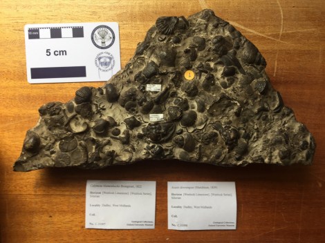
A piece of 425 million year old sea floor containing the skeletons of trilobites, brachiopods, bryozons, corals and gastropods preserved as limestone
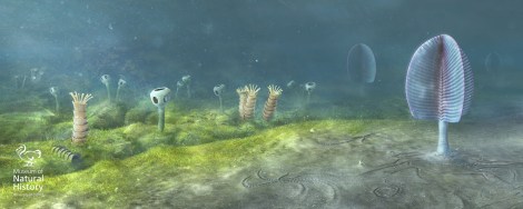
Travel back some 570 million years to a time known as the Ediacaran and the picture is very different. Although there were large-bodied creatures that were possibly animals, they were entirely soft-bodied. Then, right at the end of the Ediacaran Period, the first animals with hard skeletons evolved, creating strange tubes, stacked cones, and other bizarre forms such as Namacalathus, which resembles a baby’s rattle!
Some of the first animals with skeletons, Cloudina and Namacalathus alongside the soft-bodied Ediacaran fauna. Reconstruction based on rocks from Namibia, Southwest Africa, from 543 million years ago. Image: Mighty Fossils.
In the following few tens of millions of years, in the early part of the Cambrian Period, a whole host of animals burst onto the scene baring their ‘teeth’, hiding in their shells, and bristling their spines. In fact, we can trace the origin of almost every kind of animal skeleton to this relatively short window of the Earth’s past.
In my research, I have compiled the evidence for how and when these skeletons first appear. Three key observations have emerged. First, skeletons evolved independently many times in different animal groups. Second, there is both direct and indirect evidence, such as exceptionally preserved fossils and trace fossils, for entirely soft-bodied examples of animal groups that later evolved skeletons. And lastly, the first animal skeletons are less complex and more variable than later examples.
Added to what we know about how living animals build their skeletons, this all points to one explanation: Animal skeletons evolved independently in different groups by utilising a common ‘toolkit’ of genes, inherited from their common ancestor but used in different ways in different skeletons.
In other words, the soft-bodied ancestors of animals with hard parts had inherited all they needed to build simple skeletons that were then honed into the array of shells, teeth, spines, plates and bones we see today. For these skeletal pioneers, armed with their genetic ‘toolkit’, the environmental and ecological pressures of the early Cambrian prompted the evolution of similar, but independent, responses to their changing world – when life got hard.
Murdock, DJE. 2020. The ‘biomineralization toolkit’ and the origin of animal skeletons, Biological Reviews, is available for free here.
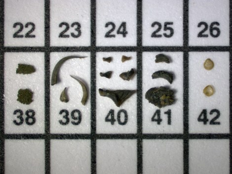
Top image: Tiny fragments of early skeletons, shells and spines, from around 510-515 million years ago.


")
")

")
")
")


")
")