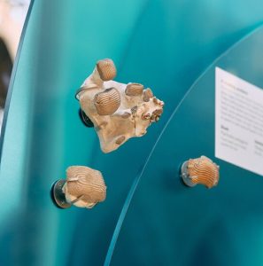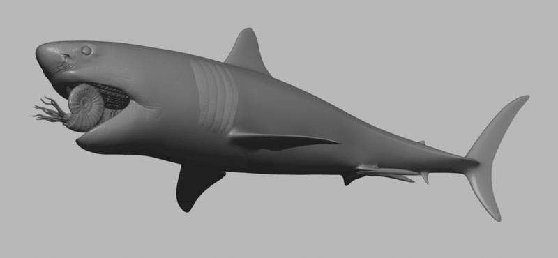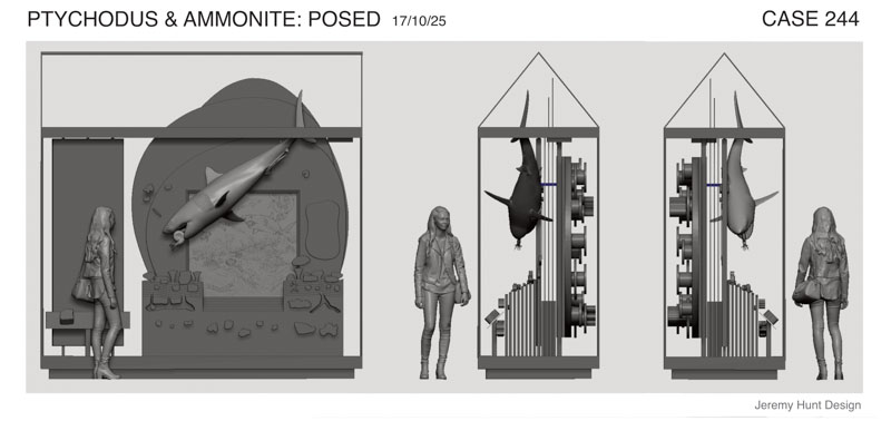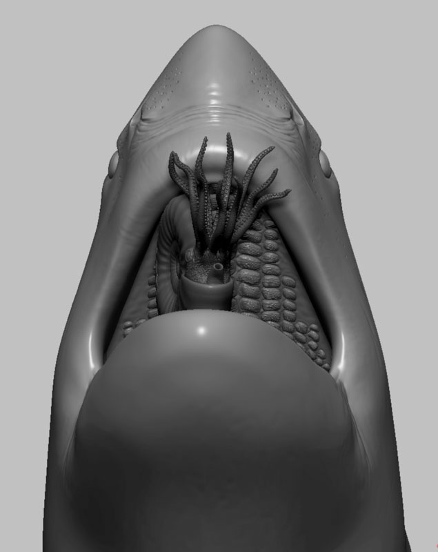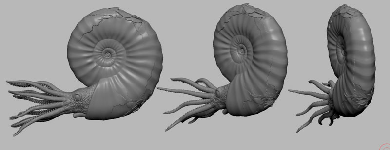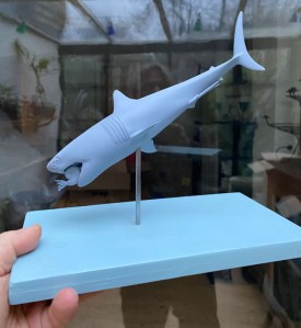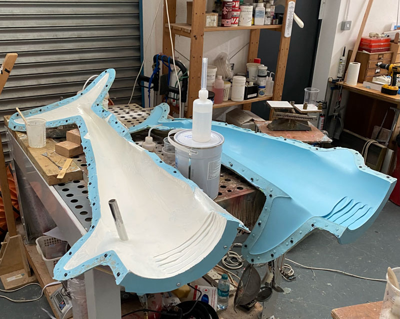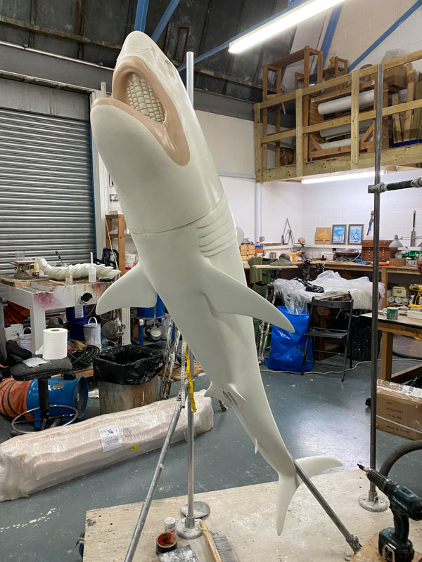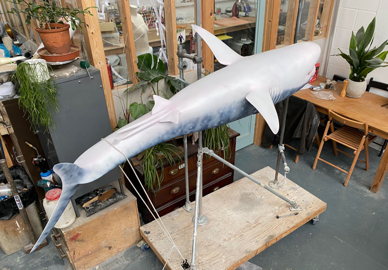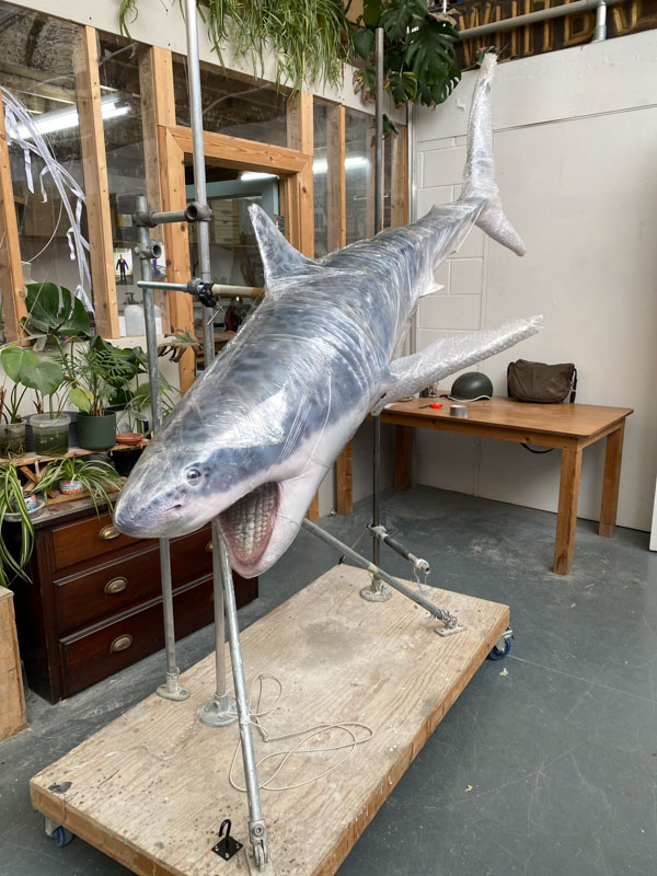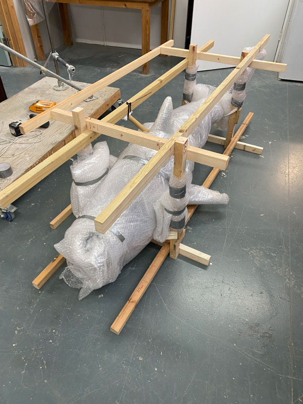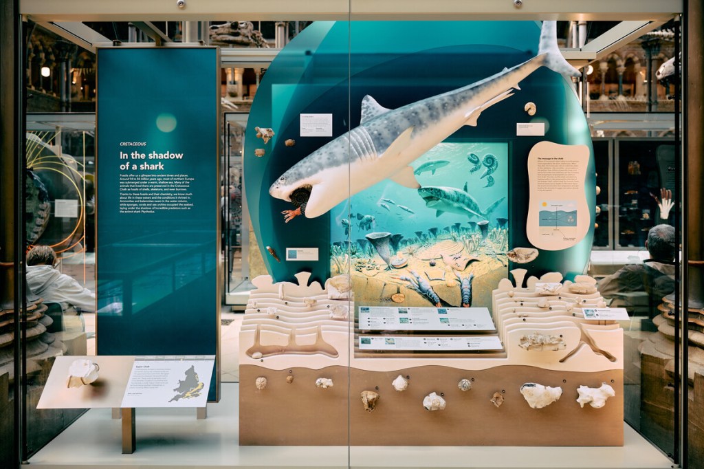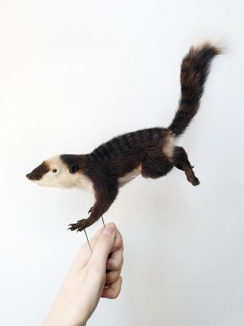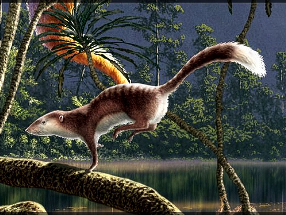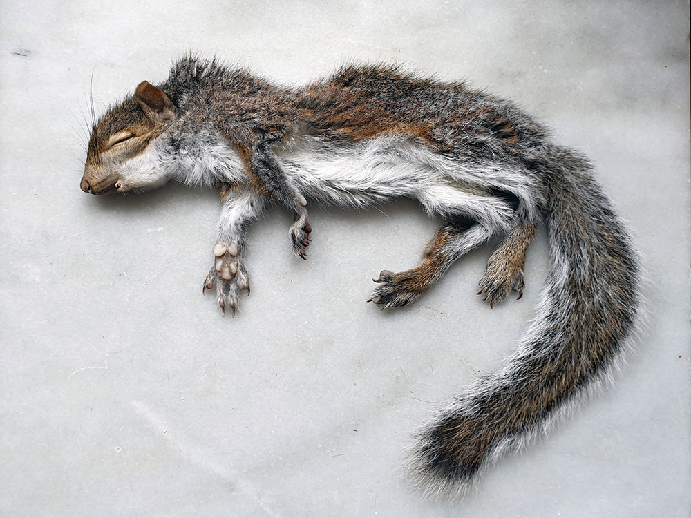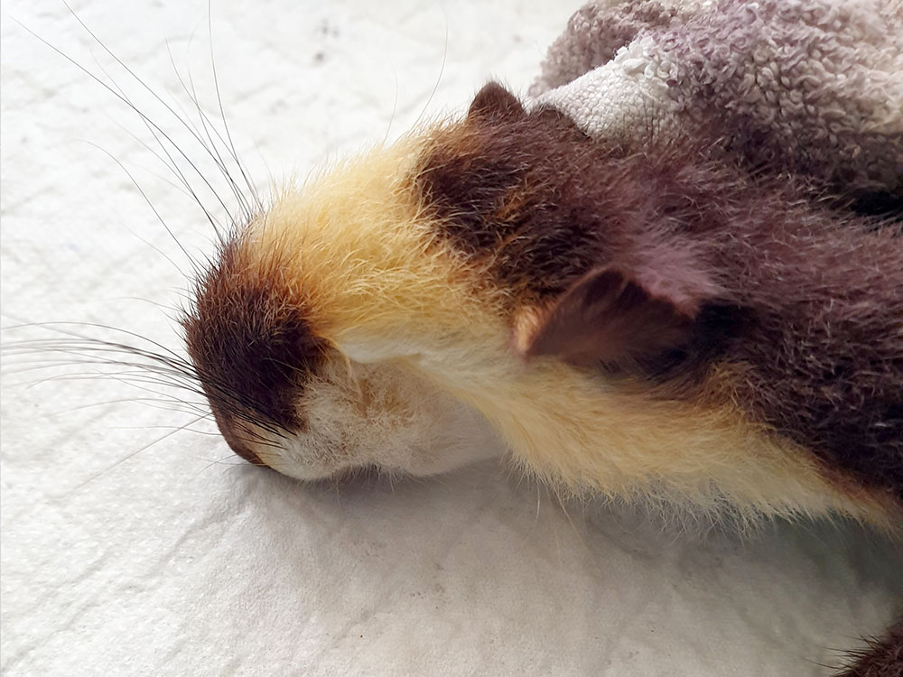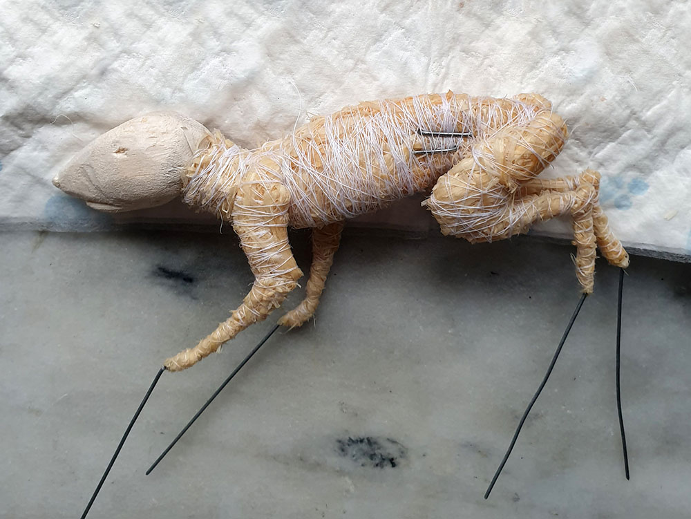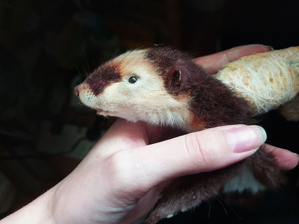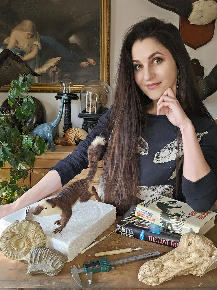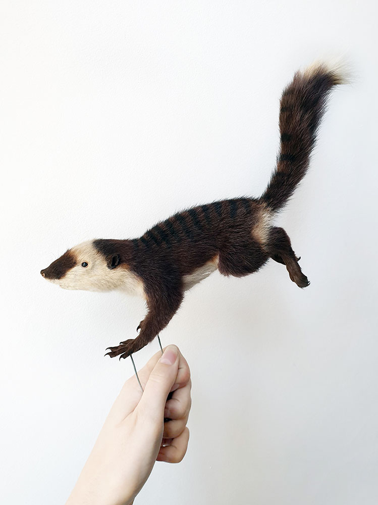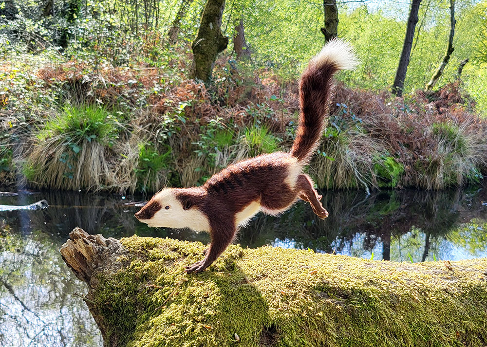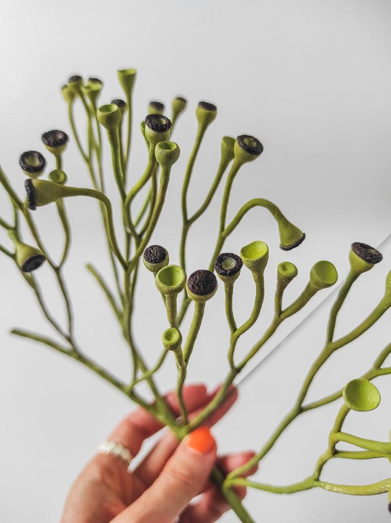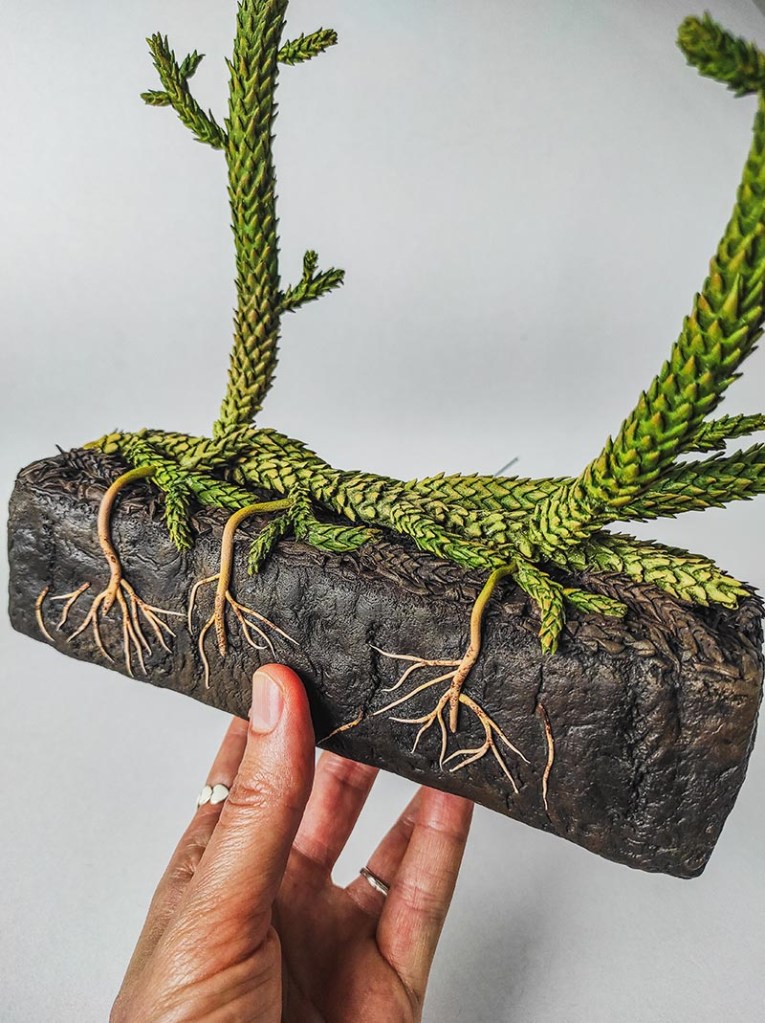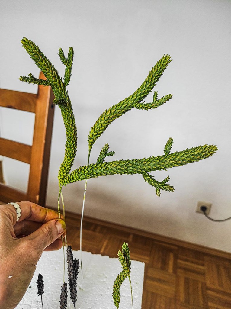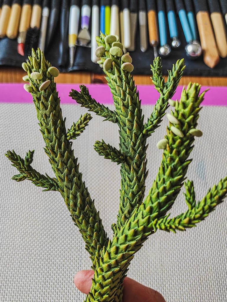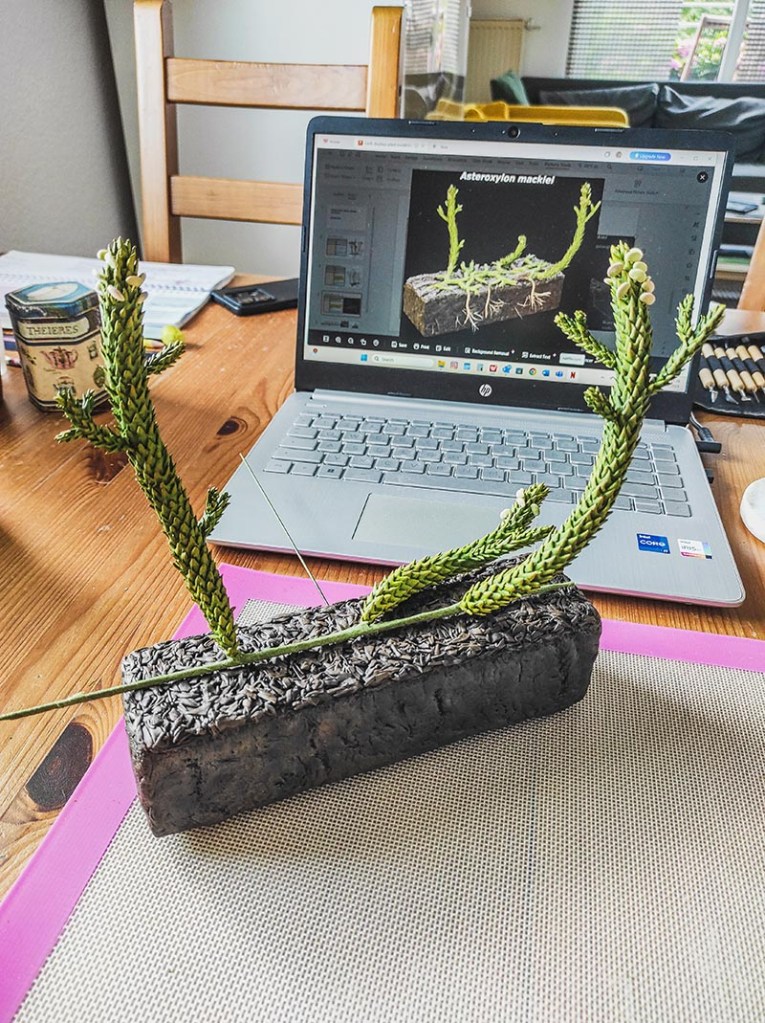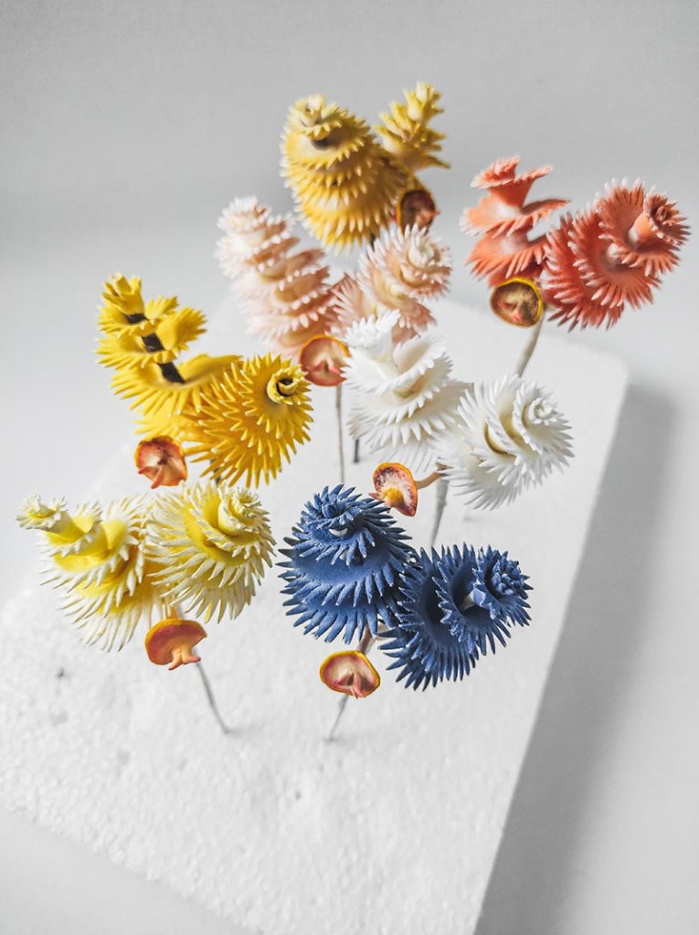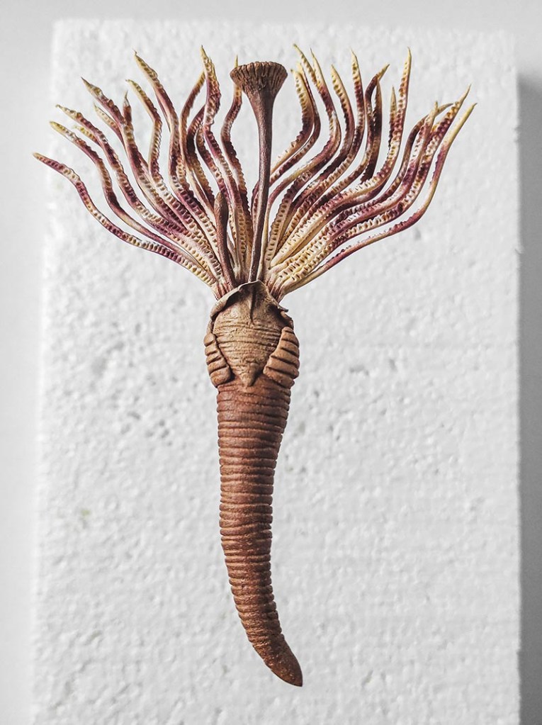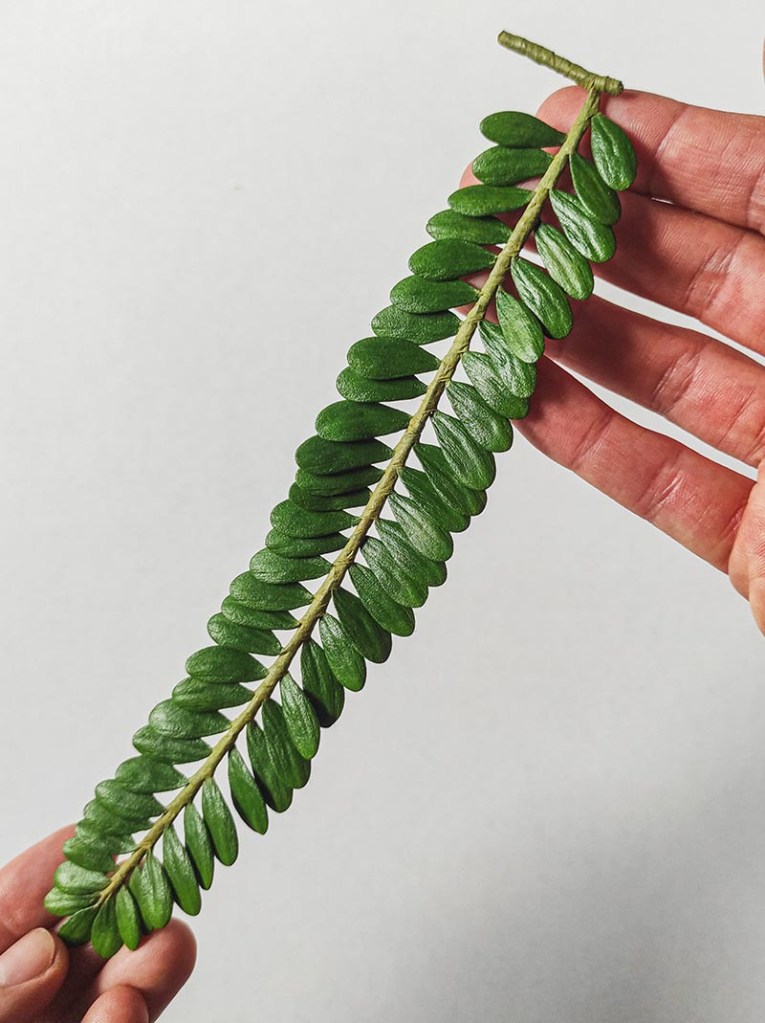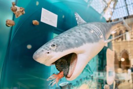
What a ride! It’s been about seven years in the making, but we have just put the finishing touches to our major redisplay project – Life, as we know it – which fully opened last week. If you haven’t been to the Museum in a while, now is a great time to come.
I have been lucky enough to have worked on this project from the very beginning, but the achievement is that of many, many people across the Museum and beyond. Over the coming weeks we are going to share, on this blog, stories from some of the skilled people who made it happen: model-makers, artists, scientists, set-builders, designers, animators, conservators – the list goes on and on.
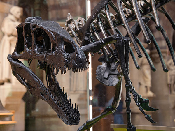
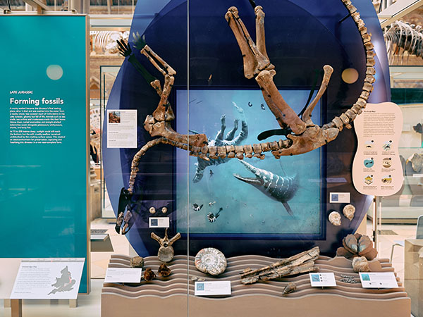
The video above gives a taster of all this. For those of us involved, it is also a nice trip down memory lane. How did we find and pin hundreds of arthropods in rings of concentric size, all presided over by a gigantic Japanese Spider Crab? Whose idea was it to suspend an articulated dinosaur upside down, sinking to its final resting place?
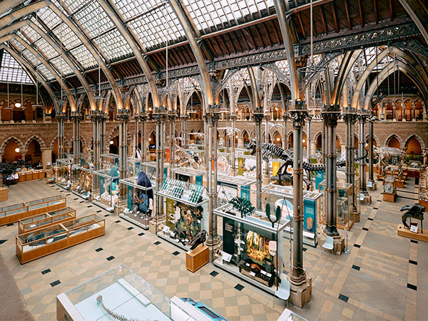
To be able to step back now and see all the new displays together is a real treat. Between them, the 48 new showcases, containing 24 new displays, show us specimen-led examples of how the natural world works, taking in big ideas in evolution, ecosystems, biodiversity, and the history of the planet.
Thanks to everyone who has worked on what is the biggest single redisplay in the Museum’s 165-year history. We hope you enjoy it as much as we have!
By Scott Billings, Life, As We Know It Redisplay Project Manager
Bethany Milne from the Museum’s Visitor Experience team has spent some time working alongside the Exhibitions team to deinstall a temporary art exhibition. She has reflected on the day – what she enjoyed and what she learnt along the way.
The deinstall of the Museum’s Deadly Six art installation is my first introduction into the world of exhibitions and an exciting one to start with. I have seen the display since it was first installed in September 2024 and I began working at the museum, watching it on my patrols of the court to make sure it stays intact and safe from all the little hands that pass through our doors every day. I didn’t imagine what it would be like to touch them myself until I found out I had the opportunity to join the team in taking them down.
On September 8th I came into the museum at 8:30am to shadow the exhibitions team as a development day. Most of the staff and contractors were already there and so I started by talking to the Head of Exhibitions, Rachel, about what needed to be done. She showed me her schedule of the day, starting at 8am, going through to 5pm, in which we would derig all the sculptures and get them set up safely out of the public areas and into storage. After, I helped set up hoarding to keep the aisle where the work was being done secluded, as the museum would be open to the public as usual around us. This aspect was an interesting learning opportunity for me as I have no earlier experience with the manual labour aspect of the work, but I enjoyed being hands-on.
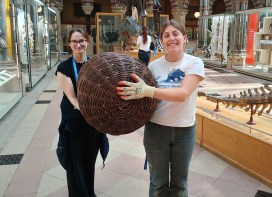
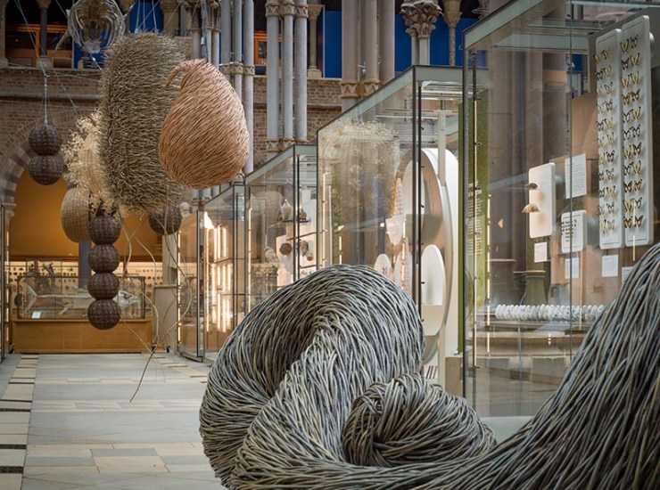
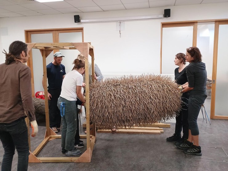
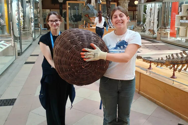
As I was doing this, the team from Outback Rigging worked on carefully lowering the suspended works to the ground and to our surprise; the take down was ahead of schedule. Most of them were removed by the middle of the day and these ones were simple to move. This was interesting to me as they seem so delicate, being large structures made of woven willow, but they proved sturdier than I thought. However, some were more difficult to take down than others, including the COVID sculpture. This was the most difficult to get down to the storage space, due to its intricate structure and its large size. Too wide to take down the stairs we usually use, Rachel organized with the Pitt Rivers to take it through their door, along the road and back into the Museum of Natural History through another door! What made this procedure more complicated was the sculpture itself, as its round construction meant there were no handholds. Luckily, the artist Issy Wilkes created some by looping some zip ties through the metal frame and, learning from the setup of the exhibition, used foam sheets to wrap around these painful to hold ties, to make the journey even easier. I felt it was an extremely rewarding process when we were finally able to put it down, as it required a lot of teamwork and shimmying about corridors to make it – I was very relieved we were able to keep it in one piece.
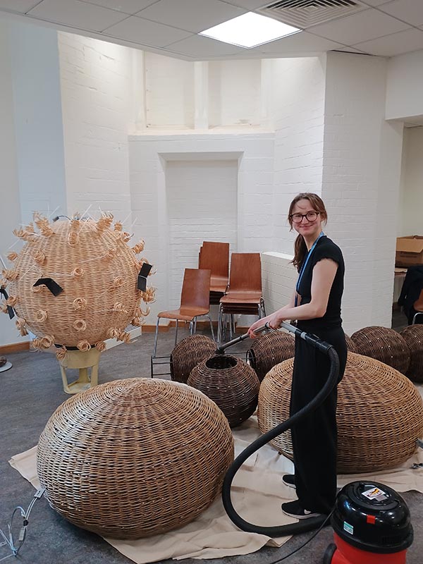
As the day went on and the sculptures were down and put away safely, I began to do smaller, but equally important tasks. The works themselves weren’t the only part of the exhibition, so I aided in removing the signage that was in the aisle that explained the meaning of each part. We also moved these down to the storage room, out of the way of the surrounding visitors and I began the task of removing the labels from inside. Another small step was vacuuming the sculptures. Despite regular conservation cleaning, hanging up in the court for so long had meant they had accumulated a lot of dust, which had to be cleaned out before they moved to their next home. These tasks were ones I wouldn‘t have thought of before the experience, but I realise that the smaller aspects are just as important as the larger ones when it comes to taking care of the exhibitions. It is our job to not only display the works as best we can, honouring their artistic intent and presenting to the audience in a way they understand, but this experience showed me how important the after care is, ensuring that they are well maintained to carry on their purpose and that the museum is returned to its original state.
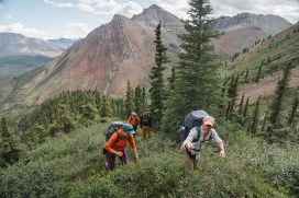
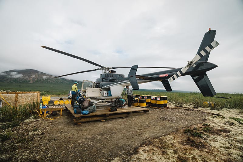
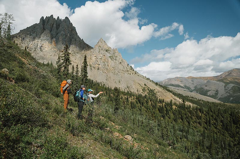
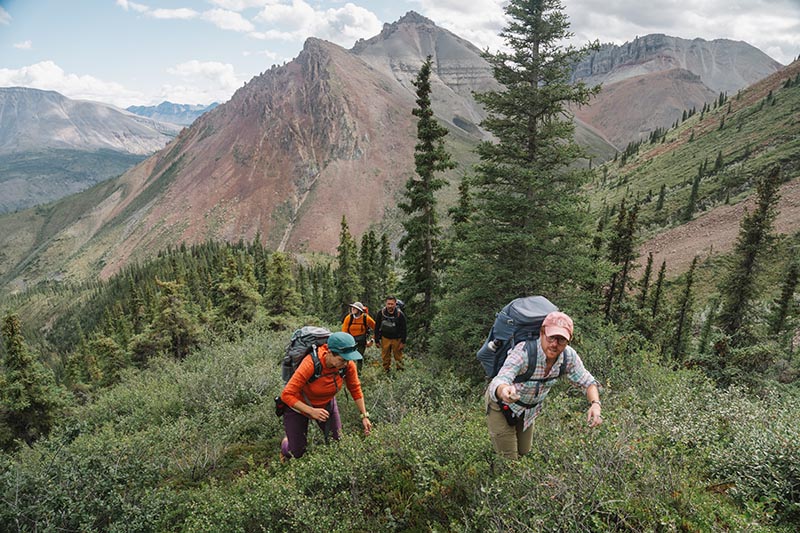
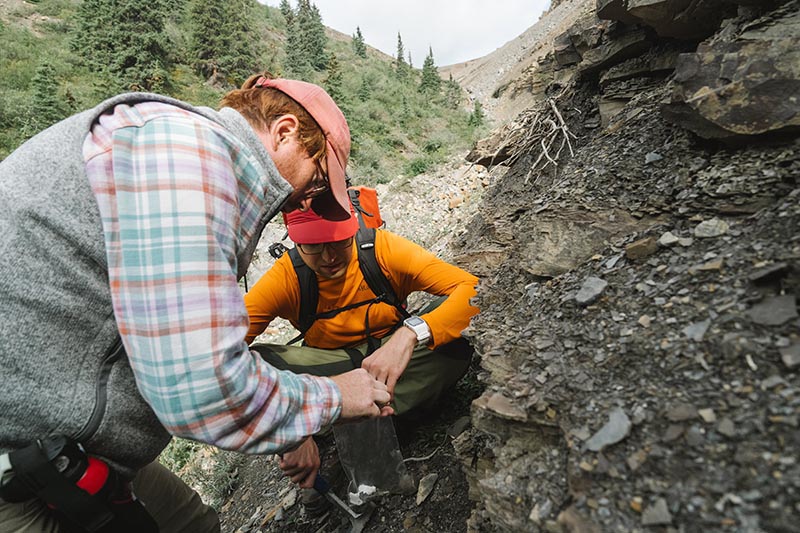
In Summer 2024, a team of palaeontologists and geologists from the University of Oxford, along with colleagues from Dartmouth College, the University of Washington, and Williams College in the USA, undertook an expedition to the Little Dal Group in the Mackenzie Mountains, Northwest Territories, Canada. Our purpose was to uncover some of the oldest fossil ecosystems that record complex life.
Photo: Robert GillPhoto: Robert Gill
Complex life comprises all organisms whose DNA is enclosed in a cell nucleus. This includes animals and plants but excludes bacteria. Today, this complex life accounts for most of the Earth’s biomass, documented biodiversity, and oxygen production. Understanding when and how it first evolved remains one of the central unanswered questions in evolutionary biology.
Photo: Robert GillPhoto: Robert Gill
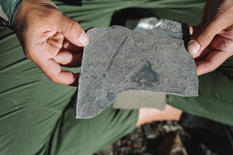
As palaeontologists, we normally use fossils to reveal the history of life. Fossils tend to preserve larger animals with hard shells or skeletons—creatures such as trilobites, ammonites, dinosaurs, and mammoths. However, the first complex organisms were microscopic and lacked such hard parts. As a result, their soft and fragile cells rarely fossilised. Put simply, we have found it a major challenge to trace the origins of complex life with fossils.
Photo: Robert GillPhoto: Robert Gill
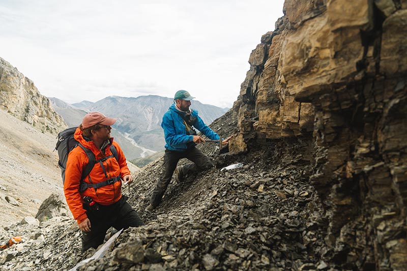
I have argued that finding rocks made up of antibacterial clay minerals holds the key. These minerals can slow the decay of organic cells long enough for them to survive as fossils. The Little Dal Group contains ~900-million-year-old rocks that are rich in just such clays, making it a prime target for new fossils that might help us unravel the origins of biological complexity.
Photo: Robert GillPhoto: Robert Gill
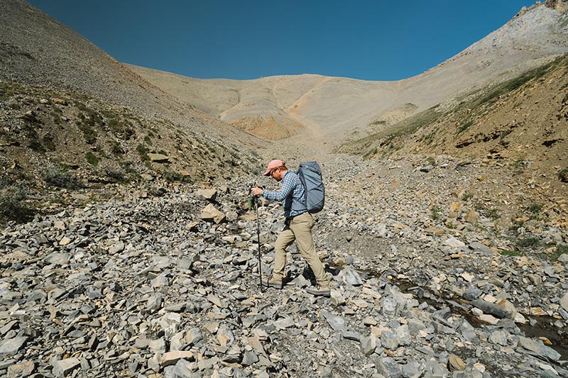
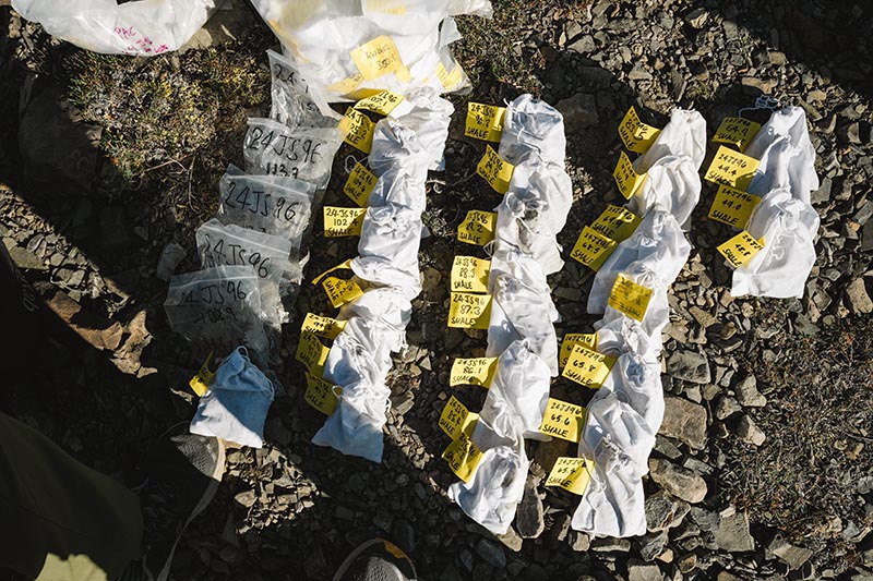
I was joined in Canada by my DPhil student, George Wedlake, from the Department of Earth Sciences. Together we spent two weeks collecting over 100 rock samples. The samples record an ancient tropical sea not unlike the Bahamas today, where early complex life likely flourished.
Back in Oxford, at the Museum of Natural History, George and I are now examining the samples; dissolving the rocks with hydrofluoric acid to extract and study the tiny fossils. We hope these new fossils will transform our understanding of how complex life first took hold on our planet.
Our fieldwork was funded by a Royal Society University Research Fellowship and by the Oxford NERC Environmental Science Doctoral Training Partnership. It was conducted under permit and with the support of the Sahtú Dene people.
Imagine a drop of ancient resin. Inside is an insect, trapped for 53 million years, so well preserved it looks like it might twitch back into life. These amber fossils offer us a breathtaking glimpse into long vanished ecosystems. But there’s a catch: the most revealing details, like delicate mouthparts or microscopic genitalia, are sealed away under the resin’s glossy surface. Cutting into them would destroy what makes them precious.
Enter the SOLEIL synchrotron.
At SOLEIL, near Paris, a group of researchers led by OUMNH’s own Dr Corentin Jouault are preparing to shine one of the world’s brightest X-ray beams through over 100 blocks of Oise amber, each containing a fossilised insect no larger than a fingernail. The goal is to see inside without cracking them open. Using a technique called in-line phase-contrast synchrotron microtomography (a bit of a mouthful, so let’s call it “supercharged 3D X-rays”), the team hopes to reveal anatomy invisible to conventional CT scanners. Think of it as upgrading from grainy black-and-white TV to ultra-high-definition.
The impressive imaging setup of the ANATOMIX beamline at SOLEIL Synchrotron. In the foreground, a rotating platform holds the amber sample in the path of the X-ray beam, turning it a full 360° so that hundreds of 2D X-ray images can be captured from every angle. In the background are the scintillators, which transform invisible X-rays into visible light, and above them, the high-resolution camera that records these images. All the data are then processed by powerful computers to reconstruct a detailed 3D model of the fossil trapped in amber.
Dr Corentin Jouaultcarefully positioning a piece of Eocene Oise amber (approximately 53 million years old, from France), containing an undescribed extinct ant species, mounted on a scanning electron microscope stub, in front of the X-ray beam for a high-resolution scan (pixel size ≈ 1.3 μm).
Why does this matter? Well, these insects lived during the Early Eocene, around 53 million years ago, when flowering plants had taken over the world in what scientists call the Angiosperm Terrestrial Revolution (ATR). This upheaval transformed landscapes and diets alike, and insects, already an evolutionary success story, had to adapt. Some lineages thrived by exploiting new blooms, while others dwindled. By studying the fine details of insect mouthparts, researchers can track how feeding strategies shifted in tandem with the rise of flowers.
The plan is ambitious. Each piece of amber will be scanned in full at a resolution fine enough to spot features a few micrometres across (a micrometre is one-thousandth of a millimetre — about one-hundredth the width of a human hair). 20 chosen specimens will then be magnified further still for an even sharper look, down to less than half a micrometre per pixel. In other words, one pixel will cover an area 140 times smaller than the thickness of a human hair.
The resulting datasets will therefore be enormous, so the team has lined up banks of high-powered computers and even machine-learning tools to speed up the laborious task of reconstructing the fossils in 3D.
What emerges won’t just be pretty pictures. These models could rewrite parts of insect evolutionary history. For example, they may uncover the earliest records of certain insect families, refine the timeline of insect diversification, and provide the raw data needed to estimate how extinction and speciation rates shifted during the ATR. In short, these pieces of amber become not just a window into the past, but a testbed for some of the biggest questions in evolutionary biology.
And there’s a democratic twist: all the scans, reconstructions, and 3D models will be made freely available in open repositories. That means anyone, from entomologists to curious hobbyists, could spin, zoom, and explore these ancient insects in digital space.
So, next time you spot an insect hovering around a flower, think of its ancestors locked in amber, their secrets now teased out by beams of light brighter than the sun. The synchrotron doesn’t just illuminate fossils, it illuminates how deep the ties between bugs and blooms really go.
Excavations at Oxfordshire’s ‘dinosaur highway’ continued this summer to uncover Europe’s longest sauropod dinosaur trackway.
The 2025 Dewars Farm trackway site, drone photography. Credit: Richard Butler | University of Birmingham
Following the success of the excavation last summer, which featured on BBC Two’s Digging For Britain, a new area of Dewars Farm quarry near Bicester has been uncovered. Teams from the Universities of Oxford, Birmingham and Liverpool John Moores joined forces for a week-long dig in the hot, dry summer of 2025. Hundreds more individual prints from four trackways were identified and documented, including Europe’s longest sauropod dinosaur trackway at some 220 metres from the first to the last exposed footprint.
The four new trackways found at the 2025 Dewars Farm site were each made by sauropod dinosaurs, large-bodied long-necked herbivores like Cetiosaurus, that made their way along an exposed mudflat on the edge of a lagoon some 166 million years ago – during the Middle Jurassic Period.
Cleaning Europe’s longest sauropod dinosaur trackway. Credit: Richard Butler | University of Birmingham
The excavation was made possible through the continued collaboration with the quarry operators Smiths Bletchington, Dewars Farm and Duns Tew Quarry Manager Mark Stanway, and his staff. As in 2024, a team of more than 100 people worked at the site, co-led by Oxford University Museum of Natural History (OUMNH) and University of Birmingham (UoB) and joined by collaborators from Liverpool John Moores University. Over seven days, the teams battled against a much drier, harder surface than the previous year focusing on a set of around 80 very large (up to 1m long) sauropod prints, that ran approximately north-south across the entire site.
Part of the 2025 excavation team and the fantastic Smiths Bletchington staff. Credit: Emma Nicholls | OUMNH
In addition to the largest sauropod trackway, three others were uncovered, one of which is a continuation of prints first found in 2022. Although not continuously exposed, it may prove to be an even longer trackway once all the data is pieced together. Smaller finds included marine invertebrates, plant material and a crocodile jaw. New for 2025, systematic sampling of the sediments that both underlie and fill the prints was undertaken, the analysis of which is underway.
More of the footprint surface is likely to be exposed over the coming years, and a full description of the significance, new scientific discoveries and potential for future preservation of the site is expected soon.
Sets of sauropod dinosaur footprints, each up to 1 metre long. Credit: Emma Nicholls | OUMNH
You’re 18 years old, an orphan, and your uncle and guardian has just been thrown in debtor’s prison. You have no other family around you. What do you do? Well, if you’re John Phillips (1800–1874), you attempt to run a lithographic printing business out of your uncle’s house; extraordinary for one so young and doubly so as lithography was still a relatively new technology. According to Michael Twyman, in 1819 “the number of lithographic printers in London could still be counted on one hand.”
Engraving vs Lithography
At the time, mass-printed drawings were still commonly engraved on copper plates. It was a time-consuming process. A skilled engraver could take over 40 hours to complete a ten-inch plate and any errors were costly to correct.1
Dale copperplate
Invented just two decades earlier in 1798 by German playwright Aloys Senefelder, lithography made the process of printing cheaper and simpler. Drawing on a prepared stone surface with waxy ink or chalk and using the repelling properties of fat and water to print the finished design, the need for precise etching was eliminated and errors could be wiped away rather than having to burnish and re-etch a metal plate.
Lithographic stone
Phillips’ Early Experiments
With few works to reference or professionals to consult, Phillips had taken what information he could find and applied himself to testing and recording different ink to chalk ratios for optimal drawing and printing. He even tinkered with existing press designs, trying to invent a more efficient machine. The results of his experiments are preserved today in his notebooks, held here at the Museum.
Phillips’ press design sketches
Phillips set up shop in his uncle’s house at 15 Buckingham Street and advertised in a circular, “I take the liberty of soliciting Your Attention to my method of Drawing and Printing from Stone. The Process is much cheaper, and is far more expeditious than the common method of Engraving on Copper.”
Phillips’ advert
Family, Fossils and Geology
Having arrived in London in 1815 to live with his uncle, William Smith—later hailed as the father of English Geology—Phillips had spent much of his youth bouncing from relative to boarding school to family friend. Most recently he had stayed with Smith’s friend the Reverend Benjamin Richardson, an avid naturalist and geologist.
William Smith had just published A Delineation of the Strata of England and Wales with part of Scotland…, the first geological map of England and Wales. Smith showed that the relative positioning of specific fossil species in strata could be used to identify the same strata across the UK — a remarkable achievement, but one that nearly ruined him financially. His finances were so desperate that when his nephew arrived he was in the middle of negotiating the sale of his fossil collection to the British Museum.
These same fossils, arranged according to the strata in which they were found, had formed the basis of his great work. Selling them was a huge blow, both personally and professionally; he had collected them himself whilst travelling as a land surveyor, consulted them frequently in his studies, and given access to any scientist wishing to see them. It also meant they would need to be formally catalogued to be useful.
Luckily his newly arrived nephew had a talent for drawing and a keen interest in shells and geology; Phillips had by then begun his experiments with lithographic printing, conscious of the enormous cost savings possible for publications, both for his uncle and his own work on fossil conchology. He became the person Smith needed to assist in drawing, arranging, and cataloguing the fossils for publication before they were crated up and sent away. This led to the publication of Strata Identified by Fossils (1816) and Stratigraphical System of Organized Fossils (1817). It was then that Phillips began experimenting with lithographic printing, conscious of the enormous cost savings possible for publications, both for his uncle and his own work on fossil conchology.
Phillips’ test print for Fossil Conchology
Notebooks, Inventions and Later Life
Phillips’ notebooks are filled with detailed observations of natural history from his travels across the UK. They also show his fascination with all things mechanical; doodling whimsical contraptions meant to solve the day-to-day problems of the active naturalist.
Phillips’ camp chair
With an interest in geology and a fascination with technology, it’s hardly surprising that when Phillips found himself with no support and no money he would see if his interest in lithography or “stone writing” could help with his pressing financial problems. But did it work? Did John Phillips make any money?
While we hold Phillips’ personal and research papers, sadly no record of sales or responses survive. What we do know is that after William Smith was released from debtor’s prison the house was repossessed and Smith, along with his wife and nephew, left London to eke out a living as itinerant mineral surveyors.
Phillips’ experiment notes
Phillips remained a lifelong advocate for science and technology and went on to become a well-respected geologist, the first keeper of the Museum, and only the second Professor of Geology in Oxford. Tragically, after a convivial dinner on 23 April 1874 at All Souls College Phillips slipped and fell down a flight of stone steps, falling into a coma never to awake.