A.R. WALLACE’S ARCHIVE NOW AVAILABLE ONLINE


“In all works on Natural History, we constantly find details of the marvellous adaptation of animals to their food, their habits, and the localities in which they are found.”

– A.R. Wallace

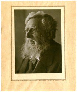
2023 marks a number of important anniversaries in the UK: it has been 75 years since the founding of the NHS and the arrival of the Empire Windrush in London, and 100 years since the first outside broadcast by the British Broadcasting Company. Importantly for the Museum, it is also the 200th anniversary of the birth of Alfred Russel Wallace (1823-1913), the trailblazing biologist, geographer, explorer, and naturalist.

Wallace was one of the leading evolutionary thinkers of the nineteenth century and is most well-known for independently developing the theory of natural selection simultaneously with Charles Darwin. The publication of Wallace’s paper “On the Tendency of Varieties of Depart Indefinitely from the Original Type” in 1858 prompted Darwin to quickly publish On the Origin of Species the following year. He was a pioneer in the field of zoogeography and was considered the leading expert of his time on the geographical distribution of animal species. He was also one of the first scientists to write a serious exploration of the possibility of life on other planets.

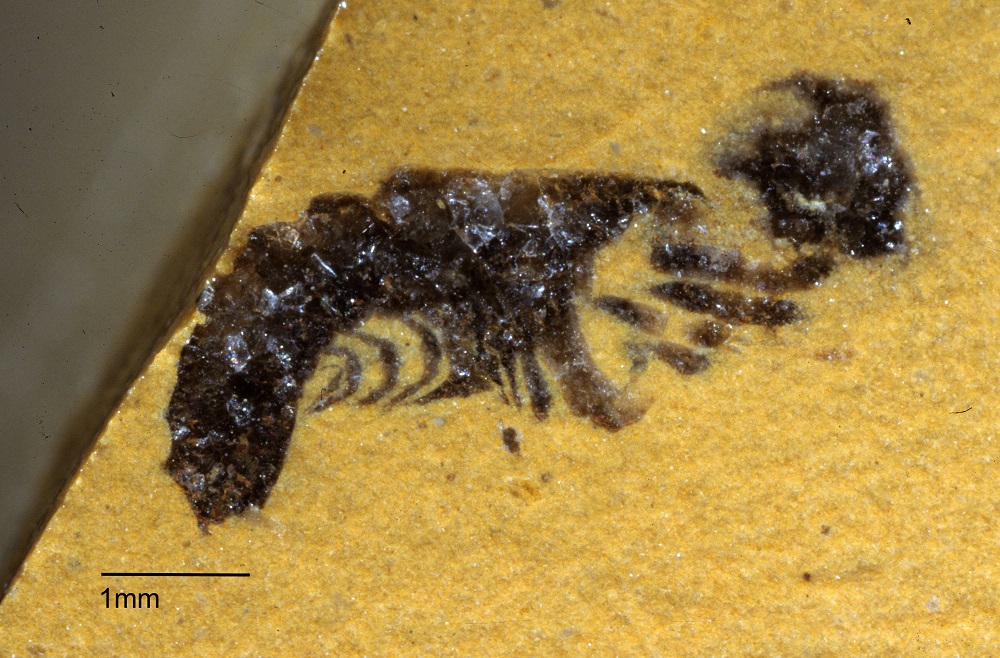
Wallace undertook extensive fieldwork in the Amazon River basin and the Malay Archipelago. He spent four years in the Amazon from 1848-52 but unfortunately lost much of his collection when the ship he returned to Britain on caught fire. Afterwards, he spent eight years in the Malay Archipelago (1854-62), building up a collection of 125,660 specimens including 109,700 insects, many of which are currently housed at Oxford University Museum of Natural History. In fact, we now hold one of the largest collections of Wallace specimens in the country.

In addition to entomological specimens, OUMNH holds a large and varied archival collection relating to Wallace. The archive includes original insect illustrations sent to Wallace by contemporary entomologists, photographs, and even obituaries. By far the largest portion of the collection is 295 letters of correspondence, of which 285 were penned by Wallace himself. The bulk of Wallace’s letters were written to fellow scientists, including the chemist and naturalist Raphael Meldola and the evolutionary biologist Edward Bagnall Poulton.

We are happy to announce that, in celebration of Wallace’s 200th year, we are making the entire Wallace correspondence available to browse online!

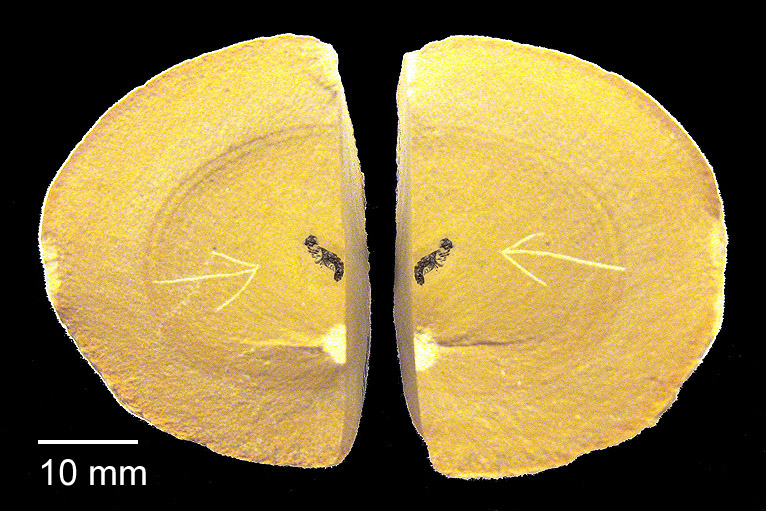
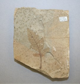
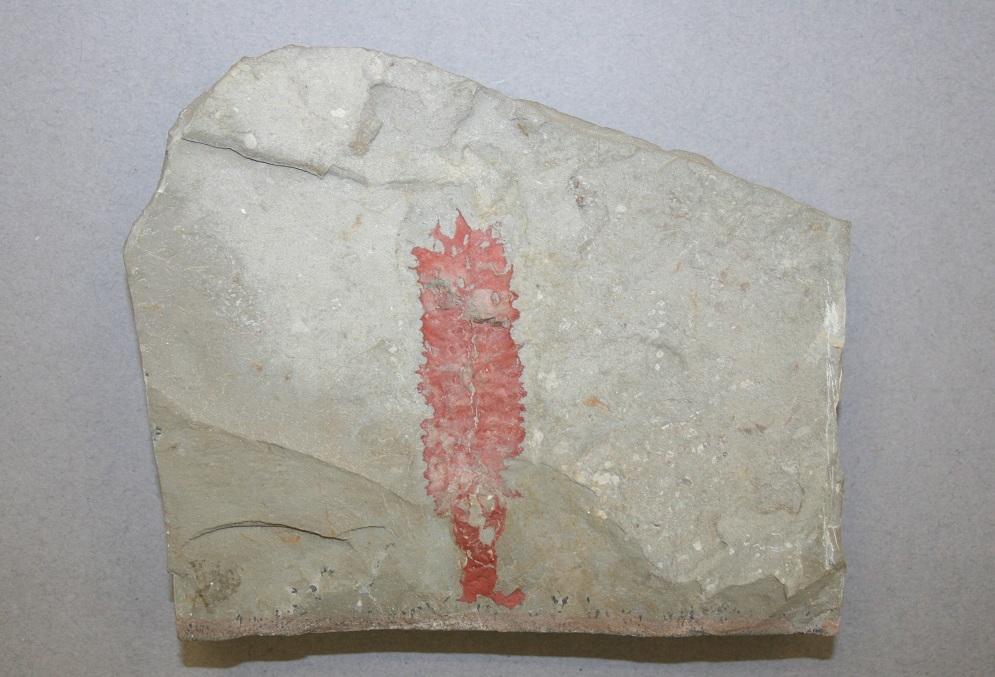
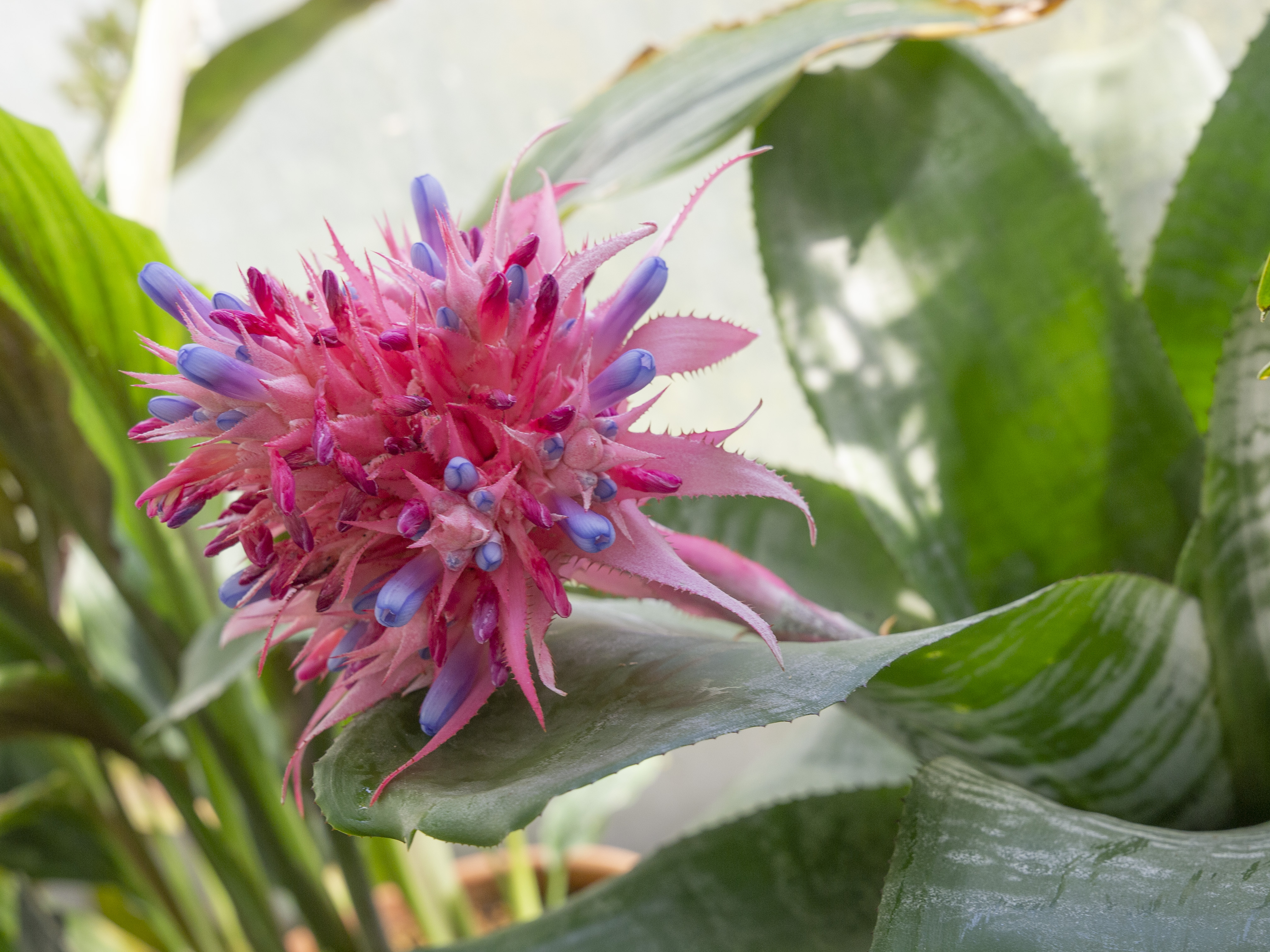
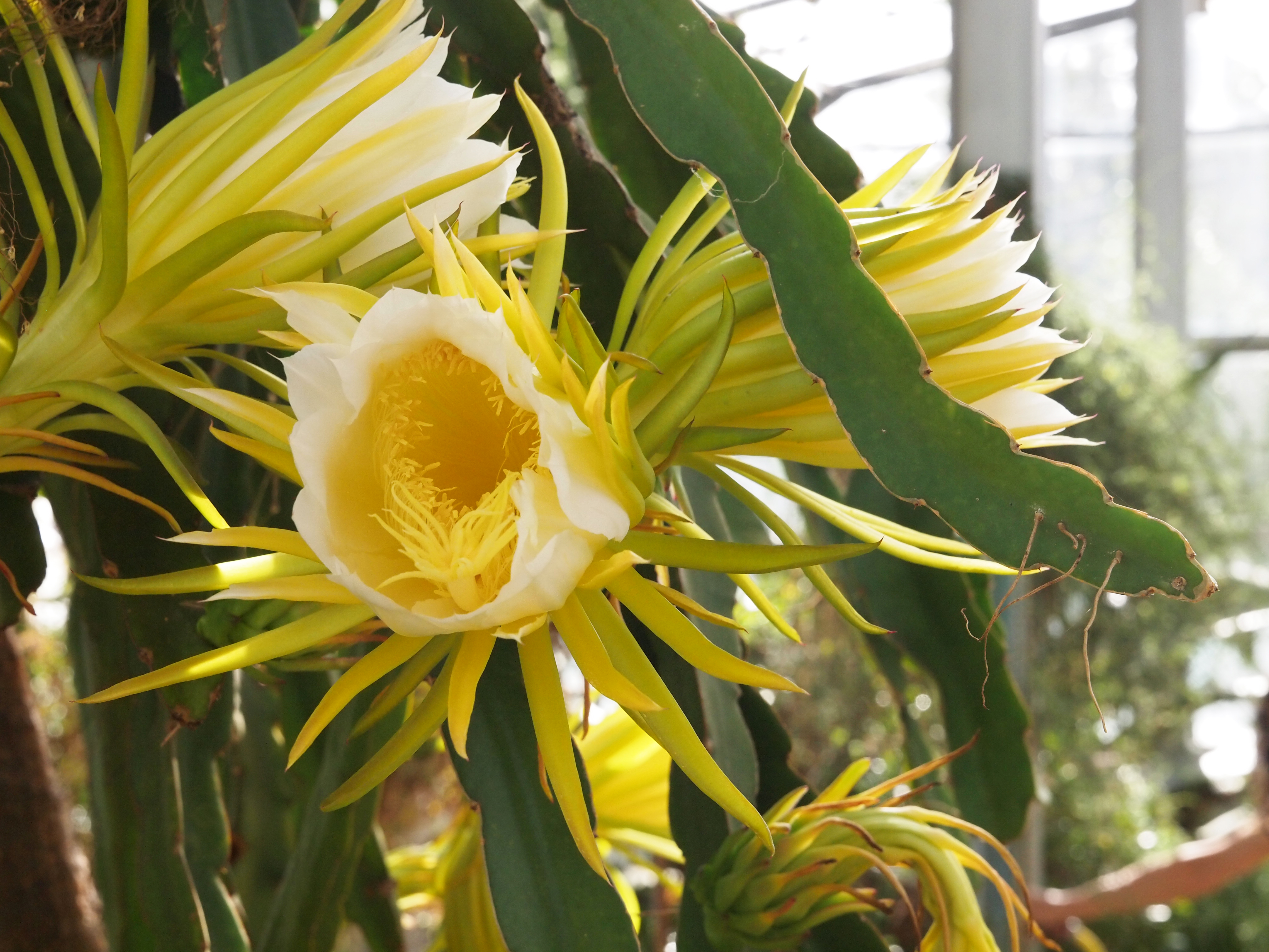
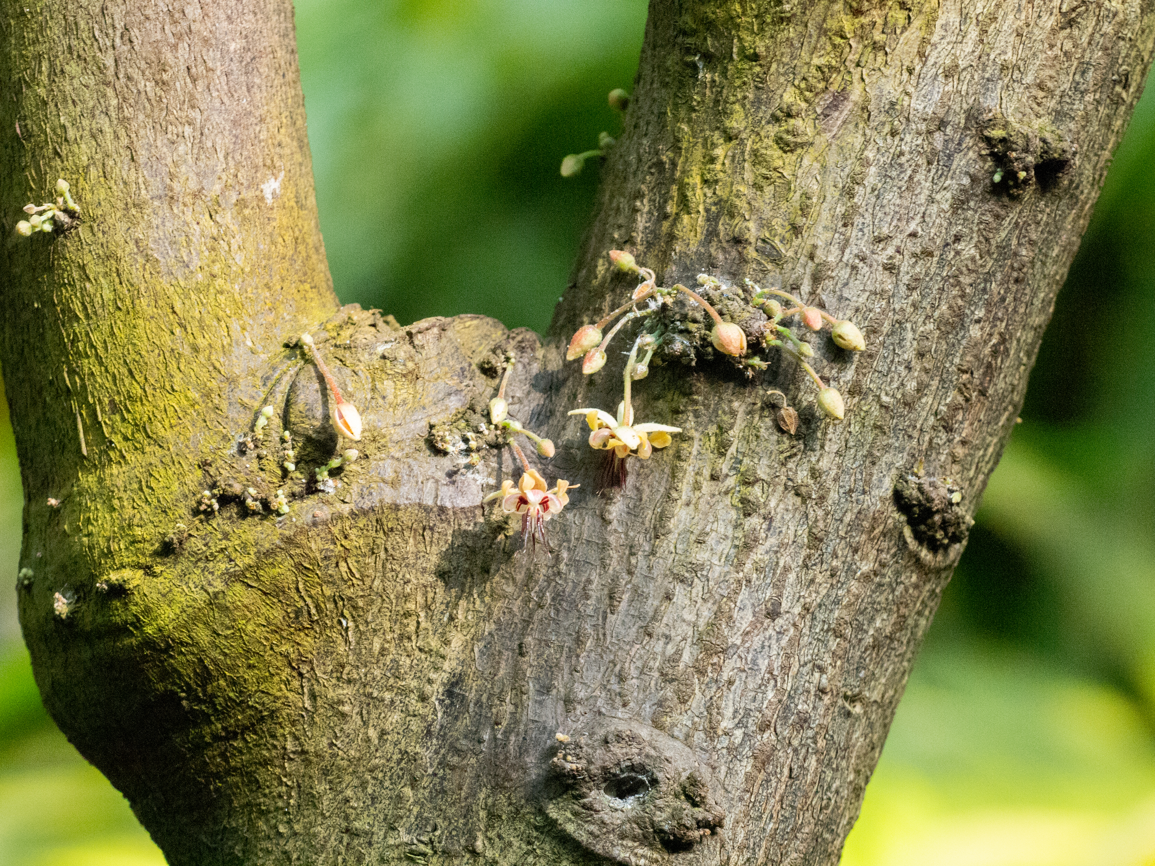
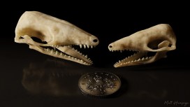
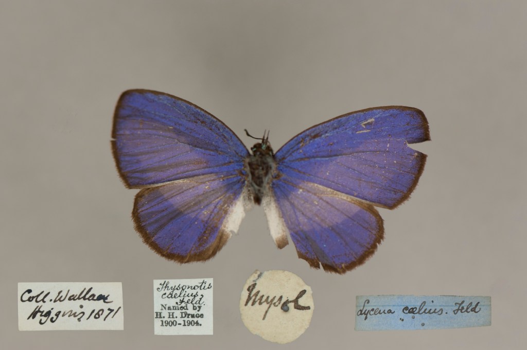
Several of the letters in the collection can be connected to the Wallace entomological collections held at OUMNH, providing us with invaluable insights into the history of these specimens. For example, you can read this 1896 letter from Wallace to Poulton in which Wallace discusses the changing of hands of his entomological collections, from Samuel Stevens to Edmond Higgins following Stevens’ retirement in 1867. The Museum subsequently acquired some of Wallace’s entomological specimens through Edmond Higgins, including the two beautiful examples shown above.

These letters are a potential treasure trove of information about Wallace and his collections, and we hope they will be of great interest to researchers in the field, as well as to the public. Interested? Learn more about Alfred Russel Wallace or explore his archive online.


Article by Matthew Barton, Digital Archivist at OUMNH

Beyond Buckland

DISCOVERING YORKSHIRE’S ANCIENT BEASTS


By Susan Newell

Susan Newell is a doctoral student researching the teaching collections of William Buckland, the first Professor of Geology at Oxford who taught from 1813 to 1849. She reminds us here about Buckland’s role 200 years ago in interpreting the important Pleistocene discoveries being celebrated this year, and the way that Mary Morland, a talented local naturalist, and many others, contributed to making this new knowledge.


This year marks the 200th anniversary of a great advance in our understanding of the geological past… a story which begins in the nineteenth century, with the discovery of a bone-filled cave in Kirkdale, Yorkshire. 

Uncovered by local quarrymen in 1821, the discovery of the Kirkdale cave and its contents of mysterious bone was the source of much intrigue. When news of the discovery reached William Buckland, Professor of Geology at Oxford University, he decided to travel up North to visit the site. However, by the time Buckland arrived at the cave, local collectors had scooped up most of its contents. Nonetheless, he was able to retrieve and examine some of the cave’s remaining material, which led him to an astonishing conclusion — Yorkshire must once have been home to hyaenas, elephants, hippopotamus and rhinoceros, and what was now known as the Kirkdale cave was once a hyaenas’ den.

W. B. Conybeare, lithograph, ‘The Hyaena’s Den at Kirkdale near Kirby Moorside in Yorkshire, discovered A.D. 1821’. Reproduced by kind permission of Christ Church, Oxford.
This light-hearted reconstruction of the hyaenas’ den shows Buckland illuminating the scene, in every sense. It is thought to be the first visual reconstruction of the pre-human past.

Central to Buckland’s theories were some small white balls that he had found amongst the debris in the cave. Buckland sent these balls to William Wollaston, a celebrated chemist based in London, for analysis.  He also asked Wollaston to visit the zoo at Exeter Exchange in London and show the balls to the hyaena’s keeper there.  Together with the results from Wollaston’s chemical analyses, the keeper confirmed Buckland’s hypothesis — the balls were droppings from animals very similar to modern hyaenas. Meanwhile, the anatomist William Clift was able to identify the bones from the Kirkdale cave as belonging to other extinct species related to those found living in tropical countries today. Buckland concluded that the cave must have been a den for ancient hyaenas, who would drag parts of the dead animals they had found (or killed) inside and, after feeding on them, leave piles of bones and droppings behind.

In order to strengthen his theory, Buckland discussed the behaviour of hyaenas in the wild with army officers connected to Britain’s colonial expansion in India. These officers also sent Buckland fresh specimens captured by local people. When a travelling menagerie visited Oxford in 1822, Buckland took the opportunity to experiment; feeding bones to a hyaena and noting that the teeth marks matched those on the fossilised bones from the cave.

Buckland’s findings were something of a shock to his contemporaries. When lecturing, he employed several different methods to try and convince his audiences that his theories were true. This included presenting fossil specimens and bones from living species for comparison, and showing maps, diagrams and drawings. Mary Morland contributed some of these illustrations, including large drawings of living animals, and technical drawings of bones that were later engraved for use in Buckland’s publications. Mary’s Kirkdale drawings seem to have been the first that she produced for William before the couple married in 1825.

Fossil hyaena jaw in the Museum’s collection, possibly the one featured in the engraving alongside it. Engraving is by James Basire after a drawing by Mary Morland. Published in William Buckland’s article in the Royal Society’s journal (1822) on the Kirkdale cave discoveries. [1]

Buckland’s work on the Kirkdale cave was revolutionary, not least because he was the first to make a scientific study of a cache of bones of this type.  Although similar bones from ‘tropical’ species had previously been found in Northern Europe, people thought that they had been washed up by a catastrophic flood, believed by many to be the biblical Noah’s Flood.  Modern analysis has now allowed us to deduce that the bones date to an Interglacial period when Britain was joined to Europe and had a hot climate, about 120,000 years ago.  

Here at the Museum, Buckland’s collections and archives are as much of a treasure trove as the Kirkdale cave. It is through accessing these archives that we can learn about the surprising range of people who contributed to the emergence of new scientific knowledge from the Kirkland cave — quarrymen, collectors, zookeepers, chemists, anatomists, colonial officers in India, workers in India, and artists like Mary Morland. To find out more about the incredible legacy of the Kirkdale Cave, look out for ‘Kirkdale200 – Lost Beasts of the North’, a symposium organised by the Yorkshire Fossil Festival, 12th March 2022.

Mary Morland, watercolour and gouache, lecture illustration of a hippopotamus, signed ‘MM’.
Hippopotamus bones were found at Kirkdale cave in Yorkshire, but as there were no living hippos to be seen in Britain at the time, this drawing would have been a valuable teaching aid.

[1] William Buckland, ‘Account of an Assemblage of Fossil Teeth and Bones of Elephant, Rhinoceros, Hippopotamus, Bear, Tiger, and Hyaena, and Sixteen Other Animals; Discovered in a Cave at Kirkdale, Yorkshire, in the Year 1821: With a Comparative View of Five Similar Caverns in Various Parts of England, and others on the Continent’, Phil. Trans., 2 (1815-30), 165-167.

Reconstructing the Cretaceous with Bones and Amber

A double window into the past

Post by Dr Ricardo Pérez-de la Fuente, Deputy Head of Research

Nature is wonderfully imperfect, and the data that we can gather from it is even further from perfection. Fossil localities, even those providing exceptionally well-preserved fossils, are inaccurate records of the past. Fossils can form from a variety of matter including organisms, their remains, or even traces of their activity. Yet not all of the material that can get fossilised at a particular site actually will. Among other factors, biases in the fossil record result from the nature of the materials responsible for fossilisation – usually sediments which are in the process of turning into rocks. In most cases, fossil localities offer us only a single ‘window of preservation’ – a skewed geological record of the ancient ecosystem that once existed there.


In 2012, a rich vertebrate bone bed was documented at the Ariño site in Teruel, Spain. Since then, researchers have unearthed more than 10,000 individual fossil bones, from which they have discovered new species of dinosaurs, crocodiles, and turtles. Plant fossils were also found, including pollen grains and amber, which is fossilised resin. Although amber was known to occur in this locality, this sort of material had remained unstudied… until recently.

Over the summer of 2019, I joined my colleagues to carry out amber excavations in the Ariño site – an open-pit coal mine that has an almost lunar appearance due to the dark carbonate-rich mudstone rocks and the total lack of vegetation. The scorching heat during a very hot summer was a bit maddening, but I did try to enjoy my yearly dose of sun before returning to the UK!


Resin pieces can be transported significant distances by runoff water before depositing on their final burial location, where they slowly transform into amber. However, we found amber pieces that had not moved from their original place of production. These large, round-shaped pieces preserved delicate surface patterns that would have been polished away even by the slightest transport. The resin that produced these amber pieces was formed by the roots of the resin-producing trees, and resembles sub-fossil resin my colleagues found in modern forests from New Zealand.

Large amber piece produced by roots (left) and assemblage of smaller amber pieces (right) from Ariño (Teurel, Spain).

The small amber pieces from Ariño contain an unusual abundance of fossils. These pieces come from resin produced by the branches and trunk of the resin-producing trees. From the almost one kilogram of amber we excavated, we identified a total of 166 fossils. These include diverse insects such as lacewings, beetles, or wasps, and arachnids such as spiders and mites. Even a mammal hair strand was found!1


We now know that the Ariño site provides two complementary windows of preservation — a bone bed preserving a rich variety of vertebrate animals, and amber with abundant inclusions. Aside from Ariño, only three localities that preserve both dinosaur bone beds and fossiliferous amber have been reported in Western France, Western Canada, and North Central United States. However, in these cases, either the bone bed or the amber have offered a much more modest abundance and diversity of fossils. Some of the fossils from these localities also show signs of significant transport, which means that the organisms could have inhabited different, distant areas even though they fossilised together. This makes Ariño unique because it offers two valuable ‘windows of preservation’ from the same ecosystem.

Thanks to all this evidence and other data, we have been able to reconstruct an ancient terrestrial ecosystem – a 110-million-year-old coastal swamp – with unprecedented detail and accuracy.2 The inherent incompleteness of the fossil record will always remain a headache for palaeontologists… but localities like Ariño make the data that we can recover from the past a bit more complete.

Reconstruction of the coastal swamp forest of Ariño, in the Iberian Peninsula, from 110 million years ago. Author: José Antonio Peñas. Source: Álvarez-Parra et al. 2021.

If you want to learn more about amber excavations, check out this post on Excavating Amber.


1Álvarez-Parra, Sergio, Ricardo Pérez-de la Fuente, Enrique Peñalver, Eduardo Barrón, Luis Alcalá, Jordi Pérez-Cano, Carles Martín-Closas et al. “Dinosaur bonebed amber from an original swamp forest soil.” Elife 10 (2021): e72477.

2Álvarez-Parra, Sergio, Xavier Delclòs, Mónica M. Solórzano-Kraemer, Luis Alcalá, and Enrique Peñalver. “Cretaceous amniote integuments recorded through a taphonomic process unique to resins.” Scientific reports 10, no. 1 (2020): 1-12.

Coloured digital models of animals in strange shapes

Revealing Exceptional fossils, one layer at a time

Around 120 years ago, William Sollas, Professor of Geology at the University of Oxford, developed a special technique for grinding down and imaging certain kinds of fossils. Sollas was based at the Museum at the time, and the process he pioneered is still used here today, as our Palaeobiology Technician Carolyn Lewis explains to mark the anniversary of Sollas’ birthday on 30 May.

Rock face with geologists hammer
Site of the Herefordshire Lagerstätte, showing the nodules embedded in soft volcanic ash.

Here at the Museum, I work on a collection of exceptionally well-preserved fossils from the Silurian Herefordshire Lagerstätte. They were deposited on the seabed 430 million years ago when the animals were buried by a volcanic ash flow. The fossils range in size from less than a millimetre up to a few centimetres, and represent a diverse collection of marine invertebrates that includes sponges, echinoderms, brachiopods, worms, molluscs and a wide variety of arthropods.

These Herefordshire Lagerstätte fossils are unusual in that many of them have preserved soft tissues in remarkable detail, including eyes, legs, gill filaments, and even spines and antennae only a few microns in diameter. The key to this extraordinary preservation is that as the fossils developed, calcium carbonate nodules formed around them, protecting and preserving the fossils since the Silurian Period.

Usually, only the hard parts of fossil invertebrates are preserved – the carapace of trilobites or the shells of brachiopods, for example – so the Herefordshire material provides us with a great opportunity to work out the detailed anatomy of these early sea creatures.

Split rock nodule showing fossil of Offacolus kingi inside.
Close-up of the fossil of Offacolus kingi

But the problem we face is how to extract the specimen from the rock nodule without losing the information it contains. The fossils cannot be separated from the surrounding rock by dissolution, because both fossil and nodule are made mainly of calcium carbonate, so would dissolve together. And they are too delicate to be extracted mechanically by cutting and scraping away the surrounding nodule. Even high resolution CT scans cannot, at present, adequately distinguish between the fossils and the surrounding rock material.

To get round this problem we use a method of serial grinding and photography based on the technique developed by William Sollas in the late 19th century. We grind the fossils in increments of 20 microns then photograph each newly ground surface using a camera mounted on top of a light microscope. This generates hundreds of digital images of cross sections through the specimen.

Then, using specially developed software we convert the stack of two-dimensional images into a 3D digital model that can be viewed and manipulated on screen to reveal the detailed form of the animal. These 3D models are artificially coloured to highlight different anatomical structures and can be rotated through 360o, virtually dissected on screen, and viewed stereoscopically or in anaglyph 3D.

Although our method of serial grinding is still fairly labour intensive, it is far less laborious and time-consuming than the process used by William and his daughter Igerna Sollas. Compared to the photographic methods of the early 20th century, where each photographic plate required long exposure and development times, digital photography is almost instant, enabling us to grind several specimens simultaneously.

Grid of images show a fossil at different stages of grinding down
Sequential serial grinding images of an ostracod

Computer software also allows us to create 3D virtual models rather than building up physical models from layers of wax. Yet despite our modern adaptations, we are using essentially the same technique that William Sollas developed here at the Museum 120 years ago. And using this technique to study the fossils of the Silurian Herefordshire Lagerstätte has yielded a wealth of new information that opens up a unique window into the evolution and diversification of early life in our oceans.

The Evolution of Plants

To mark Plant Appreciation Day today, Lauren Baker and Chris Thorogood of the University of Oxford Botanic Garden and Arboretum take us on a quick tour of the evolution of plants: from primitive water-dwelling algae to the colonisation of land, and the eventual success of angiosperms – the flowering plants.

The Earth formed around 4.6 billion years ago, and around 2.7 billion years ago the very first plants evolved. These were the algae, a diverse group that live mainly in water. The ancestor of all modern algae – and the first organisms to photosynthesise – were cyanobacteria. Green algae evolved from these cyanobacteria and are the ancestors to all modern plants.

We owe the air we breathe to plants. With the production of oxygen through photosynthesis came a drastic climatic shift around 2.4-2.0 billion years ago. Known as the Great Oxygenation Event, it dramatically increased oxygen and decreased carbon dioxide in the atmosphere.

Non-flowering plants

Jump ahead 1.5 billion years and the evolution of plants really takes off. To leave the water, plants needed to develop protection from drying out. The group that colonised the land is called the bryophytes, and includes the liverworts, hornworts and mosses.

Bryophytes are simple plants that lack true roots or ‘plumbing’ vascular tissue such as xylem or phloem. Bryophytes may have evolved from green algae in shallow, fresh water and developed the ability to survive on land when these pools dried out: 470 million years on, you can still see many bryophytes growing in damp habitats today.

A living bryophyte: Marchantia species growing in the Carnivorous House at Oxford Botanic Garden

The first vascular plants appear around 430 million years ago. One of the earliest examples was Cooksonia, consisting of a simple branching stalk without leaves.

Lycophytes, which evolved around 350 million years ago, also have vascular systems that enable water and nutrients to be moved around the plant. This drove the evolution of more complex, multicellular plants.

The ability to pump water allowed lycophytes to grow to heights of 45 m and they formed vast forests. Their remains also make up the coal, oil, and natural gas we use for energy today. More than 1,200 species of lycophytes exist now, grouped into three orders: the club mosses, quillworts and spike mosses.

A ‘living fossil’ that can be seen growing at the Botanic Garden is Equisetum, commonly called the horsetail. Horsetails evolved around 350 million and although the species alive today are herbaceous, extinct horsetails such as Calamites once formed large trees. The fossilised remains of Calamites in the collections of the Museum show the vascular tissues that would have carried water and nutrients up the vast trunk of the tree.

​Cycads also evolved around the same time as the lycophytes and horsetails. They could easily be confused with palms, but unlike palms they are not flowering plants. Cycads belong to a group of plants called the gymnosperms, a name that literally means ‘naked seed’, and refers to the plants’ reproduction with seeds that are not encased in an ovary. Cycads can survive for over 1,000 years and are very slow growing. Today, the majority of the 200 surviving species are threatened with extinction.

​Another ancient and unusual group of gymnosperms that evolved alongside cycads and lycophytes are Gnetophytes, which include plants such as Ephedra, Welwitschia, and Gnetum. There are about 40 living species of Gnetum, and they are tropical evergreen trees, shrubs and lianas. Before DNA sequencing technology, they were believed to be the closest living relatives of flowering plants due to the sugary sap they produce to attract pollinating insects, like the nectar produced by flowers.

Fossils of Ephedra date back as long as 120 million years ago. They are pollinated by both wind and insects, and are found across all continents except for Australia. With small, scale-like leaves they are highly adapted to arid environments, growing in sandy soils with direct sun exposure.

But perhaps the most familiar gymnosperms are the conifers. Conifers include the world’s oldest tree, the bristlecone pine, and the world’s largest tree, the giant Sequoia. There are over 615 species of conifers, most belonging to the pine family, Pinaceae.

Flowering plants​

The evolution of flowering plants – the angiosperms – 125 million years ago, was the start of a global botanical competition with gymnosperms, and it changed the appearance of our planet forever. The fossil record shows the earliest flowering plants bloomed alongside the dinosaurs, and probably looked something like a magnolia.

Magnolia stellata blooming at Oxford Botanic Garden

Unlike the gymnosperms, the angiosperms reproduce with flowers and their seeds are contained within protective ovaries. Despite their relatively late emergence, the diversity of flowering plant species was accelerated by their evolution alongside insect pollinators. Today, of the roughly 350,000 known plant species, 325,000 are flowering plants.

Rare Jurassic mammal fossil from Scotland is new species

By Elsa Panciroli, Research Fellow

This week my colleagues and I announced the discovery of a new species of mammal from the time of dinosaurs. It is one of two rare skeletons we’re studying from the Isle of Skye in Scotland. These mouse-like animals lived in the Middle Jurassic (166 million years ago), and tell us about the evolution of mammals in the time of dinosaurs.

The two fossils belong to Borealestes serendipitous and Borealestes cuillinensis. B. serendipitous was the first Jurassic mammal ever found in Scotland, known originally from pieces of fossil jaw found on Skye in 1971. In our new paper, we describe the skull of a partial skeleton of this species, found in 1972 by the original discoverer of the site, Dr Michael Waldman and his colleague Prof Robert Savage. But this exceptional fossil lay unstudied for over 40 years. Only now is it giving up its secrets thanks to powerful synchrotron X-ray scans, which reveal the anatomy in incredible detail.

The other fossil skeleton was found in 2018 by my colleague Prof Richard Butler. After taking it back to the lab and CT-scanning it, we realised it was a new species. We named it Borealestes cuillinensis in honour of the Cuillin mountain range on Skye (Gaelic: An Cuiltheann), a stunningly jagged set of peaks that overlooks where the discovery was made.

The fossil jaw of new species, Borealestes cuillinensis, moments after its discovery. By Elsa Panciroli

Most ancient mammals are only known from a few teeth and jaws, so these skeletons are exceptionally rare. They are currently the most complete Jurassic mammals described from the UK.

The Middle Jurassic is an important time in animal evolution, because it marks an increase in the diversity of lots of different groups. Just afterwards, in the Late Jurassic, there are many new species of mammals, amphibians, small reptiles and dinosaurs, which flourish into the Cretaceous period. All of this diversity began in the Middle Jurassic, but fossils from that time are rare, making it difficult to unpick the causes of these changes. This means that any material from that time period is extremely important to our understanding of the course of evolution, and the drivers of animal diversity.

Fieldwork team on the Isle of Skye: (L to R) Roger Benson (University of Oxford), Richard Butler (University of Birmingham), Elsa Panciroli (OUMNH and National Museums Scotland), Stig Walsh (National Museums Scotland).

Our team have been carrying out fieldwork and research on Skye for the last decade. It includes researchers from National Museums Scotland and the universities of Oxford and Birmingham. We are working on many more exciting fossils from the island, so keep an eye out for the next discovery!

Read the paper ‘New species of mammaliaform and the cranium of Borealestes (Mammaliformes: Docodonta) from the Middle Jurassic of the British Isles’ published today in the Zoological Journal of the Linnean Society.

Top image: Digital reconstruction of two Jurassic mammal skulls. (c) Matt Humpage