Excavating amber

First amber excavation in the El Soplao outcrop, Cantabria, N Spain in 2008. Credit IGME-UB.

By Dr Ricardo Perez-De-La Fuente, Research Fellow

Amber, or fossilised plant resin, is a unique material to learn about the history of life on Earth. Its incredible preservation and ability to capture life “in action” are well known thanks to the Jurassic Park saga, but fewer people know where amber is found, what it looks like in the field, and how it is gathered.

Cretaceous amber, about 130 to 70 million years old, is the oldest amber that provides abundant fossils, specifically insects and spiders. Ecosystems drastically changed during this period due to global greenhouse conditions and the diversification of flowering plants, among other factors. Amber from that time has been discovered in Lebanon, Spain, France, Myanmar, eastern United States, Canada, and northern Russia.

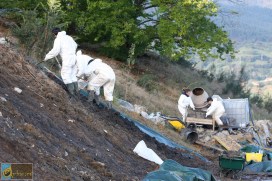
My research team and I carry out regular amber excavations in northern Spain, working in teams of six to ten people. The outcrops that we excavate are often located next to roads and highways because amber is typically uncovered during roadworks. Excavations take place during the summer or fall to try and minimise the risk of rain, and we usually embark on one field trip each year.

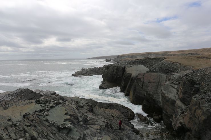
The goal is to recover as much amber as possible – usually a few kilograms – from the muddy and sandy sediments. These materials were transported downstream tens of million of years ago by heavy rain and river swellings from the forests where the resin was produced, before being finally deposited in near-shore areas.

Manual extraction of amber. Credit IGME-UB
Manual extraction of amber in the El Soplao outcrop, Cantabria, northern Spain in 2008. Credit: IGME/UB.

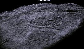
I find amber excavations quite romantic. In the field, amber has a dull appearance that makes it difficult to distinguish from rocks or woody remains. This is due to an opaque crust resulting from oxidation in the sediments and other processes.

This outer layer makes detecting potential fossils inside the amber highly unlikely while the excavation is ongoing. So, in the field we just gather as many amber pieces as possible, and hope for the best.

Only when amber is polished – or shows broken surfaces – does its distinct yellowish to reddish shine emerge, and any possible fossils within become evident. Some ambers are highly fossiliferous, while others are very poor in fossils.

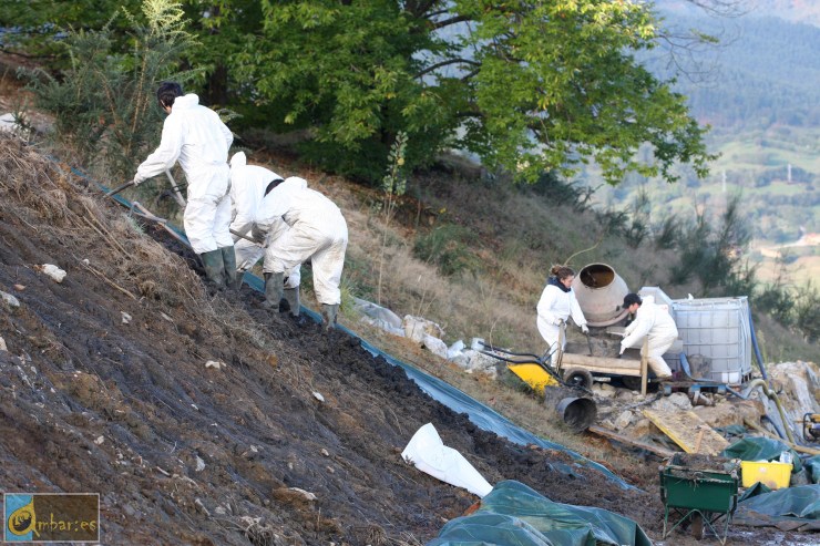
Amber can be gathered by hand using regular tools such as hammers. However, the most efficient method to extract amber from soft sediments is with concrete mixers! This rather unsophisticated piece of equipment provides the best way to recover medium quantities of amber in the field.

We charge water and amber-bearing sediments into the mixer, and after stirring for a while amber floats to the top because it is less dense than muddy water. Then, the surface of the water containing the amber is poured into sieves, which separates even the tiniest pieces.

Amber pieces recovered in a sieve after washing
Amber pieces recovered in a sieve after having been “washed” from their sediment. First amber excavation in the La Manjoya outcrop, Asturias, northern Spain in 2017.

After fieldwork, many hours will be spent looking for fossils within the amber and preparing them. Gathering raw amber is just the first part of a process in unearthing the secrets held within – fragments of encapsulated time.

Top image: First amber excavation in the El Soplao outcrop, Cantabria, N Spain in 2008. Credit: IGME/UB.

Through the looking glass

With our Life, As We Know It redisplay project now underway, our Senior Archives and Library Assistant Danielle Czerkaszyn takes a behind-the-scenes look at how we captured the contents of the current displays for the Museum’s archive.

The archive here holds a unique collection of natural history books, journals and documents covering a wide range of subjects related to the Museum’s collections and research. It also contains papers and objects on the history of the building, providing an institutional memory of Oxford’s ‘University Museum’ since its foundation in 1860.

From an archive perspective it was really important to document the current layout of the cases, their specimens and text before they were removed from the court to make way for the new showcases in the first phase of our redisplay work.

The museum in late 2019

The displays as we know them – with exhibitions on the Oxfordshire dinosaurs, Alice in Wonderland, the Oxford Dodo, and more – were last changed in 2000. For the last 20 years visitors to the Museum would remember their first time being wowed by the Megalosaurus jaw – the world’s first scientifically-described dinosaur – or charmed by the Dodo made famous in Lewis Carroll’s Alice Adventures in Wonderland.

Although after 20 years it is time for a change, the stories and information in the displays are too good to be forgotten. So before anything was removed we began to build the archive for the future.

A display of the fossilised remains of Megalosaurus
The previous display on Megalosaurus: The First Dinosaur

The best way to capture all the information of the displays was through high resolution photography, but this was not as straightforward as we hoped.

The first two obstacles to good photographs are pretty obvious to anyone looking at the cases: glass causes huge amounts of glare; and each case has a big dividing line down the centre where the two sliding glass doors meet, cutting what should be a lovely seamless image into two halves.

To avoid glare and the solve the problem of the dividing line, our photographer Scott opened each individual side of the case, photographed two or three images of the display, and then stitched the separate photos together using Photoshop.

Each case was photographed in two or three segments
The segments were then stitched back together and adjusted for exposure and colour balance to create the final image

Another obstacle to taking good photographs of the displays came from the Museum itself. Some of our larger display furniture, such as the glass case for the Atlantic Bluefin Tuna or the huge T. rex plinth – got in the way of a nice straight shot. Because these items are so large and heavy they were impossible to move, so we had to improvise and do our best.

Capturing the displays before the current cases were removed allowed us to keep an archival record of their contents

Thankfully, we managed to get shots of all 24 displays before they were removed and so a record of each case now rests with the Museum’s archive. If anyone wants to know what the display cases in the court looked like from 2000 to 2020, they will now be able to look back at the images in the archive and recall the magic of the Oxford Dodo exhibit that perhaps first made them fall in love with the Museum.

Our new displays are now in development, and will include some beautiful presentations of the diversity of life, looking at the importance and fragility of biodiversity and human impact on the environment. These new exhibits will show how the biological processes of evolution combine with the geological processes of our dynamic Earth to give rise to the immense, interconnected variety of the natural world.

We look forward to telling you more about that here as the project progresses.

The Life, As We Know It redisplay project is supported by a generous grant from FCC Communities Foundation.

Uncovering ancient threads

By Dr. Frankie Dunn, Research Fellow

Some of the very oldest complex, macroscopic communities on Earth appear in the fossil record about 570 million years ago and record the presence of a group of organisms – the rangeomorphs – with an unfamiliar body plan that, at their ultimate extinction, was lost from life’s repertoire.

Rangeomorphs are characterised by a strange frondose branching anatomy, where large primary branches host smaller branches which themselves host smaller branches again. This arrangement appears to maximise the surface-area to volume ratio of the organism, rather like a lung or a gill would today.

The smallest known rangeomorphs are less than a centimetre in length, but they grew huge and the largest records indicate they could stand more than two metres tall. There is no evidence to suggest that rangeomorphs were able to move around, rather, they lived stuck to the sea floor in the deep ocean, far below the reach of light.

Despite this strange set of characters, there is growing consensus that rangeomorphs likely represent very ancient records of animal life. However, they lived at such a remote time in Earth’s history that they do not possess any direct living descendants. Given all this, it may not be a surprise to hear that we know relatively little about how these organisms made their living and came to dominate the ancient seafloors.

Fig A
The UNESCO world heritage site Mistaken Point in Newfoundland, Canada, is one of the sites on which we find exceptionally preserved rangeomorph fossils. Photo: Alex Liu.

In order to better understand them, my co-author Alex Liu and I travelled to Newfoundland, Canada to explore the rocks which host these remarkable fossils and over the past few years we have made an unexpected discovery. We found that fine filamentous threads connect rangeomorph fronds of the same species, in some cases over many meters, though they are typically between two and 40 centimetres long.

N3
An undescribed rangeomorph fossil with filamentous connections at the base of the frond. We find that this species of rangeomorph can be connected to each other over meters! Photo: Alex Liu. 

It is possible that these filaments were involved in clonal reproduction, like strawberry plants today, but they may have had additional functions such as sharing nutrients or providing stability in strong ocean currents.

The discovery of the filaments means that we have to reconsider how we define an individual rangeomorph, and may help us understand how rangeomorphs (seemingly) rapidly colonised deep-sea environments. Either way, some reassessment of the palaeobiology of these unique organisms is certainly required!

More information:

  • Read the full research paper here.


Top image: Beothukis plumosa, a rangeomorph from Newfoundland showing the intricate branching anatomy of rangeomorphs. Photo: Alex Liu.