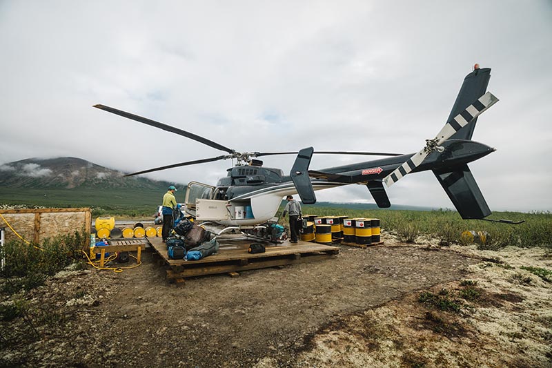
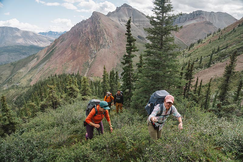
In Summer 2024, a team of palaeontologists and geologists from the University of Oxford, along with colleagues from Dartmouth College, the University of Washington, and Williams College in the USA, undertook an expedition to the Little Dal Group in the Mackenzie Mountains, Northwest Territories, Canada. Our purpose was to uncover some of the oldest fossil ecosystems that record complex life.
Photo: Robert GillPhoto: Robert Gill
Complex life comprises all organisms whose DNA is enclosed in a cell nucleus. This includes animals and plants but excludes bacteria. Today, this complex life accounts for most of the Earth’s biomass, documented biodiversity, and oxygen production. Understanding when and how it first evolved remains one of the central unanswered questions in evolutionary biology.
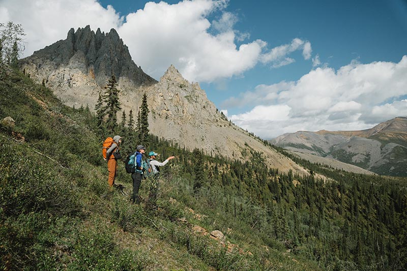
Photo: Robert GillPhoto: Robert Gill
As palaeontologists, we normally use fossils to reveal the history of life. Fossils tend to preserve larger animals with hard shells or skeletons—creatures such as trilobites, ammonites, dinosaurs, and mammoths. However, the first complex organisms were microscopic and lacked such hard parts. As a result, their soft and fragile cells rarely fossilised. Put simply, we have found it a major challenge to trace the origins of complex life with fossils.
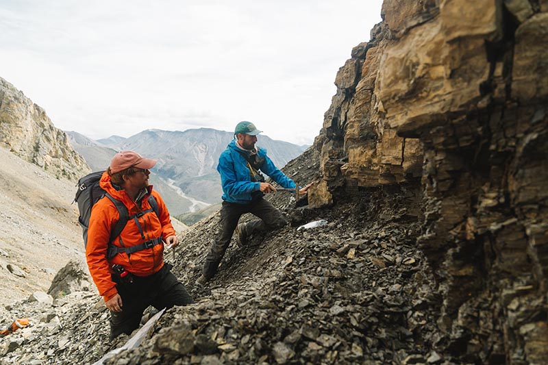
Photo: Robert GillPhoto: Robert Gill
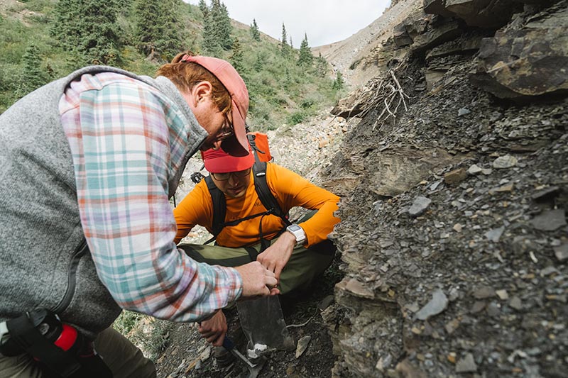
I have argued that finding rocks made up of antibacterial clay minerals holds the key. These minerals can slow the decay of organic cells long enough for them to survive as fossils. The Little Dal Group contains ~900-million-year-old rocks that are rich in just such clays, making it a prime target for new fossils that might help us unravel the origins of biological complexity.
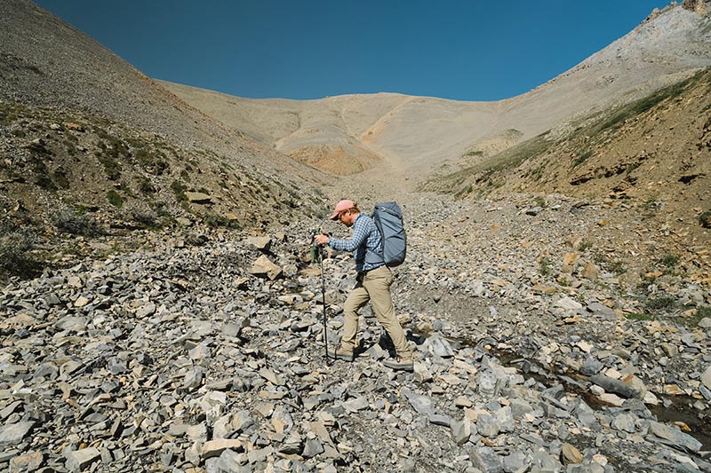
Photo: Robert GillPhoto: Robert Gill
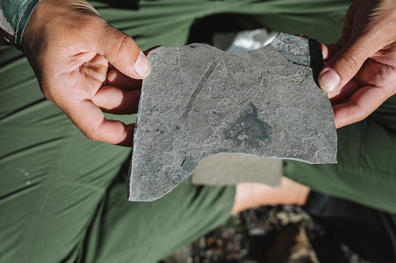
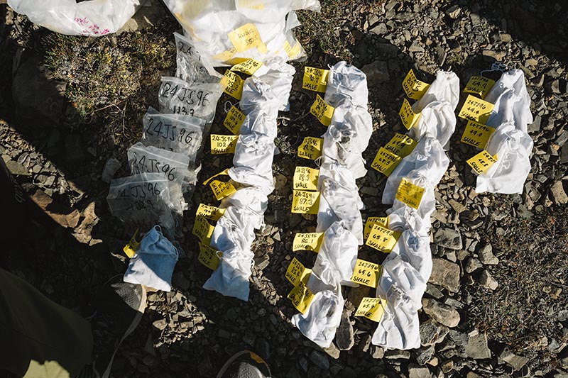
I was joined in Canada by my DPhil student, George Wedlake, from the Department of Earth Sciences. Together we spent two weeks collecting over 100 rock samples. The samples record an ancient tropical sea not unlike the Bahamas today, where early complex life likely flourished.
Back in Oxford, at the Museum of Natural History, George and I are now examining the samples; dissolving the rocks with hydrofluoric acid to extract and study the tiny fossils. We hope these new fossils will transform our understanding of how complex life first took hold on our planet.
Our fieldwork was funded by a Royal Society University Research Fellowship and by the Oxford NERC Environmental Science Doctoral Training Partnership. It was conducted under permit and with the support of the Sahtú Dene people.
Dr Emma Nicholls and colleagues discuss some of the fascinating stories behind the species and specimens featured on these stamps.
Megalosaurus stamps
Two of the stamps in the Age of Dinosaurs stamp set include artistic reconstructions of Megalosaurus by the palaeo-artist Joshua Dunlop. The animal that nineteenth-century naturalists once understood to be a lumbering long-legged lizard is now depicted as a fearsome Jurassic predator that ran on its hind legs and tore into prey with its large serrated teeth. Dunlop shows Megalosaurus wading through shallow coastal waters, preparing to pounce on Cryptoclidus¸ a plesiosaur that lived alongside Megalosaurus in Jurassic Britain. The artwork also shows Megalosaurus covered in feathers. Although we don’t have any direct evidence that Megalosaurus was a feathered dinosaur, feather-like filaments have been found among the fossils of other dinosaurs such as Sciurumimus, meaning it is highly possible that Megalosaurus had feathers too.
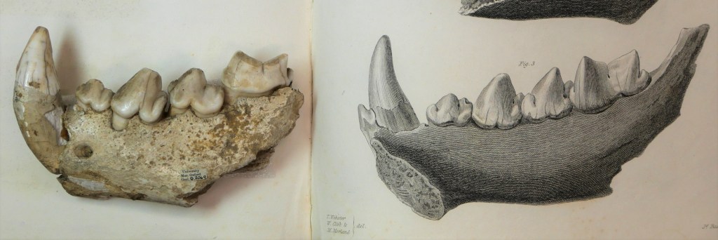
OUMNH’s famous Megalosaurus type specimen, a lower-right jawbone with teethMegalosaurus stamp from the Age of Dinosaurs series
Dapediumstamps
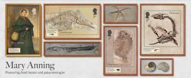
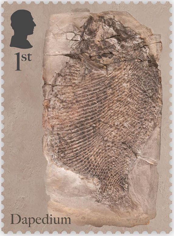
OUMNH collaborated directly with Royal Mail to help produce the Age of Dinosaurs miniature sheet, which showcases fossils collected by Mary Anning. One of the stamps in this collection features a photograph of the fossil of an extinct Jurassic fish, Dapedium, which is housed at OUMNH.
Despite Anning’s illustrious reputation, it wasn’t always known that this Dapedium specimen was connected to her — all that was known about it was that it had probably once belonged to William Buckland and had been collected from Lyme Regis.
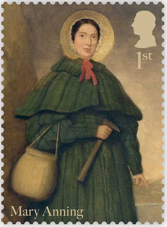
Stamp from the Age of Dinosaurs series featuring a portrait of Mary AnningStamp from the Age of Dinosaurs series featuring a Dapedium fossil from OUMNH
Although Anning is one of the most prolific fossil collectors to have worked in Lyme Regis, naturalists like Buckland often visited Anning to go “fossicking” together, or purchase fossils from her. There are very few archival records of transactions between Anning and other fossil collectors from this time, making it difficult to decipher exactly who extracted fossils such as this, found in nineteenth-century Dorset.
Fortuitously, while Dr Sue Newell was conducting research for her PhD on the Buckland Collection in 2021, she found an exciting letter in OUMNH Archive, dated 3rd September 1829. It was from a former student of Buckland’s, Beriah Botfield, and contained details of two fossils that Buckland had bought from Anning to present to the University of Oxford. Using evidence in the letter, Sue was able to work out that Botfield was referring to a Dapedium fossil which she later recognised tucked away in OUMNH’s fossil store.
Botfield had had the fossil mounted in an expensive (and very heavy!) stone frame, with “Presented by Beriah Botfield Esq. Dapedium politum. Lyme, Dorset” beautifully inscribed on the front surface. At the time, the identity of Anning as the fossil’s original finder, identifier, preparator and vendor, was probably common knowledge and, typically, Botfield did not consider these facts important enough to record on his presentation frame.
The Dapedium fossil is a near-complete example of this Jurassic fish, in which scale patterns and delicate fin structures are preserved in breathtaking detail. Dapedium is the first OUMNH object to grace a Royal Mail stamp – an ideal choice given its scientific and historic importance.
Visit the Museum to see the Dapedium fossil as well as temporary displays about the new stamp collection.
CELEBRATING THE RECENT ACQUISITION OF AN IMPORTANT ARCHIVE
By Danielle Czerkaszyn, Librarian and Archivist and Grace Exley, AHRC Doctoral Student
200 years since the first scientific description of a dinosaur, the Museum has welcomed a significant archival collection relating to the man who introduced us to Megalosaurus, William Buckland (1784-1856). The archive contains over 1,000 items including letters, notebooks, family papers, prints, and artworks. It joins the Museum’s existing Buckland archive, as well as more than 4,000 geological specimens, and helps fill in the knowledge gaps surrounding the life and work of Oxford’s first Reader in Geology and Mineralogy. Not only is there the potential to learn more about Buckland’s early life as a student at Christ Church, there is also material relating to the wider Buckland family, including his son, the zoologist Francis Trevelyan Buckland, and wife, the naturalist Mary Buckland (née Morland, 1797-1857).
Among the 70 letters in the archive that are addressed to Mary, there is correspondence from chemist William Wollaston, Scottish polymath Mary Somerville, and a lively letter from John Ruskin, explaining to Mary his disgust at all things marine:
“I dont [sic] doubt that those double natured or no-natured salt water things are very pretty alive, but they disgust me by their perpetual gobbling and turning themselves inside out and on the whole I think for purple and rose colour & pretty shape, I may do well enough with convolvulus’s [sic] & such things which dont [sic] eat each other up, backwards & forwards all day long.”
Ruskin was clearly teasing his friend, as molluscs happened to be Mary’s specialist subject!
Mary Buckland’s notebook, recently acquired by OUMNH
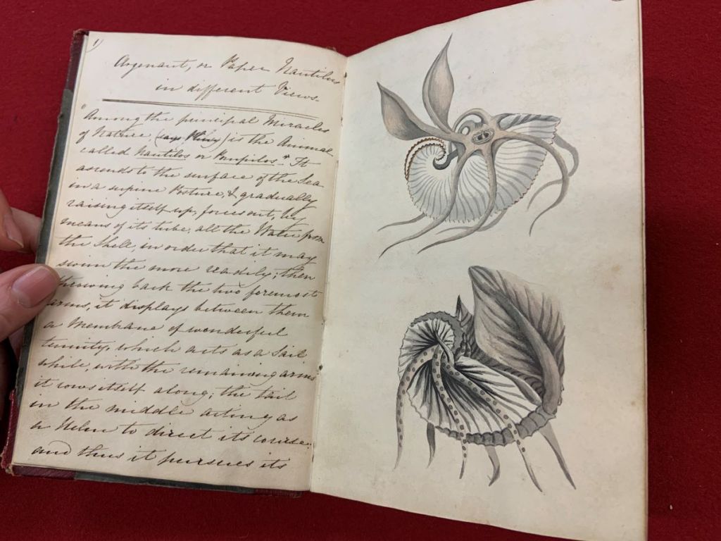
The collection also contains two sketchbooks belonging to Mary, one of which dates from June 1817, seven years before her marriage to William and contains exquisite ink and watercolour illustrations of natural history specimens.
Illustration of Megalosaurus type fossil by Mary Morland, used in the original 1824 description of the species.Fulgurite, or, as Mary called it a ‘vitrified sand tube’. These form when lightning strikes the ground, particularly sandy surfaces. Similar examples are present in the remains of Mary’s mineral collection, held by OUMNH.
The sketchbook gives us a rare glimpse into how a nineteenth-century woman learned about natural history. The book contains copied passages from natural history texts, enabling us to trace what Mary was reading. Her interests spanned geology and mineralogy, and she also included pieces on zoological curiosities and even polar exploration. She read and copied extracts from a variety of sources, some of which – George Shaw’s Zoological Lectures, for example – were intended to suit a lay-audience (as Shaw put it, intended as a “familiar discourse with Lady-Auditors”). However, other elements of her reading were probably never intended for a woman like Mary — she also copied passages from the Transactions of the Geological Society even though women could not join as Fellows until 1919. As archival materials relating to women are often sparse, this is a truly rare and incredibly valuable insight into how Mary used her connections to access resources and the techniques she used to teach herself about natural history.
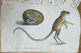
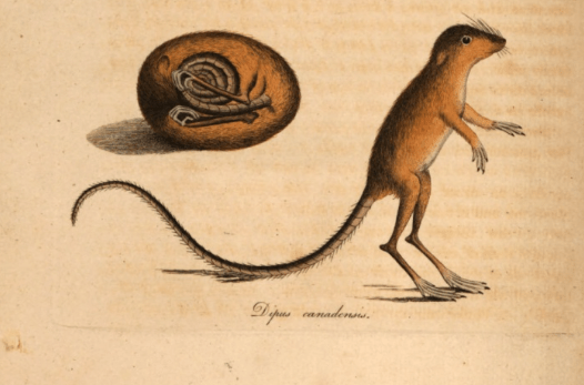
Perhaps the most striking feature of the notebook is its intricate, exquisite illustrations. These, done in watercolour, ink, and pencil, are reproductions of the figures from the works Mary copied out. A favourite in the sketchbook is the “Canadian Jumping Mouse”, a long-tailed rodent described in a piece in the Transactions of the Linnaean Society by Major General Thomas Davies in 1797. There are also many representations of molluscs (detailed enough to repulse Ruskin), mineral specimens, and occasional fold-out geological sections. As we flick through the book, we can see Mary experimenting with media and techniques — not only developing as an artist but also honing her skills as a scientific illustrator.
The skills and knowledge Mary developed in her natural history notebook were crucial to her later collaboration with William, as well as her own independent work as a draughtswoman before her marriage. In 1824, when Buckland presented the jaw of Megalosaurus to the Geological Society, it was “M. Morland” who provided the painstakingly detailed plates. Research has begun to uncover the extent of Mary’s work as a naturalist and illustrator, and now, with the help of the materials in the newly acquired archive, we can explore the origins of her skills. The archive is currently in the hands of a Paper Conservator, Anna Español Costa, to ensure the material is kept in the best condition for many years to come. Items from the archive will feature in the Breaking Ground exhibition, opening in October 2024.
Our fundraising campaign saw us receive generous support from the National Heritage Memorial Fund, Arts Council England/V&A Purchase Grant Fund, Friends of the National Libraries, Headley Trust, and other private donors. Additionally, in late 2022, we launched the Buckland Papers Appeal, asking members of the public to help us meet our target to purchase the archive. Thank you to all our funders and members of the public who responded to our call. We could not have raised the money so quickly without your support and we are now thrilled to share the archive with you all.
Looking through the collections at OUMNH never gets boring, but sometimes a drawer will open up to reveal something even more eye-catching than the fossils usually found inside. Whilst working on the Museum’s Jurassic marine reptiles a few weeks ago, I came across something particularly surprising: a jewel-green box with a fantastic piece of art on the front. I was instantly intrigued and reminded of all the other times I had encountered a holder as fascinating as the specimen inside it.
Storage in museum collections is an ongoing pursuit of balance between ideal environmental conditions, specimen accessibility, and efficient use of space. This balance applies to all levels of storage: from building to room, cabinet to specimen tray. OUMNH’s Earth Collections are stored in conservation-grade, acid-free boxes or trays made of plastic or cardboard. These boxes are sometimes layered with low-density foam or ‘plastazote’ which can be carved to fit the specimen and keep it from being jostled or damaged. Holders with lids can also provide a micro-environment for specimens to help minimise their exposure to changes in humidity and temperature. The use of these standard materials not only helps protect specimens from degradation but can also deter pests from harbouring in collections spaces.
However, historical collections like those at OUMNH may retain holders that are not standard use. Sometimes, a clean and empty plastic Ferrero Rocher box is the perfect size for that small mammal skeleton that needs storing! Other times, an unusual holder might have been the only thing a field collector had on hand to transport a specimen to the Museum.
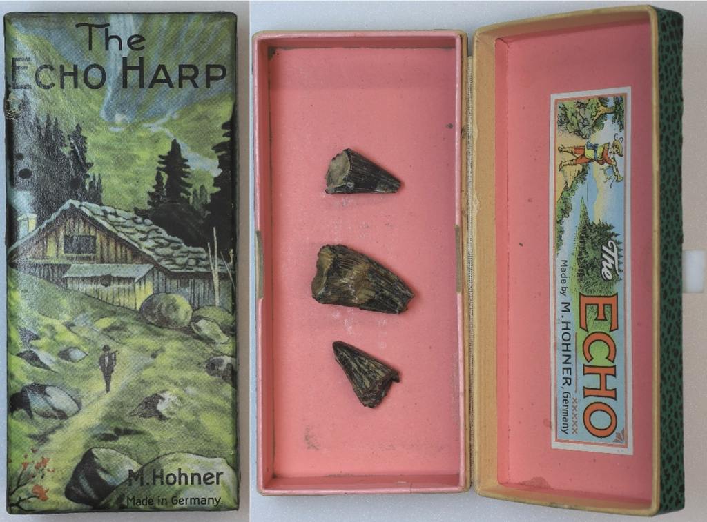
A harmonica box containing pliosaur teeth, a marine reptile that lived during the Jurassic (145.5 million – 201.6 million years ago).
One example of an unusual specimen holder is this ‘Echo Harp’ box by pre-eminent German harmonica manufacturer Hohner, likely from the 1960s. The box no longer holds a harmonica, but instead accompanies pieces of Jurassic pliosaur teeth from Weymouth, Dorset. Pliosaurs were a kind of carnivorous marine reptile related to plesiosaurs, with four flippers, and long tails and necks. If they hadn’t gone extinct in the Cretaceous-Paleogene extinction event 66 million years ago, perhaps they would have come to appreciate the harmonica and its artistic packaging!
Aside from their artistic value, museums may sometimes retain unusual holders because they contain primary source information on the specimen. One such example is a ‘Bryant and May’s Patent Safety Matches’ box in our Earth collections, bearing a packaging design from the early 1900s. The box actually houses a chicken tarsometatarsus bone excavated from “High St. New Schools” in Oxfordshire and is accompanied by a label which describes the particular layer of gravel the specimen was found in — important information for any archaeological or palaeontological find. Although the specimen is stored alongside Pleistocene fossils (10,000 – 2.6 million years ago), chickens did not originate in the UK, so the bone is likely from much more recent times. Someone still must have thought it was important enough to keep in its own special holder!
A Tate and Lyle sugar bag containing a Jurassic specimen, with handwriting on the outside describing the stratigraphy the fossil was found in.
Similarly, this ‘Tate and Lyle Granulated Sugar’ paper bag features a handwritten original notation in blue pen on the outside. The bag originally contained a specimen found in a collection of Jurassic gastropods and bivalves from Somerset, with the handwriting describing the fossil’s stratigraphic information. The bag also features a recipe for cinnamon apples on the reverse, which we have yet to try!
A wooden box and the Quarternary fossils (up to 2.6 million years ago) it originally housed. An accompanying letter describes the delivery of the fossils to William Buckland, Oxford University’s First Reader in Geology.
In addition to primary source information, original holders may also provide specimens with provenance. This ovular wooden box filled with organic stuffing material originally contained Quarternary fossil specimens found in Peak’s Hole, Derbyshire. The Museum archive also holds a handwritten letter describing the specimens inside the package and how they were found. The letter dates to 1841 and is addressed to Oxford University’s first Reader in Geology, William Buckland. The specimen holder forms part of a group of objects with such a strong interconnection, and such strong documentation, that retaining the box is a matter of course.
All in all, it’s great that we’ve come so far in the advancement of safe and stable housing for specimens. At the same time, it’s always fascinating to see what else has made its way into collections, just by nature of being able to hold things, either for a short time or a long one. Despite living in the Earth Collections – among fossils, rocks, and the geological past – these objects offer us a little bit of human history too.
Susan Newell is a doctoral student researching the teaching collections of William Buckland, the first Professor of Geology at Oxford who taught from 1813 to 1849. She reminds us here about Buckland’s role 200 years ago in interpreting the important Pleistocene discoveries being celebrated this year, and the way that Mary Morland, a talented local naturalist, and many others, contributed to making this new knowledge.
This year marks the 200th anniversary of a great advance in our understanding of the geological past… a story which begins in the nineteenth century, with the discovery of a bone-filled cave in Kirkdale, Yorkshire.
Uncovered by local quarrymen in 1821, the discovery of the Kirkdale cave and its contents of mysterious bone was the source of much intrigue. When news of the discovery reached William Buckland, Professor of Geology at Oxford University, he decided to travel up North to visit the site. However, by the time Buckland arrived at the cave, local collectors had scooped up most of its contents. Nonetheless, he was able to retrieve and examine some of the cave’s remaining material, which led him to an astonishing conclusion — Yorkshire must once have been home to hyaenas, elephants, hippopotamus and rhinoceros, and what was now known as the Kirkdale cave was once a hyaenas’ den.
W. B. Conybeare, lithograph, ‘The Hyaena’s Den at Kirkdale near Kirby Moorside in Yorkshire, discovered A.D. 1821’. Reproduced by kind permission of Christ Church, Oxford. This light-hearted reconstruction of the hyaenas’ den shows Buckland illuminating the scene, in every sense. It is thought to be the first visual reconstruction of the pre-human past.
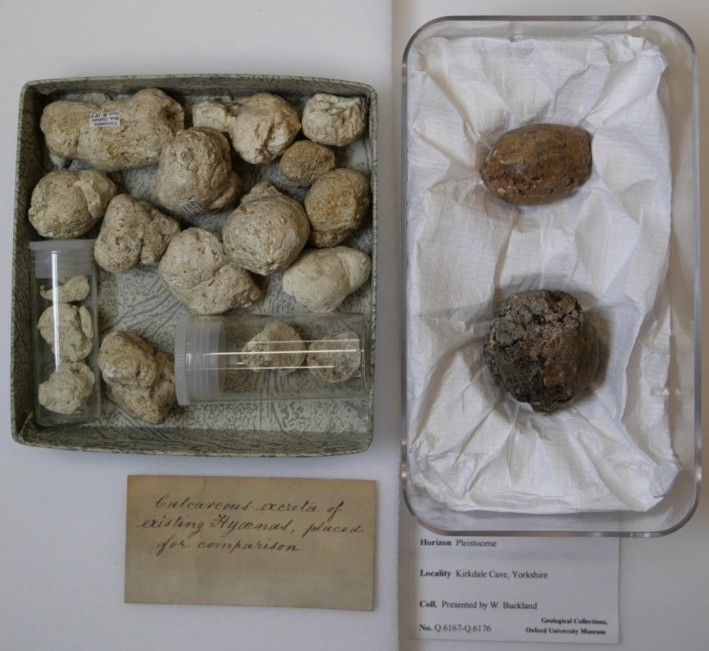
Central to Buckland’s theories were some small white balls that he had found amongst the debris in the cave. Buckland sent these balls to William Wollaston, a celebrated chemist based in London, for analysis. He also asked Wollaston to visit the zoo at Exeter Exchange in London and show the balls to the hyaena’s keeper there. Together with the results from Wollaston’s chemical analyses, the keeper confirmed Buckland’s hypothesis — the balls were droppings from animals very similar to modern hyaenas. Meanwhile, the anatomist William Clift was able to identify the bones from the Kirkdale cave as belonging to other extinct species related to those found living in tropical countries today. Buckland concluded that the cave must have been a den for ancient hyaenas, who would drag parts of the dead animals they had found (or killed) inside and, after feeding on them, leave piles of bones and droppings behind.
In order to strengthen his theory, Buckland discussed the behaviour of hyaenas in the wild with army officers connected to Britain’s colonial expansion in India. These officers also sent Buckland fresh specimens captured by local people. When a travelling menagerie visited Oxford in 1822, Buckland took the opportunity to experiment; feeding bones to a hyaena and noting that the teeth marks matched those on the fossilised bones from the cave.
Left: “Small white balls” of hyaena excrement. The left box probably contains samples collected from the hyaena in the travelling menagerie which visited Oxford, and the right contains fossilized samples collected by Buckland from Kirkdale Cave, now part of the Museum’s Collections. Right: Comparison of two shinbones – one fed to a hyaena by Buckland, and the other collected from Kirkdale cave. The similarity of the gnaw-marks suggest that hyaenas were once present in Kirkdale. On display in the Museum.
Buckland’s findings were something of a shock to his contemporaries. When lecturing, he employed several different methods to try and convince his audiences that his theories were true. This included presenting fossil specimens and bones from living species for comparison, and showing maps, diagrams and drawings. Mary Morland contributed some of these illustrations, including large drawings of living animals, and technical drawings of bones that were later engraved for use in Buckland’s publications. Mary’s Kirkdale drawings seem to have been the first that she produced for William before the couple married in 1825.
Fossil hyaena jaw in the Museum’s collection, possibly the one featured in the engraving alongside it. Engraving is by James Basire after a drawing by Mary Morland. Published in William Buckland’s article in the Royal Society’s journal (1822) on the Kirkdale cave discoveries. [1]
Buckland’s work on the Kirkdale cave was revolutionary, not least because he was the first to make a scientific study of a cache of bones of this type. Although similar bones from ‘tropical’ species had previously been found in Northern Europe, people thought that they had been washed up by a catastrophic flood, believed by many to be the biblical Noah’s Flood. Modern analysis has now allowed us to deduce that the bones date to an Interglacial period when Britain was joined to Europe and had a hot climate, about 120,000 years ago.
Here at the Museum, Buckland’s collections and archives are as much of a treasure trove as the Kirkdale cave. It is through accessing these archives that we can learn about the surprising range of people who contributed to the emergence of new scientific knowledge from the Kirkland cave — quarrymen, collectors, zookeepers, chemists, anatomists, colonial officers in India, workers in India, and artists like Mary Morland. To find out more about the incredible legacy of the Kirkdale Cave, look out for ‘Kirkdale200 – Lost Beasts of the North’, a symposium organised by the Yorkshire Fossil Festival, 12th March 2022.
Mary Morland, watercolour and gouache, lecture illustration of a hippopotamus, signed ‘MM’. Hippopotamus bones were found at Kirkdale cave in Yorkshire, but as there were no living hippos to be seen in Britain at the time, this drawing would have been a valuable teaching aid.
[1] William Buckland, ‘Account of an Assemblage of Fossil Teeth and Bones of Elephant, Rhinoceros, Hippopotamus, Bear, Tiger, and Hyaena, and Sixteen Other Animals; Discovered in a Cave at Kirkdale, Yorkshire, in the Year 1821: With a Comparative View of Five Similar Caverns in Various Parts of England, and others on the Continent’, Phil. Trans., 2 (1815-30), 165-167.
Nature is wonderfully imperfect, and the data that we can gather from it is even further from perfection. Fossil localities, even those providing exceptionally well-preserved fossils, are inaccurate records of the past. Fossils can form from a variety of matter including organisms, their remains, or even traces of their activity. Yet not all of the material that can get fossilised at a particular site actually will. Among other factors, biases in the fossil record result from the nature of the materials responsible for fossilisation – usually sediments which are in the process of turning into rocks. In most cases, fossil localities offer us only a single ‘window of preservation’ – a skewed geological record of the ancient ecosystem that once existed there.
In 2012, a rich vertebrate bone bed was documented at the Ariño site in Teruel, Spain. Since then, researchers have unearthed more than 10,000 individual fossil bones, from which they have discovered new species of dinosaurs, crocodiles, and turtles. Plant fossils were also found, including pollen grains and amber, which is fossilised resin. Although amber was known to occur in this locality, this sort of material had remained unstudied… until recently.
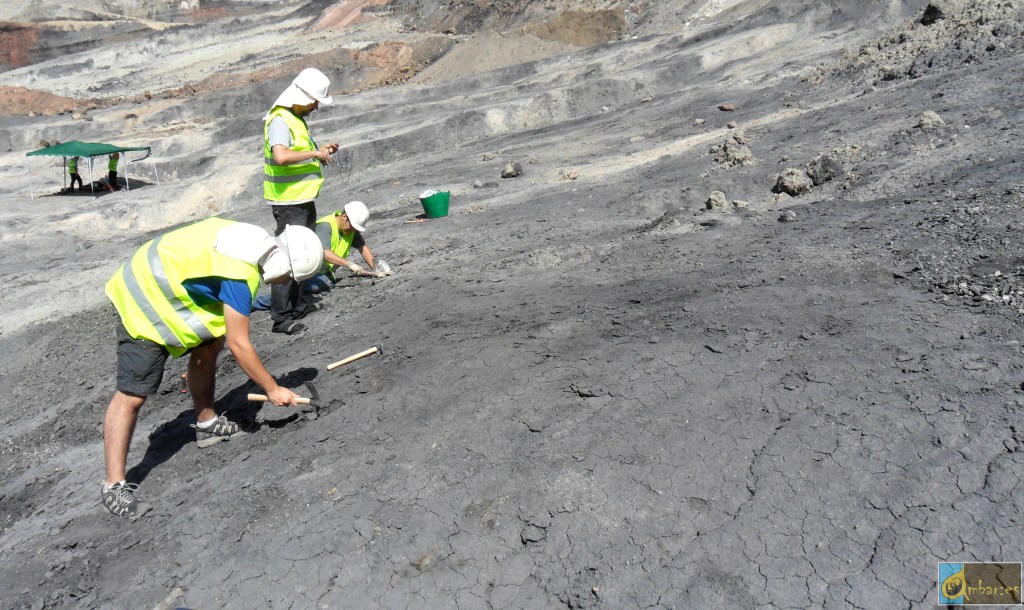
Over the summer of 2019, I joined my colleagues to carry out amber excavations in the Ariño site – an open-pit coal mine that has an almost lunar appearance due to the dark carbonate-rich mudstone rocks and the total lack of vegetation. The scorching heat during a very hot summer was a bit maddening, but I did try to enjoy my yearly dose of sun before returning to the UK!
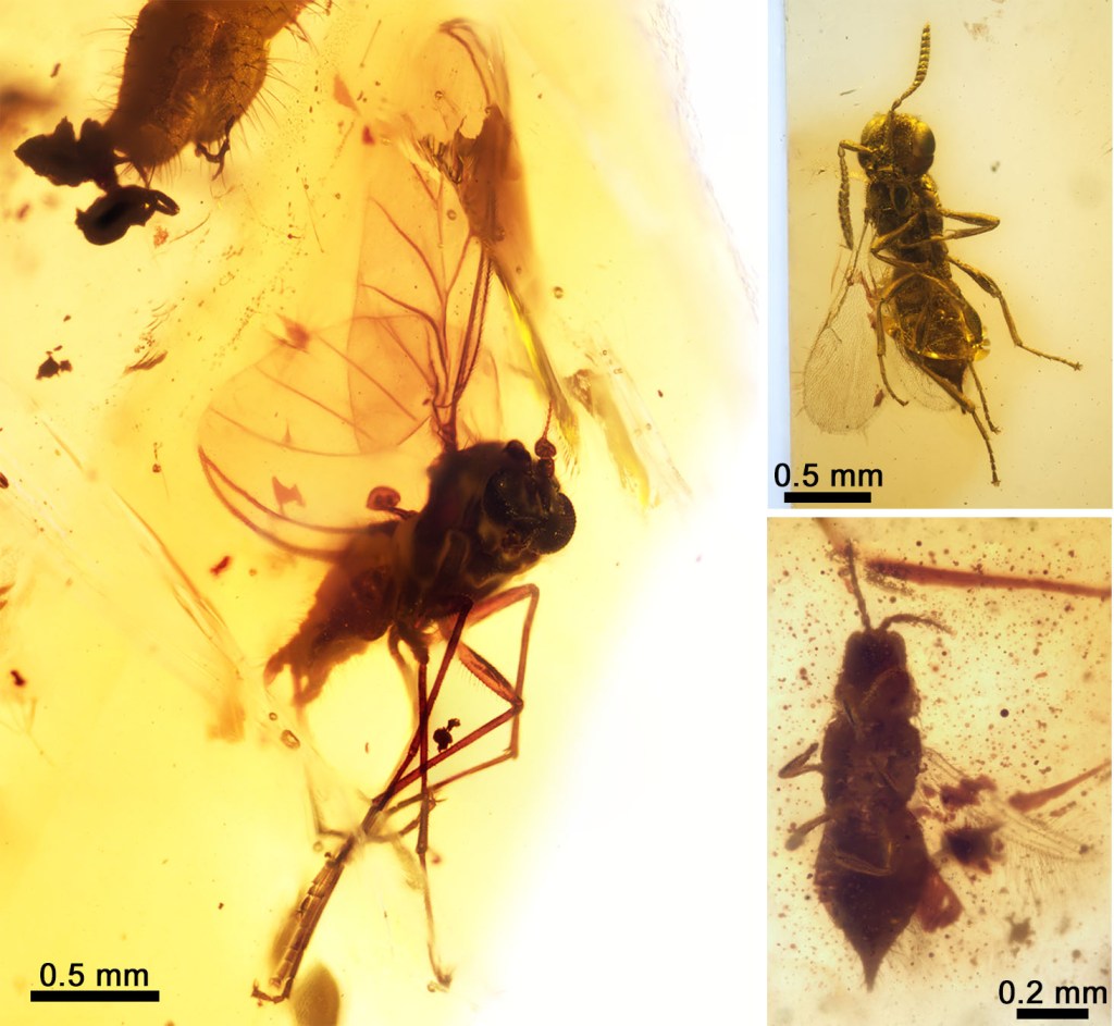
(Left) Manual extraction of amber from Ariño during the 2019 summer campaign. (Right) A selection of fossil insects found in Ariño amber: a true fly belonging to an extinct family (left); a platygastroid parasitoid wasp (top right); and a thrips (bottom right). Modified from Álvarez-Parra et al. 2021.
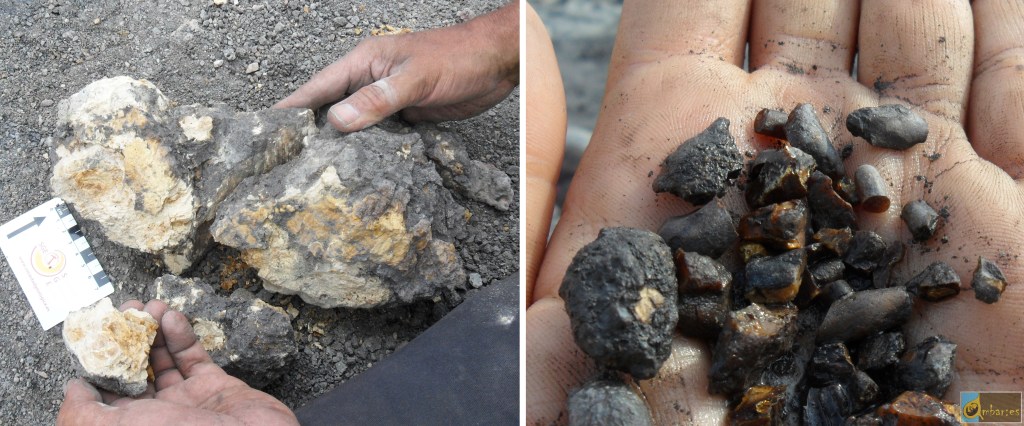
Resin pieces can be transported significant distances by runoff water before depositing on their final burial location, where they slowly transform into amber. However, we found amber pieces that had not moved from their original place of production. These large, round-shaped pieces preserved delicate surface patterns that would have been polished away even by the slightest transport. The resin that produced these amber pieces was formed by the roots of the resin-producing trees, and resembles sub-fossil resin my colleagues found in modern forests from New Zealand.
Large amber piece produced by roots (left) and assemblage of smaller amber pieces (right) from Ariño (Teurel, Spain).
The small amber pieces from Ariño contain an unusual abundance of fossils. These pieces come from resin produced by the branches and trunk of the resin-producing trees. From the almost one kilogram of amber we excavated, we identified a total of 166 fossils. These include diverse insects such as lacewings, beetles, or wasps, and arachnids such as spiders and mites. Even a mammal hair strand was found!1
We now know that the Ariño site provides two complementary windows of preservation — a bone bed preserving a rich variety of vertebrate animals, and amber with abundant inclusions. Aside from Ariño, only three localities that preserve both dinosaur bone beds and fossiliferous amber have been reported in Western France, Western Canada, and North Central United States. However, in these cases, either the bone bed or the amber have offered a much more modest abundance and diversity of fossils. Some of the fossils from these localities also show signs of significant transport, which means that the organisms could have inhabited different, distant areas even though they fossilised together. This makes Ariño unique because it offers two valuable ‘windows of preservation’ from the same ecosystem.
Thanks to all this evidence and other data, we have been able to reconstruct an ancient terrestrial ecosystem – a 110-million-year-old coastal swamp – with unprecedented detail and accuracy.2 The inherent incompleteness of the fossil record will always remain a headache for palaeontologists… but localities like Ariño make the data that we can recover from the past a bit more complete.
Reconstruction of the coastal swamp forest of Ariño, in the Iberian Peninsula, from 110 million years ago. Author: José Antonio Peñas. Source: Álvarez-Parra et al. 2021.
1Álvarez-Parra, Sergio, Ricardo Pérez-de la Fuente, Enrique Peñalver, Eduardo Barrón, Luis Alcalá, Jordi Pérez-Cano, Carles Martín-Closas et al. “Dinosaur bonebed amber from an original swamp forest soil.” Elife 10 (2021): e72477.
2Álvarez-Parra, Sergio, Xavier Delclòs, Mónica M. Solórzano-Kraemer, Luis Alcalá, and Enrique Peñalver. “Cretaceous amniote integuments recorded through a taphonomic process unique to resins.” Scientific reports 10, no. 1 (2020): 1-12.