Hedgehog Awareness Week

For Hedgehog Awareness Week, Zoology Collections Manager Mark Carnall and Museum Librarian and Archivist Danielle Czerkaszyn discuss these prickly and charming creatures.

The 2-8 May is Hedgehog Awareness Week, which give us an excuse, not that one were needed, to talk about these charismatic mammals. Although the West European hedgehog (or common hedgehog if you’re in Europe, these vernacular names get very confusing when geography and language is taken into account), Erinaceus europaeus, is probably the hedgehog that springs to mind to many of our readers, there are nearly twenty living species of hedgehog and many fossil species are known.

Hedgehog specimen at OUMNH

In terms of evolutionary relationships they share a family with the moonrat and the rather wonderful gynmures, distinctly un-hedgehog-like relatives.

Their characteristic spikes that run across the back of hedgehogs are modified hairs which are periodically replaced and each individual hedgehog has around 7000 spines at any one time, varying slightly with age and size. Behaviourally, they are competent climbers (and have a built in shock-absorbing coat should they fall) and surprisingly perhaps, all species are thought to be competent swimmers.

Although much loved across their native range, Erinaceus europaeus, is considered a pest species in New Zealand where it was deliberately introduced as a form of biological control, by acclimatisation societies and possible as pet animals. They have now spread to all but the highest parts of New Zealand threatening native species of birds, amphibians, reptiles and directly competing with native mammal species.

In 2020, Erinaceus europaeus was added to the Red List for British Mammals as vulnerable across the lists for Great Britain, England, Scotland and Wales informed by analysis of citizen science data although there remains some uncertainty about true population levels.

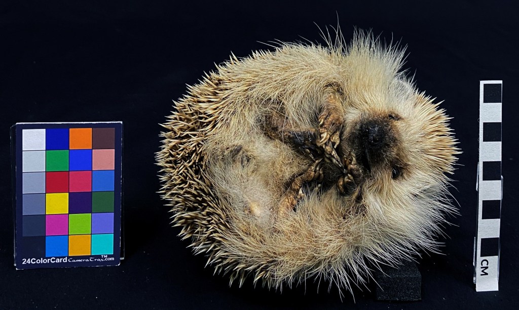
Unsurprisingly perhaps they are comparatively well represented in the collections at the Museum including specimens donated and prepared for the Museum from the 19th Century through to much more recent specimens acquired from road death animals for display. The specimen pictured above being one such relatively recent acquisition for display in the Museum’s display case on the animals featured in Alice in Wonderland.

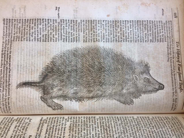
We’ll leave you with one more hedgehog from the Museum’s library and archives. Hedgehogs unusual appearance initially led to some odd beliefs about why their quills existed. For example, in his book ‘The History of Four-Footed Beasts and Serpents’ (1658) Edward Topsell wrote:

“The hedgehog’s meat is apple, worms and grapes: when he findeth them upon the earth, he rolleth on them until he hath fylled up all his prickles, and then carrieth them home to his den.”

– Edward Topsell

One of the most common questions about hedgehogs is how do they mate? The answer is of course, very carefully.

The astounding story of the fake butterfly specimen Papilio ecclipsis – would you be fooled?

For April Fool’s Day, our Senior Collections Manager Darren Mann recounts the story of an elegantly fake butterfly – Papilio ecclipsis – asking whether it was a piece of scientific fraudulence or practical joke that went awry.

James Petiver, a 17th-century London apothecary, was renowned for having one of the largest natural history collections in the world. Petiver (1665-1718) published some of the first books on British insects and created common names for some of our butterflies.

Volume 3, Plate II of Jones Icones – the two lower images are of Papilio ecclipsis

On plate 10 of his Gazophylacium naturae et artis — an illustrated catalogue of British insects (1702) he figured a unique butterfly that “exactly resembles our English Brimstone Butterfly were it not for those black Spots, and apparent blue Moons in the lower wings”. It was given to him by his late friend and butterfly collector William Charlton (1642-1702). This butterfly was later named Papilio ecclipsis by the father of taxonomy himself, Carl Linnaeus, in his 1763 work Centuria Insectorum Rariorum, and it became known as the Charlton Brimstone or the blue-eyed brimstone.

Petiver’s collection was purchased by Sir Hans Sloane (1660–1753), who later donated his entire ‘cabinet of curiosity’ to the nation, becoming the foundation for the Natural History Museum, London, originally part of the British Museum. It was here that wine merchant and naturalist William Jones (1745-1818) examined and later figured Petiver’s specimen in his Icones, an unpublished masterpiece of some 1,500 watercolour images of butterflies.

Jones’ Icones, held in the Museum’s archive, is the subject of numerous articles and is still examined by butterfly specialists the world over. Many of the specimens figured by Jones are no longer in existence, being ravished by pests or lost over time, so all that remains of these butterflies are the painted images within.

A drawer of British butterflies from the cabinet of William Jones. The Common Brimstone butterfly is the fourth from the right on the top row

When visiting London, Danish entomologist Johann Christian Fabricius (1745-1808) studied the paintings that Jones made and described over 200 species of butterfly new to science. Fabricius also visited the British Museum where he examined Petiver’s specimen of ecclipsis. In Entomologia systematica (1793) Fabricius revealed the enigmatic ecclipsis to be no more than a painted and “artificially spotted” specimen of the Common Brimstone (Gonepteryx rhamni). So, the dark spots and blue eyes were merely artistic licence, but whose?

Iconotypes, published by Thames & Hudson, will be available from October 2021

Petiver’s specimen, seen by both Jones and Fabricus in the British Museum in the late 18th century, had mysteriously disappeared by the following century. It is said that when Dr. Gray (1748-1806), Keeper of National Curiosities at the Museum, heard of the deception he became so enraged that he “indignantly stamped the specimen to pieces.”

It is still unclear whether this was an example of scientific fraud by Charlton, or if it was intended as a practical joke that went awry.

There remain two specimens of ecclipsis in the collection of the Linnean Society. Although it is uncertain who created these, it is believed that these replicas were made by none other than our very own William Jones, as he was one of the few who had the artistic skills to undertake such work. The forthcoming publication of Iconotypes, showing Jones’ Icones in all its splendour, will hopefully demonstrate how he had both the knowledge and the skill to recreate these fascinating fakes.

Links and References
Salmon, M., Marren, P., Harley, B. (2001) The Aurelian legacy: British butterflies and their collectors. University of California Press.
The Linnean Society https://www.linnean.org/
Vane-Wright, R. I. (2010) William Jones of Chelsea (1745–1818), and the need for a digital online ‘Icones’. Antenna. 34(1), 16–21

Crunchy on the outside

By Susie Glover, HOPE Learning Officer

We have an ambitious project underway at the Museum, to preserve a unique and scientifically important collection of over one million British insects. It’s called HOPE for the Future, after the Hope Entomological Collections, and we are keen to shout about how these specimens can help us understand biodiversity, habitats and ecologies.

The learning team behind the project are today launching a new blog for young people interested in entomology. Intriguingly, it’s called Crunchy on the Outside, but please don’t confuse this with the similar, but fundamentally different, mid-’90s advertising campaign for the Dime bar.

A high magnification image of an insect, face-on, showing large proboscis and brown body.
One of many weird and wonderful specimens from our collection, the Acorn Weevil (Curculio glandium).
Here is a peek at some of the tools of the trade, used to move and mend specimens like this moth.

Crunchy will be crammed full of interesting insect info, fun things to make and do, a peek behind the scenes at the Museum, and news from people, past and present, who work in the field of entomology. The odd bad joke may also worm its way in (What do butterflies sleep on? Cater-pillows).

The blog will also be a platform for young people to have their say, about the topics covered on Crunchy itself, as well as on the activity of the Museum. It will give them first dibs on access to related events too. You can check it out, follow, and share at crunchyontheoutside.com.

HOPE for the Future is funded by the National Lottery Heritage Fund.

Wax models of magnified mites mounted on a black board

Of parasites, dinosaurs, and other model animals

Elaine Charwat has been on a journey into the attic storerooms behind the scenes of the Museum to discover 19th-century wax models of parasites. A strange occupation you might think, but it’s all part of her doctoral research programme with the Arts and Humanities Research Council to learn about the use of models and replicas in science, past and present. In the podcast above Elaine meets Mark Carnall, Zoology Collections Manager at the OUMNH, who talks about the differences between models and the thousands of specimens he looks after, and Dr Péter Molnár, Assistant Professor of Biological Sciences at the University of Toronto, who offers important insights into current research using mathematical models.

Different types of models and replicas are everywhere in the Museum, and they tell us much about the organisms they represent or reconstruct, but even more about processes in research and science. Made to communicate and produce data, these larger-than-life objects are as fascinating as their subjects…

Top image: Wax models of Sarcoptes scabiei (itch mite) produced by Rudolf Weisker, Leipzig (Germany), probably late 1870s or early 1880s. These models are listed as having been on public display at the Museum in 1911, labelled: “Sarcoptes scabiei: enlarged wax models, male & female + mouth parts”.

On the trail of the evolution of mammals

Woman sitting on top of a large, layered rock formation

Elsa Panciroli recently joined the Museum research team as an Early Career Leverhulme Research Fellow. Elsa is a Scottish palaeontologist, whose studies focus on the early evolutionary origins of mammals, working extensively on fossils from the Isle of Skye. Here she tells us how her work will combine studies of mammal evolution with stunning new fossil finds from Scotland.

We are mammals. This means we share a common ancestor with creatures as different as hippos, opossums and platypuses. All of us are united in one taxonomic group by a suite of characteristics in our bodies, but principally, that we feed our young on milk. Every mammal from a baboon to a blue whale produces milk for their offspring, and this makes us unique among animals alive on Earth today.

Wareolestes rex is a Middle Jurassic mammal, illustrated here by Elsa Panciroli

But not all mammals bring their young up in the same way; raising a kitten is nothing like raising a kangaroo or a platypus. Kittens are born stumbling around with their eyes closed, while platypus babies are laid in eggs – yes eggs – and when they hatch they look like little scampi. Both are underdeveloped at birth or hatching, but that’s nothing compared to kangaroos. They leave the womb only millimetres in length, and wriggle their way like living jellybeans toward a teat in the marsupial pouch, where they latch on. Only after two months of milk-drinking are they able to hop for themselves and leave the pouch.

The different ways that mammals are born and grow is a huge area of scientific research. But there are still some major questions to answer about the evolution of these growth patterns. When did the ancestors of mammals stop laying eggs? Were they born defenceless, or able to fend for themselves? How quickly did they grow up and how long did they live?

The Rock Hyrax (Procavia capensis) is a terrestrial mammal native to Africa and the Middle East

Over the next three years at the Museum, I’ll be looking for evidence in the fossil record to help us try and answer some of these questions. I’ll study living mammals to understand how they are born and grow, combining this information with data from some of the amazing fossils being found on the Isle of Skye. With collaborators in South Africa I’ll try and work out how the ancestors of mammals developed, and what this means for the bigger picture of the origin of mammals as a group.

Alongside my main research I hope to share lots of stories about our fossil past through the museum’s fantastic public engagement programme. I’m also very active on social media, and I write about science for online and in print publications. So if you see me on your next visit to the building, or find me online, feel free to ask about my research! I look forward to seeing you, and sharing more about the elusive and exciting origins of mammals – and ourselves.

Follow Elsa on Twitter at twitter.com/gssciencelady.

High-tech insect origami

By Dr Ricardo Pérez-de la Fuente, Research Fellow

Earwigs are fascinating creatures. Belonging to the order Dermaptera, these insects can be easily recognised by their rear pincers, which are used for hunting, defence, or mating. But perhaps the most striking feature of earwigs is usually hidden – most can fly with wings that are folded to become 15 times smaller than their original surface area, and tucked away under small leathery forewings.

With protected wings and fully mobile abdomens, these insects can wriggle into the soil and other narrow spaces while maintaining the ability to fly. This is a combination very few insects achieve.

I have been working on research led by Dr Kazuya Saito from Kyushu University in Japan, which presents a geometrical method to design earwig wing-inspired fans. These fans could be used in many practical applications, from daily use articles such as fans or umbrellas, to mechanical engineering or aerospace structures such as drone wings, antennae reflectors or energy-absorbing panels!

Dr Saito came to Oxford last year for a six-month research stay at Prof Zhong You’s lab, in the Department of Engineering Science at the University of Oxford. He introduced me to biomimetics, an ever-growing field aiming to replicate nature for a wide range of applications.

Biological structures have been optimised by the pressures of natural selection over tens of millions of years, so there is much to learn from them. Dr Saito had previously worked on the wing folding of beetles, but now he wanted to tackle the insect group that folds its wings most compactly – the earwigs.

He was developing a design method and an associated software to re-create and customise the wing folding of the earwig hind wing, in order to use it in highly compact structures which can be efficiently transported and deployed. Earwigs were required!

Here at the Museum we provided access to our insect collections, including earwig specimens from different species having their hind wings pinned unfolded. These were useful to inform the geometrical method that Saito had been devising.

Dr Saito was also interested in learning about the evolution of earwigs and finding out when in deep time their characteristic crease pattern established. Some fossils of Jurassic earwigs show hints of possessing the same wing structure and folding pattern of their relatives today.

However, distant earwig relatives that lived about 280 million years ago during the Permian, the protelytropterans, possessed a different – yet related – wing shape and folding pattern. That provided the chance to test the potential and reliability of Saito’s geometrical method, as all earwigs have very similar wings due to their specialised function.

The geometrical method turned out to be successful at reconstructing the wing folding pattern of protelytropterans as well, revealing that both this extinct group and today’s earwigs have been constrained during evolution by the same geometrical rules that underpin the new geometrical design method devised by Dr Saito. In other words, the fossils were able to inform state-of-the-art applications: palaeontology is not only the science of the past, but can also be a science of the future!

We were also able to hypothesise intermediate extinct forms – somewhere between protelytropterans and living earwigs – assuming that earwigs evolved from a form closely resembling the protelytropterans.

As a collaboration between engineers and palaeobiologists, this research is a great example of the benefits of a multidisciplinary approach in science and technology. It also demonstrates how even a minute portion of the wealth of data held in natural history collections can be used for cutting-edge research, and why it is so important to keep preserving it for future generations.

Soon these earwig-inspired deployable structures might be inside your backpacks or used in satellites orbiting around the Earth. Nature continues to be our greatest source of inspiration.

Original paper:  Saito et al. (2020). Earwig fan designing: biomimetic and evolutionary biology applications. Proceedings of the National Academy of Sciences of the United States of America.