In our Bacterial World exhibition we offer a selection of ten bacteria that have changed the world, some in profound ways. In this series of short fact-file posts we present one of the ten each week. This week’s bacteria are…
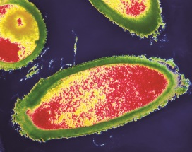
Lactobacillus acidophilus
– the gut-guzzlers
Where they live Lactobacillus acidophilus is one of the hundreds of species of bacteria that live in your gut. This particular species is found all through the gut from your mouth to your anus.
Why they are important
In your gut, this species digests lactose in milk, splitting it into the simpler sugars glucose and galactose. People suffering from diseases such as HIV and cancer tend to have abnormal levels of Lactobacillus in their gut – either too many bacteria, or too few.
How they are named Lacto is Latin for milk and bacillus refers to the rod shape of these bacteria. Acidophilus means ‘acid-loving’ in Latin – this species makes sure that its home remains slightly acidic by releasing its own acid, which helps to keep other bacteria at bay.
How they work Not only does Lactobacillus acidophilus produce sugar from milk, but it may also produce tryptophan – an essential nutrient that we cannot produce ourselves.
Top image: Coloured transmission electron micrograph of the Gram-positive rod-shaped bacteria Lactobacillus acidophilus. Copyright: Science Photo Library
In our Bacterial World exhibition we offer a selection of ten bacteria that have changed the world, some in profound ways. In this series of short fact-file posts we present one of the ten each week. This week’s bacteria are…
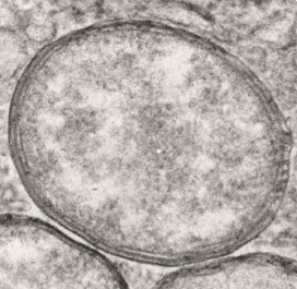
Wolbachia
– the man-killers
Where they live Up to 60 percent of insect species are infected with the bacterium Wolbachia, as are other species such as nematode worms.
Why they are important Wolbachia selectively kills off males in many species of insect and alters the sex ratio of the population to its own advantage. However, some species of insect rely on it for protection against other threats.
How they are named
The bacteria take their name from Simeon Burt Wolbach, who along with Marshall Hertig co-discovered Wolbachia in 1924 in a mosquito.
How they work Infected female insects pass the Wolbachia to their offspring – so the bacteria do everything they can to ensure females survive. Their strategies include killing male larvae, making males infertile, and rendering females able to reproduce without males.
Top image copyright: Joshua Blight (University of Oxford) & Steven Sinkins (University of Glasgow)
You have probably heard of sea cucumbers. If you’re lucky, you might have seen one, if not in the wild, then perhaps in a nature documentary like Blue Planet or the children’s cartoon Octonauts. If you’re less lucky, you might have eaten one – they are most commonly described as slippery and bland in taste!
Despite their appearance, sea cucumbers are actually marine animals most closely related to sea urchins, rather than to worms or slugs. Over the past century palaeontologists have uncovered a range of ancient fossil relatives of modern sea cucumbers that allow us to piece together the story of how they evolved from armoured ‘tanks’ into the naked slug-like forms we see today. One such fossil is described in a new paper by my colleagues and I, just published in the journal Proceedings of the Royal Society B.
The fossil in question is 430-million-years-old, and it comes from a site of exceptionally-preserved fossils in England called the Herefordshire Lagerstätte. Herefordshire has produced many exciting discoveries over the years, from prehistoric parasites to an ancient ‘kite runner’. The new fossil is the first of its kind from this deposit.
Like all fossils from Herefordshire, the specimen was preserved in an egg-shaped nodule of rock. Because the rock has the same chemical composition as the fossil, it could not be studied with modern imaging methods such as CT scanning. Instead, it had to be studied by painstakingly grinding away the fossil, a few hundredths of a millimetre as a time, with photographs taken of each exposed surface using a digital camera. This allowed us to build up a dataset of hundreds of slice images through the fossil, which were digitally reconstructed as a 3-D ‘virtual fossil’ on a computer.
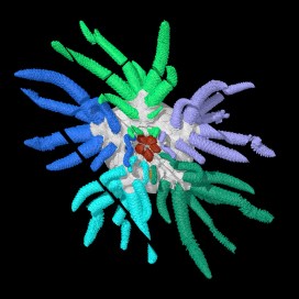
The 3D computer reconstruction revealed a very peculiar animal, about 3 cm wide, with 45 tentacle-like ‘tube feet’ and a large mouth surrounded by five teeth. The animal had a skeleton made up of numerous hard plates, which were composed of the mineral calcite. After studying this fossil and comparing it to other similar ones from the same time period, we were able to identify it as a species new to science. We named the species Sollasina cthulhu, for its resemblance to monsters from the Cthulhu universe created by author H.P. Lovecraft.
One of the most useful things about our 3D computer reconstruction was that it enabled us to study the inner features of the fossil, as well as the parts visible on the outer surface. This revealed internal soft parts that had never previously been described in this group of fossils. In particular, it allowed us to see an internal ring-like structure within the main body cavity.
3D reconstruction of Sollasina cthulhu. Left-hand image shows part of lower surface. Right-hand image shows same view with outer surface partly transparent to reveal inner ring (in red). Credit: Imran Rahman, Oxford University Museum of Natural History
We interpreted this inner ring as part of the water vascular system – the system of fluid-filled canals used for feeding and movement in modern sea cucumbers and their relatives, such as sea urchins and starfish. In life, the ring was connected to the large tube feet, which were filled with seawater. Most of these tube feet were used for crawling over the seafloor, with those nearest the mouth used for capturing food. The teeth could cut and crush food items, which were then eaten by the animal.
Life reconstruction of Sollasina cthulhu. Credit: Elissa Martin, Yale Peabody Museum of Natural History
To work out the evolutionary relationships of Sollasina cthulhu, we assembled a list of characteristics for various fossil and modern sea cucumbers and sea urchins. We analysed this matrix using several computational methods to determine how these different animals were related to one another. The results confirmed that Sollasina cthulhu and closely-related forms were ancient relatives of modern sea cucumbers. This allowed us to reconstruct the early evolution of sea cucumbers, back to their shared common ancestor with sea urchins, over 450 million years ago. Our study demonstrates this was a story of loss, with fossil sea cucumbers becoming progressively less armoured as they evolved into modern forms.
This discovery has greatly improved our understanding of sea cucumber evolution, but several questions remain. One intriguing question is when and how did sea cucumbers lose their teeth, and did these evolve into any features seen in living sea cucumbers? Future study of existing and new fossil sea cucumbers and sea urchins will help to answer this and other intriguing questions.
In our Bacterial World exhibition we offer a selection of ten bacteria that have changed the world, some in profound ways. In this series of short fact-file posts we present one of the ten each week. This week’s bacteria are…
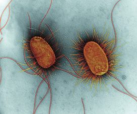
Escherichia coli
– the medicine-manufacturers
Where they live Millions of Escherichia coli live harmlessly in your gut, keeping more dangerous bacteria at bay. A few strains cause food poisoning.
Why they are important E. coli can act as a protein factory, accepting genes from other species and reproducing them. By combining DNA from more than one source, scientists can manipulate E. coli so that it manufactures human insulin.
How they are named Escherichia coli’s name reflects its discoverer, Theodor Escherich, and the fact that he found it in the human colon.
How they work Bacteria often contain plasmids, extra DNA rings that confer particular properties. Researchers can introduce genes into E. coli using plasmids, enabling the bacteria to make all kinds of biotechnology products from foods to medicines.
Top image: Coloured transmission electron micrograph (TEM) of two Escherichia coli bacteria. E. coli are Gram-negative bacilli (rod-shaped) bacteria. Long flagellae (thin thread-like structures) are used by the bacteria to move themselves. The spiky filaments on the sides of the bacteria are pili, thin strands of protein used when two bacteria conjugate (transfer DNA). E. coli is a normal inhabitant of the human intestine. However, under certain conditions its numbers may increase, causing infection. Magnification: x17,200 at 10 centimetres high. Copyright: Science Photo Library