A shark’s tale

Following the completion of our Life, As We Know It redisplay project earlier this year, we are taking a look at some of the exceptionally skilled people who helped to bring it all together. In this article, model maker Jeremy Hunt and our Vertebrate Palaeontology Collections Manager Emma Nicholls take a deep dive into what we believe is the world’s first 3D reconstruction of the extinct shark Ptychodus…

In six of our newest displays, fossil material found in and around Oxfordshire is used to reconstruct ecosystems from slices of geological time. One of these recreates a marine environment of the Cretaceous Period, between 94 and 84 million years ago, when most of northern Europe was submerged under a warm shallow sea. In these seas swam a formidable 8-metre long shark called Ptychodus.

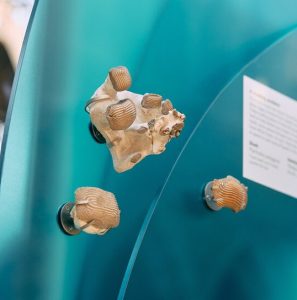
Fossils of Ptychodus teeth in the new display

Until recently, Ptychodus remained something of a mystery: teeth and vertebrae were the only remains scientists could use to work out what it looked like. Its robust, low-domed teeth are covered in deep ridges of enamel that created a mosaic in each jaw. They were capable of crushing shelled prey like a giant nutcracker. Similar teeth in modern sharks are found only in slow-moving species that prowl the seabed, so it was presumed that Ptychodus behaved in the same way.
But then in 2024 an incredible fossil with much of the animal’s soft tissue preserved was published by Dr Romain Vullo and co-authors. This new fossil revealed clues to the shapes of the fins and their position on the body. The study concluded that Ptychodus was in fact not a bottom-dwelling animal at all, but much more likely to be a fast, agile predator living higher in the water column, and perhaps related to the Great White Shark.

Armed with this recent discovery, we decided to create a three-dimensional model of the reimagined Ptychodus for our Cretaceous display. It had to be a quarter of life-size to fit it in the showcase, but would still be a huge two metres long.
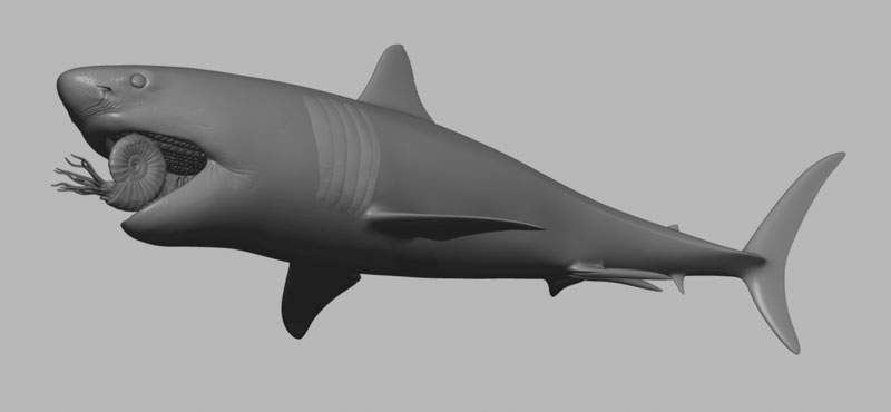
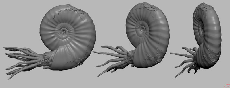
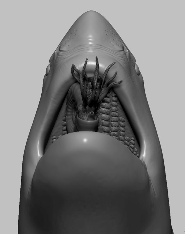
Digital 3D model of a shark, shown in grey, without any skin colouring or texture. The mouth is open and inside is an ammonite being crushed by the shark's plate-like teeth.
An early computer model of the shark and ammonite

“Working with Dr Vullo and others we began to decipher the shark’s possible appearance,” says Emma.  “Aspects to sink our teeth into included the skull shape, how it would have fed on hard-shelled free-swimming prey such as ammonites, how the mosaic of teeth sat in the mouth, as well as where and what shape the fins would have been.”

The next stage was to turn these ideas into a realistic model, and for this we turned to specialist model maker Jeremy Hunt. “When I was asked to make the reconstruction of Ptychodus I thought it was a great opportunity to incorporate modern digital techniques and processes that would give great accuracy, but also and flexibility so that adjustments could be made at later stages. This would be really difficult using a traditional clay sculpting approach,” he explains.

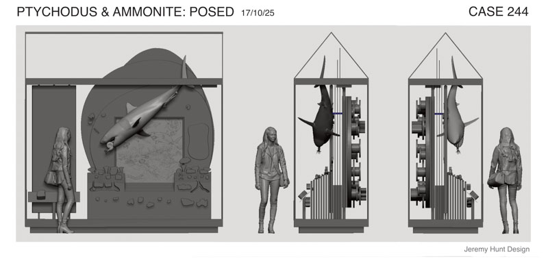
Together, Emma and Jeremy began to translate the fossil research into draft models on the computer, digitally sculpting versions on-screen. As well as being scaled down to quarter size, the position and pose of the model also needed to work inside the showcase structure and the designs of the rest of the display. Combining computer models helped us to make sure that Ptychodus would fit perfectly and that its method of support could be worked out well ahead of installation.
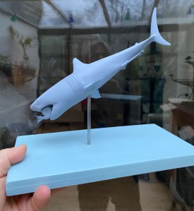
To give us a sense of how the final model would actually look, Jeremy 3D-printed little maquettes just a few centimetres long. On these we could check that the anatomy and pose were just what we needed. The maquettes also showed how the shark would be suspended in a diving angle, chomping down on an ammonite which Jeremy was modelling separately.

A hand holds out a wooden plinth, painted blue, onto which a 3D printed shark model is mounted on a vertical metal rod. The 3D print is also blue and is approximately 15cm  long.
3D-printed maquettes of the model helped us to check its form and pose

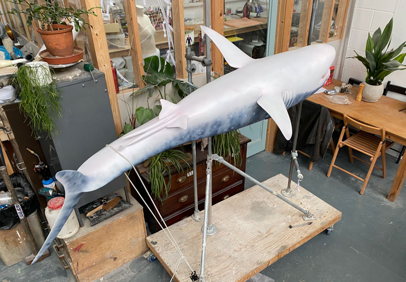
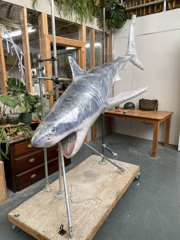
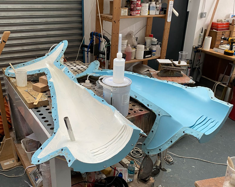
Once ready, the model was 3D-printed at its display size of two metres long, allowing Jeremy to create a silicone rubber mould to produce a fibreglass cast of the final structure. Fibreglass is both strong and light, perfect for the display. But the model really came to life when Jeremy hand-painted and airbrushed its colourings and markings based on a research by Emma and a concept drawing by palaeo-artist Julius Csotonyi.
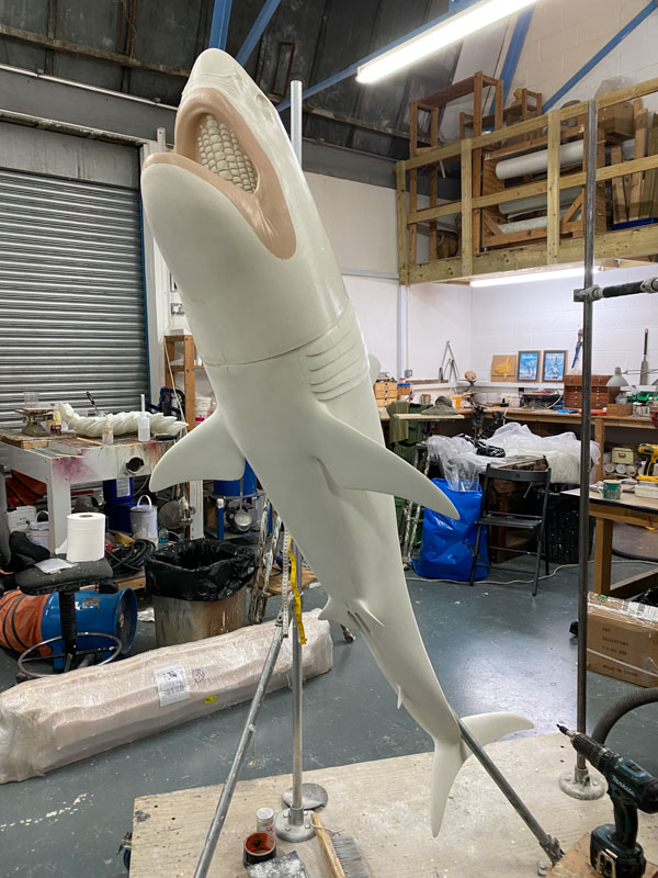
Once Ptychodus and the rest of the display were installed, the ammonite was placed in the mouth. It is held safely there by hidden strong magnets, caught permanently between those crushing plates, about to become shark food.
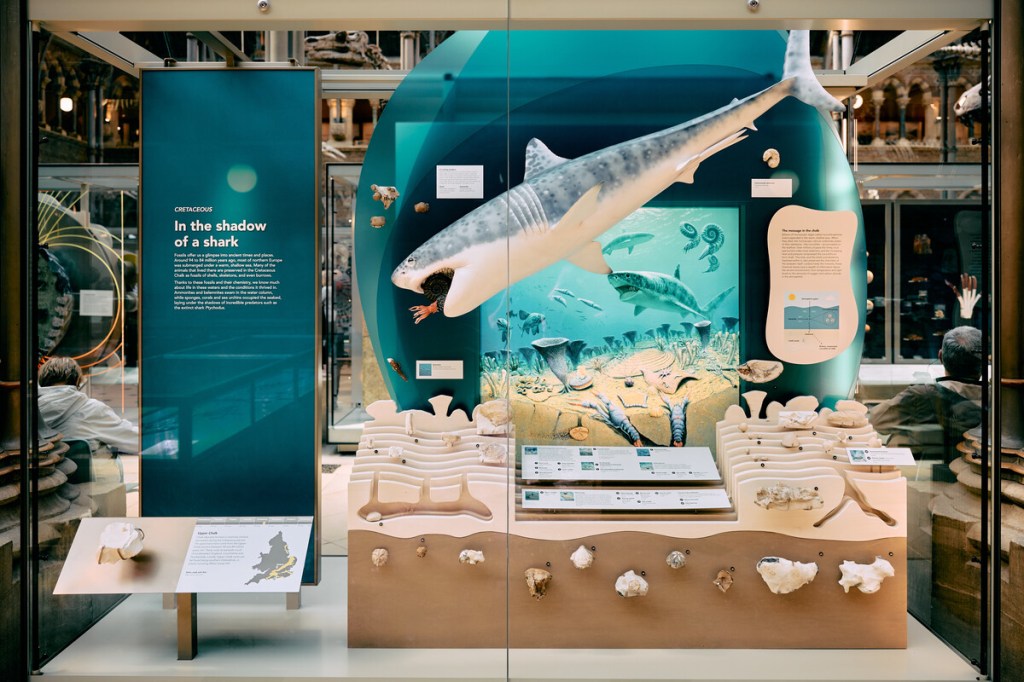
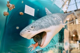
Museum exhibit showing a 2 metre long shark model against a backdrop artwork of an ocean containing drawings of other sharks, fish, ammonites and belemnites in the sea, along with corals, shellfish and lobsters on and burrowing into the seabed. Below this scene are a selection of chalk fossils. On the left the title panel is printed on blue acrylic and shows the display's name as 'In the shadow of a shark'.
The Ptychodus model in the final display at the Museum

“This was a great project to be involved with, and I hope the new displays will be enjoyed by all who visit the Museum,” says Jeremy.

Jeremy Hunt, model-maker

*

See more of Jeremy’s work at: www.instagram.com/memsculpt
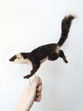
From Fossil To Flesh: Reconstructing a Phascolotherium

Following the completion of our Life, As We Know It redisplay project earlier this year, we are taking a look at some of the exceptionally skilled people who helped to bring it all together. In this article, taxidermist Alexandra Rose Cherrett tells us about the ‘unusual proposal’ that she received one day…

I was sitting at my studio desk ready to start the day with some admin. I often receive emails enquiring about commissions ranging from peoples’ pets to wild animals that have been found by the public, asking for them to be preserved after death through taxidermy. At the top of my inbox was an email with an “unusual proposal” from the Museum of Natural History in Oxford.

As a taxidermist I have been lucky enough to work on a wide range of animals over my short career, from kingfishers to vultures, hedgehogs to lemurs and many more in-between. Safe to say I was not expecting the request to taxidermy an animal that hasn’t walked the earth for 165 million years.

I was then introduced to Phascolotherium; a small, furry mammal that lived in the Mid Jurassic alongside Megalosaurus, the first dinosaur ever named and discovered, right here in Oxford, UK. A proverbial stone’s throw from the museum itself.

Now, I have always been rather obsessed with dinosaurs. My obsession and adoration for dinosaurs and wildlife has been evenly matched for as long as I can remember. Suddenly I found my Jurassic Park dreams were coming to life. An impossible task? I thought, “Life, uh…finds a way”.

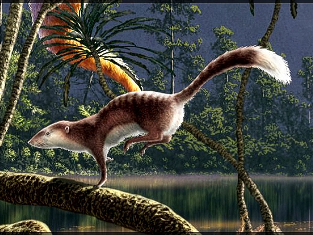
The first challenge was that only the jawbone and teeth of Phascolotherium had ever been discovered. I was given a reference image by Paleo artist, Julius Csotonyi, from which I primarily  worked.

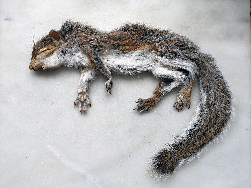
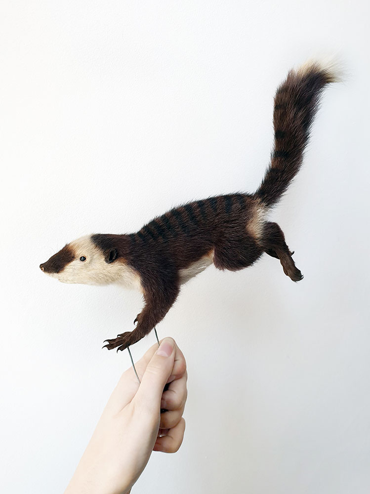
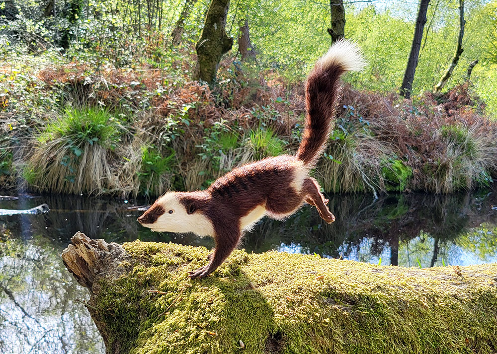
It was rodent-like in nature, with an elongated head, a lithe body and a long, plumed tail. My first thought was to use a squirrel as the base as the limb structure and body shape were so similar. I knew I had a pair of juvenile grey squirrels in my freezer that could be the perfect size, one was donated as a cat kill and the other perished at a wildlife rescue.

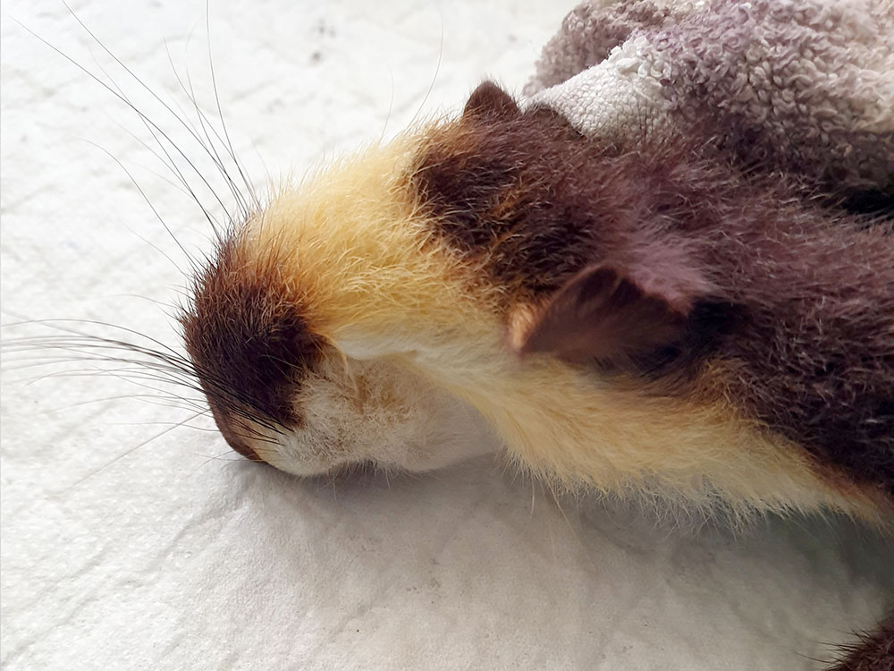
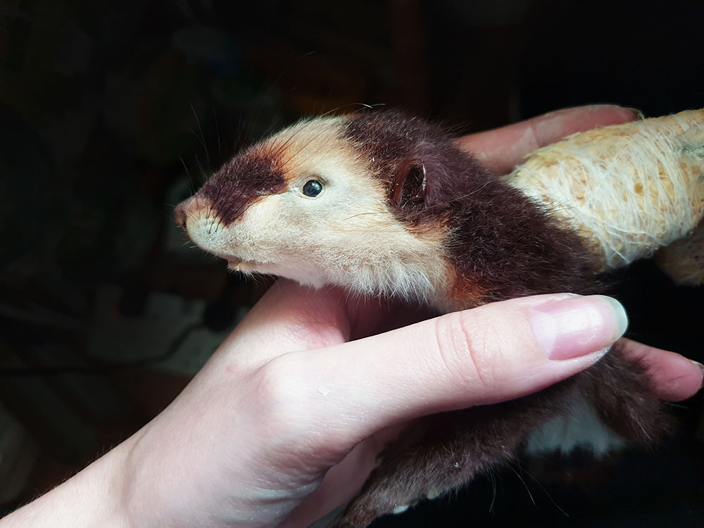
The most obvious visual changes were the colouration. The artworks showed a body of a dark reddish hue, with patches of white on the face, stomach and tip of the tail and stripes stemming down the spine. My next step of course, was Boots. A few boxes of Garnier hair dyes and peroxide bleach later and I had a completely different animal skin laid out on my desk.

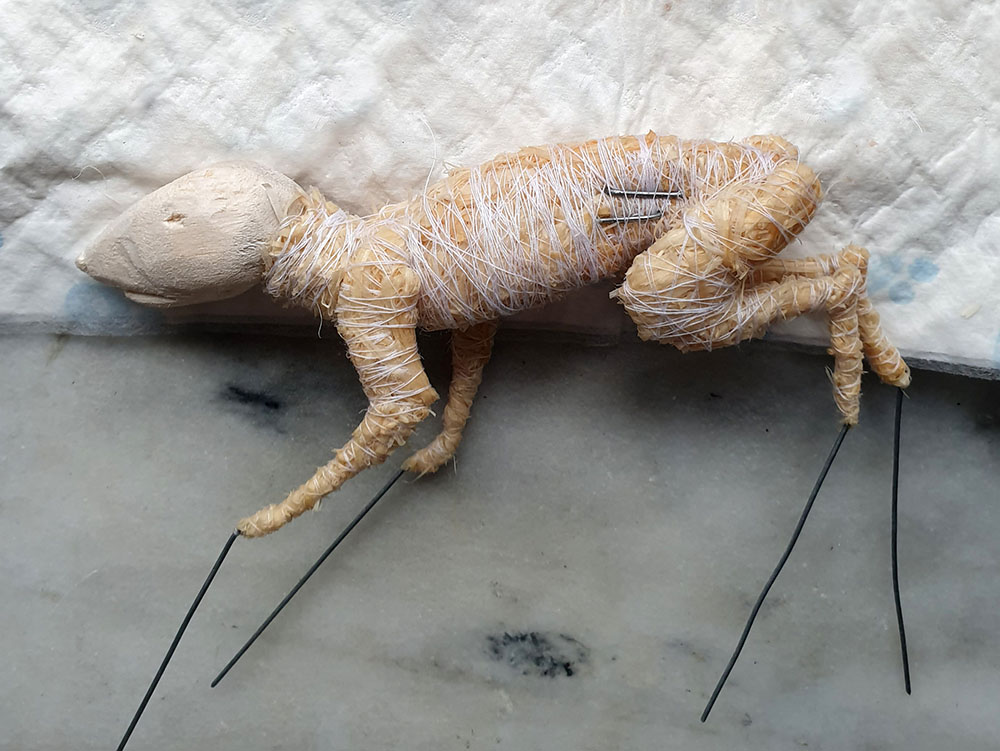
My next process was to create the body form over which the skin would be fitted. For mammal taxidermy, I usually carve or cast a head out of balsa wood or PU foam and make a bind up copy of the body and limb shape from woodwool.

With the head essentially being a non-existent shape, I copied the paleo-art and carved a small balsa block to a similar shape as a large shrew. The body was very similar to the young squirrel already so that was a simple workaround.

With the nature of its surroundings and the looming Megalosaurus, I imagined this creature was indeed, always on the run. So, an action pose was created, mid run, which highlighted its slight build and balancing tail very nicely.

When dressing the skin, the biggest alteration was the eye and ear placement. The young squirrel had eyes around 8mm, whereas Phascolotherium measured in at 4mm. Not to mention they were set further back and lower, as were the ears at two thirds their original size and shape. I made an incision across the eyelid and manipulated the skin to fit its new size and position, copying the same methodology for the ears. Almost instantly, a face that hadn’t been seen for 165 million years was right there looking back at me.

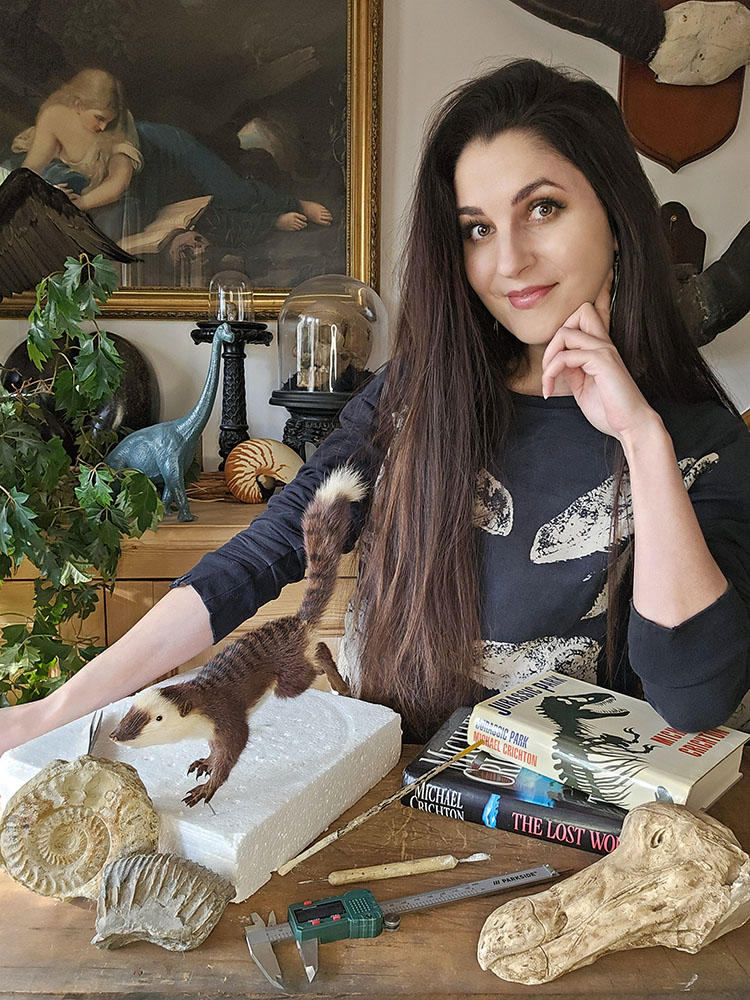
Me with the completed Phascolotherium

I have been fortunate enough to work with the Museum of Natural History on a few projects now and it’s safe to say this project has been one of my biggest challenges and greatest pleasures to date.

www.alexandrarosetaxidermy.co.uk

Instagram: @alexandrarosetaxidermy

Facebook: @alexandrarosetaxidermy

A Botanical Journey In Time

Following the completion of our Life, As We Know It redisplay project earlier this year, we are taking a look at some of the exceptionally skilled people who helped to bring it all together. In this article, model maker Inese Veismane recalls some of the weird and wonderful extinct plants that we needed for our new displays…

I remember the exact moment I saw the message from the Museum of Natural History. I had just returned home from a trip, switched my phone back on after the flight, and there it was – waiting for me to jump into the unknown. It came at a time when my personal life was shifting in a difficult direction, and in an unexpected way, this project became something that held me steady.

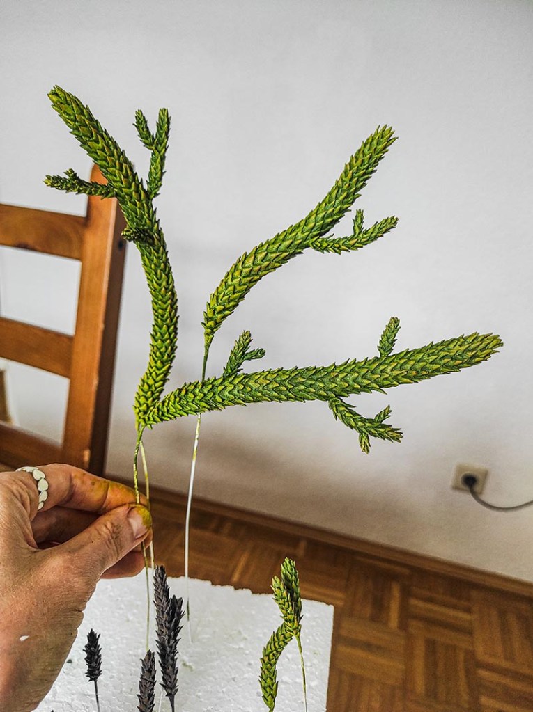
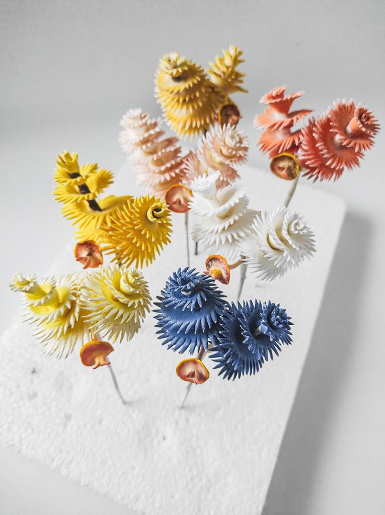
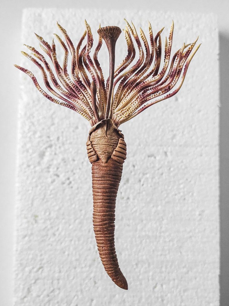
My work usually lives in the botanical world – delicate flowers, soft forms and colours, artsy details ­– so stepping into a space shaped by scientific accuracy and unfamiliar subjects felt like entering a new landscape. This time, I was asked to create not only plants but organisms like worms, including the intricate and challenging Christmas tree worms.

The commission began in 2023, and from the very start, it was clear that this would be a different kind of creative process. Instead of observing a living plant or working from my own references, I was guided by scientific material provided by the museum. There was a responsibility to honour not just the visual beauty of each subject but also its structure, proportions, dimensions, and defining characteristics.

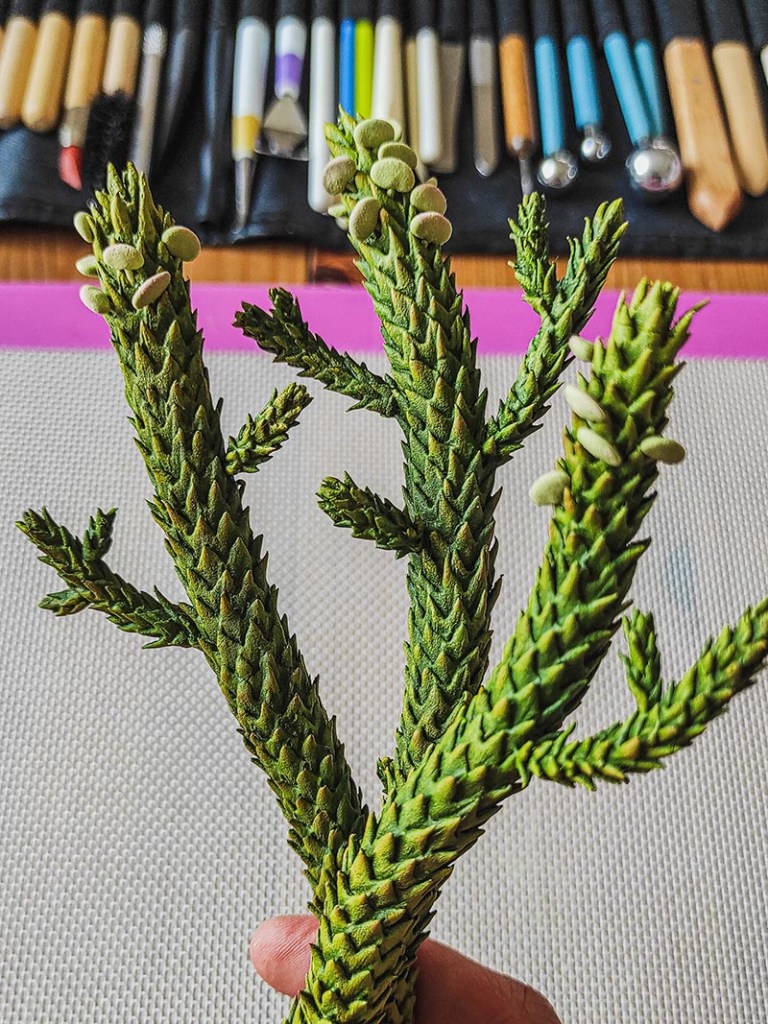
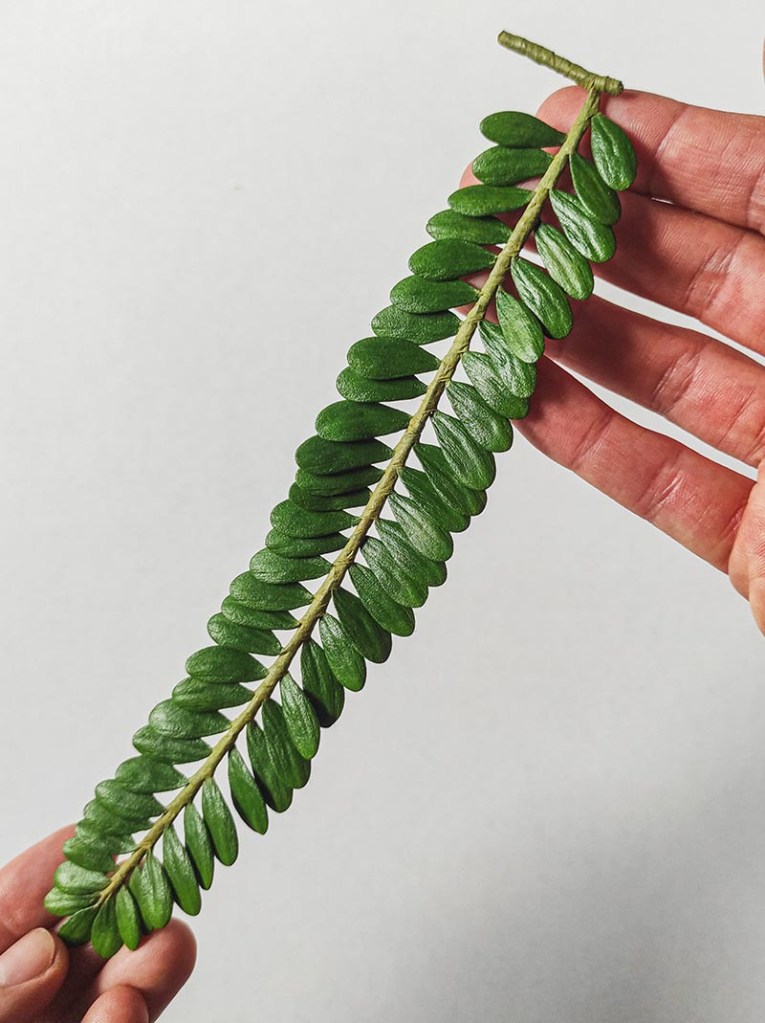
I work with air-dry clay, building each form slowly by hand. My process is intuitive but also very attentive – I spend time studying botanical forms and shapes and then transforming that into something tangible. With these models, I had to balance that intuitive, artistic approach with careful observation. It became a dialogue between art and science.

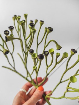
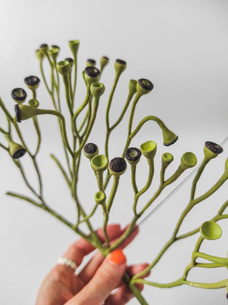
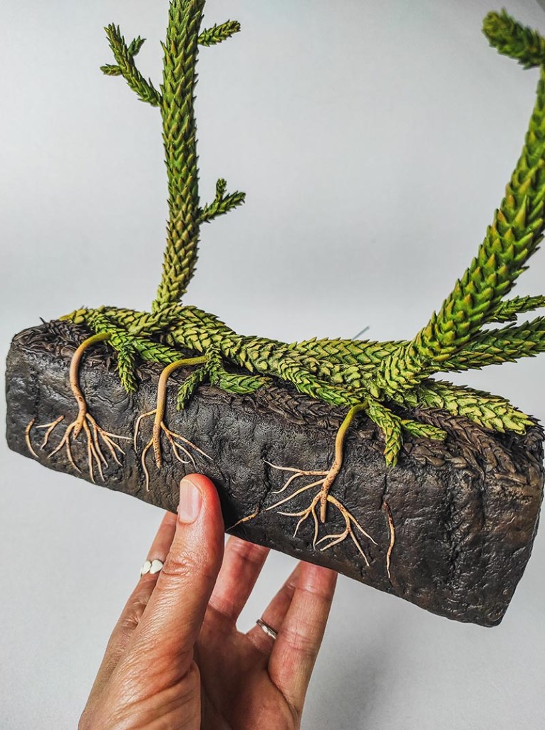
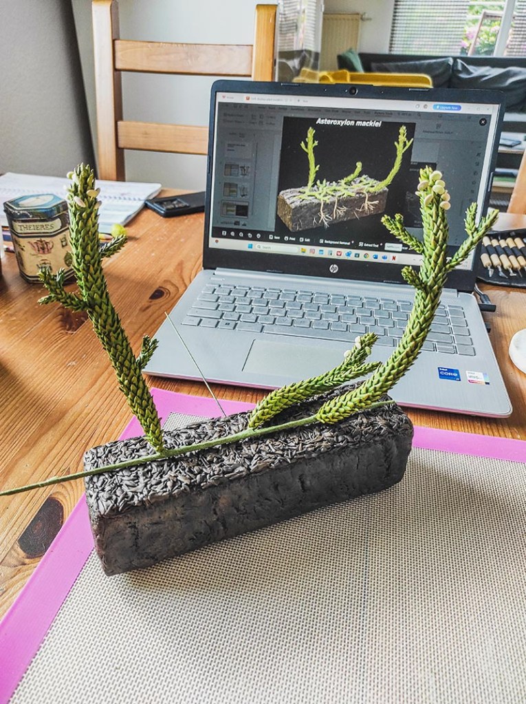
Working on the early plants became one of the most interesting parts of the project. Recreating species like Asteroxylon mackiei, Rhynia, and Cooksonia felt like reaching into a distant past. These are plants that no longer exist, known only through scientific study and careful reconstruction. There was something deeply humbling in shaping them by hand – giving form to something that once lived hundreds of millions of years ago. It felt like a journey back in time. I had to rely entirely on references and guidance while still allowing the forms to come alive through my own interpretation.

One of the most meaningful parts of this creative process has been witnessing the transformation of each piece – from a simple beginning to something that holds presence and detail. I also find importance in sharing these stages on my social media. It creates a sense of continuity – an archive of the work that can be returned to over time.

Looking back, this experience has expanded the way I see my work. It reminded me that even when we step outside of what feels familiar, the essence of creating remains the same: to pay attention, to be patient, and to trust that something will slowly take shape.

There is also something special in knowing that these handmade models will live within the Museum and become a part of a much larger story – held in a space where art and science meet.

*

See more of Inese’s work at: www.instagram.com/cooleblumen/

Through the looking glass

With our Life, As We Know It redisplay project now underway, our Senior Archives and Library Assistant Danielle Czerkaszyn takes a behind-the-scenes look at how we captured the contents of the current displays for the Museum’s archive.

The archive here holds a unique collection of natural history books, journals and documents covering a wide range of subjects related to the Museum’s collections and research. It also contains papers and objects on the history of the building, providing an institutional memory of Oxford’s ‘University Museum’ since its foundation in 1860.

From an archive perspective it was really important to document the current layout of the cases, their specimens and text before they were removed from the court to make way for the new showcases in the first phase of our redisplay work.

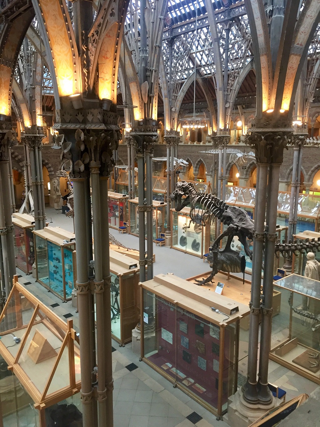
The museum in late 2019

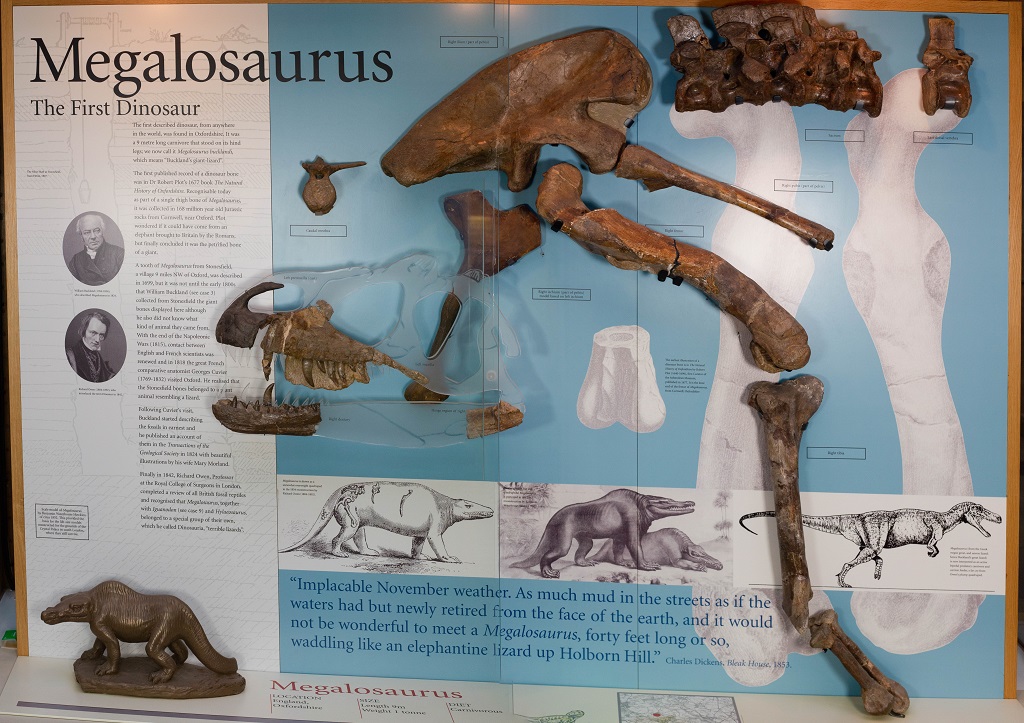
The displays as we know them – with exhibitions on the Oxfordshire dinosaurs, Alice in Wonderland, the Oxford Dodo, and more – were last changed in 2000. For the last 20 years visitors to the Museum would remember their first time being wowed by the Megalosaurus jaw – the world’s first scientifically-described dinosaur – or charmed by the Dodo made famous in Lewis Carroll’s Alice Adventures in Wonderland.

Although after 20 years it is time for a change, the stories and information in the displays are too good to be forgotten. So before anything was removed we began to build the archive for the future.

A display of the fossilised remains of Megalosaurus
The previous display on Megalosaurus: The First Dinosaur

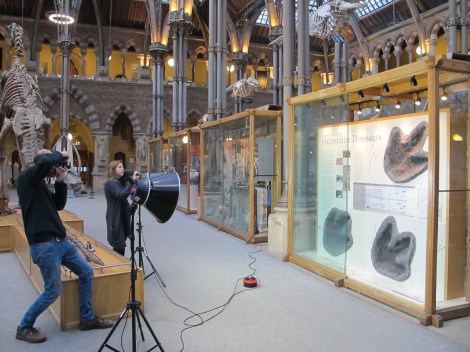
The best way to capture all the information of the displays was through high resolution photography, but this was not as straightforward as we hoped.

The first two obstacles to good photographs are pretty obvious to anyone looking at the cases: glass causes huge amounts of glare; and each case has a big dividing line down the centre where the two sliding glass doors meet, cutting what should be a lovely seamless image into two halves.

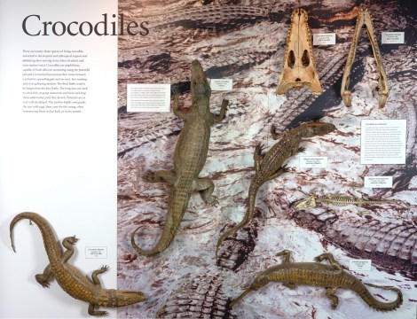
To avoid glare and the solve the problem of the dividing line, our photographer Scott opened each individual side of the case, photographed two or three images of the display, and then stitched the separate photos together using Photoshop.

Each case was photographed in two or three segments

The segments were then stitched back together and adjusted for exposure and colour balance to create the final image

Another obstacle to taking good photographs of the displays came from the Museum itself. Some of our larger display furniture, such as the glass case for the Atlantic Bluefin Tuna or the huge T. rex plinth – got in the way of a nice straight shot. Because these items are so large and heavy they were impossible to move, so we had to improvise and do our best.

Capturing the displays before the current cases were removed allowed us to keep an archival record of their contents

Thankfully, we managed to get shots of all 24 displays before they were removed and so a record of each case now rests with the Museum’s archive. If anyone wants to know what the display cases in the court looked like from 2000 to 2020, they will now be able to look back at the images in the archive and recall the magic of the Oxford Dodo exhibit that perhaps first made them fall in love with the Museum.

Our new displays are now in development, and will include some beautiful presentations of the diversity of life, looking at the importance and fragility of biodiversity and human impact on the environment. These new exhibits will show how the biological processes of evolution combine with the geological processes of our dynamic Earth to give rise to the immense, interconnected variety of the natural world.

We look forward to telling you more about that here as the project progresses.

The Life, As We Know It redisplay project is supported by a generous grant from FCC Communities Foundation.

Oxford University Museum 1860

An ever-evolving museum

Oxford University Museum 1860

As we embark on our Life, As We Know It redisplay project – the first substantial changes to the permanent exhibits in more than 20 years – our Senior Archives and Library Assistant Danielle Czerkaszyn takes a look back at 160 years of an ever-evolving museum, in the first of a series of posts around the redisplay.

On 15 June 1860, Henry W. Acland, Regius Professor of Medicine at the University of Oxford, wrote:

The Oxford Museum slowly approaches completion. The building will shortly sink into insignificance when compared to the contents it will display, and the minds it will mould.

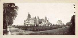
The University Museum at Oxford, as the Museum was originally known, was established to bring together scientific teaching and collections from across the University under one roof. The doors opened in June 1860, and soon after several departments moved into the building – Geometry, Experimental Physics, Mineralogy, Geology, Zoology, Chemistry, Astronomy, Human Anatomy, Physiology, and Medicine.

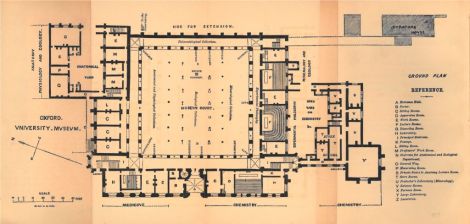
Ground floor plan 1866
Ground floor plan of the University Museum in 1866

When the University Museum opened, it was not simply a museum; each department got a lecture room, offices, work rooms and laboratories, as well as use of the library and display areas. According to Acland, a key figure in the Museum’s foundation, in 1860 the outer south aisle of the main court featured mineralogical specimens and chemical substances, while the inner aisle exhibited Oxfordshire dinosaurs.

Acland’s detailed descriptions of the central aisle highlighted zoological specimens with twelve parallel cases of taxidermy birds, four side cases of taxidermy animals, including animals on top of the cases, and six table cases down the centre showing shells, crabs, insects, corals and sponges, starfish and urchins. The inner north aisle presented reptiles and fish, while the outer aisle introduced the Ashmolean‘s zoology specimens, as well as anatomical and physiological collections.

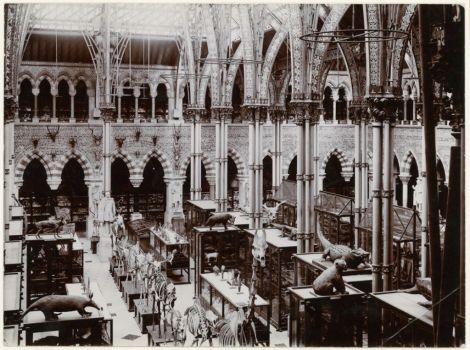
The Museum in 1890
The Museum court in 1890

Although members of the public were welcome in the Museum from the start, the departments which inhabited the building were more concerned with teaching space, research facilities and the storage of their specimens than the needs of visitors. As a result, most of the early displays and cases were arranged in a systematic manner that focused on space-saving practicalities and communicating scientific knowledge, rather than aesthetics.

Geology specimens on the walls
Geology specimens displayed on shelves on the walls

Early Dodo display case
An early display focused around the Museum’s famous dodo specimen

Tracing through old annual reports it is clear that cases in the main court have been almost constantly refreshed and updated, with displays highlighting new specimens and changes to scientific understanding, or through practical improvements to lighting, electricity points and environmental monitoring. Nonetheless, the overall layout of the cases remained the same until the early 1980s.

The Museum court, unknown date

From the early 1990s a focus on public engagement began to increase. Longer opening hours were introduced and displays were redesigned to link to both undergraduate teaching as well as the National Curriculum. Temporary exhibitions also regularly featured in the main court to increase the variety of specimens on display.

The Museum court in 1994

Megalosaurus temporary exhibition
A temporary exhibition about the Megalosaurus dinosaur in the 1990s

The turn of the millennium marked the start of a major project to update the main court displays. The central cases were reconfigured and a new set of introductory cases installed, including many themes familiar to visitors in recent years, such as exhibits on the Oxfordshire dinosaurs, Alice in Wonderland, and the Oxford Dodo.

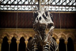
T. rex makes its presence known

These showcases were complemented by the addition of an imposing cast of ‘Stan’ the Tyrannosaurus rex in the centre aisle, positioned behind the historic Iguanodon cast. The changes were well received and attendance in the month of July 2000 was the highest ever recorded. The Museum also introduced live insects for the first time in 2000, with Upper Gallery tanks containing Madagascan Hissing Cockroaches, South American Burrowing Cockroaches, a variety of stick insects, and some large tarantulas.

The project completed in late 2005 when the displays on Evolution, the History of Life, and Invertebrate Biodiversity were installed. Touchable specimens were also given their own permanent display area, allowing visitors the opportunity to physically interact with natural history material. These and other public engagement activities were recognised when the Museum won The Guardian newspaper’s Family Friendly Museum of the Year Award for 2005.

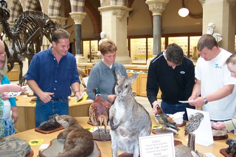
People around a table of touchable taxidermy specimens
New tables of touchable specimens were introduced for visitors in the 2000s.

The last substantial update to the fabric of the building took place in 2013, when the Museum closed for a year to fix the leaks in the glass roof. Taking advantage of the closure, a major piece of conservation work was undertaken on the seven whale specimens suspended from the roof. Having been on display for over 100 years, the whales were in need of considerable TLC.

A conservation team worked on the whale skeletons during the Museum’s closure for roof repairs in 2013.

Today, new and exciting changes are afoot as we embark on the first major changes to our permanent displays in almost 20 years. New high-end showcases will present displays under the concept of Life, As We Know It – beautiful presentations of the diversity of life, and the importance and fragility of biodiversity and human impact on the environment. The new exhibits will look at how the biological processes of evolution combine with the geological processes of our dynamic Earth to give rise to the immense, interconnected variety of the natural world.

Looking back across the decades we can see that the Museum is never static, but instead constantly changing and adapting, shifting from its foundation as a Victorian centre of academia to the accessible and engaging space we know and love today.

The Life, As We Know It redisplay project is supported by a generous gift from FCC Communities Environment.