Exploring Borings

Trace fossils are a record of life; examples are footprints, bite marks, and burrows. The earliest trace fossils are thought to have been produced by an amoeba and are about 2,000 to 1,800 million years old. A special form of taxonomy (classification) has been created for trace fossils based on behaviour as it’s rare to find a body fossil preserved at the end of its trace and different organisms may produce identical tracks. Originally five behaviours were recognised but this number has now been expanded. (To find out more a good starting point is here).

trace-fossil
A burrow preserved as a trace fossil, Thalassinoides

In this blog we are looking at boring trace fossils. No, not yawn boring! I mean the types of holes made by different animals in shells for predation, shelter and attachment. These will definitely not make you yawn. We are going to explore the three types of trace fossil that can be found in Charles Lyell’s collection. These represent two different types of behaviour and therefore taxonomy: Praedichnia, defined as “trace fossils that show evidence of predatory behaviour, such as borings and bite marks” and Domichnia defined as “dwelling structures reflecting the life position of the organism that created it”.
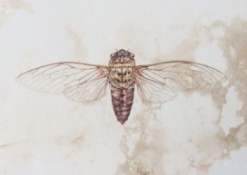
 

Gastropod Borings

Gastropods have been boring into shells in the same way for the past 100,000 years. There are two main families of predatory snails, the Naticidae and the Muricidae. They can be identified to family level by the shape of the hole they create. The predation attempts are deemed successful when the hole has fully penetrated the shell.

gastropod-boring
Pleurotoma shell that has been bored into 5 times (the fifth is on the reverse). Image taken by Sarah Joomun

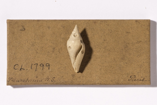
The Naticidae create holes with slanted walls by secreting acid and scraping with their radula (rasp-like structure of tiny teeth used by molluscs for feeding). They feed on bivalves, scaphopods (tusk shells), and gastropods (including other naticids). The size of the holes they create varies depending on the species. This family evolved in the Late Triassic/ Early Jurassic and occur worldwide.

naticidae
Diagram of the boring that is created by the family Naticidae

The Muricidae create holes with straight walls by using a softening secretion and scraping with their radula. They feed on bivalves, barnacles, and gastropods. This family evolved in the Early Cretaceous.
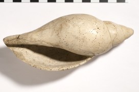
muricidae
Diagram of the boring that is created by the family Muricidae


Sponge Borings

Unlike the gastropod borings, sponges do not create their borings to feed. Instead they use the shells they have bored into for shelter. Cliona celata is a boring sponge which creates holes up to 5 mm in diameter in mollusc shells and limestone. They use acid to bore into their chosen home. If they bore into a shell the animal usually dies because it has lost its protection as it is structurally weakened.

sponge-boring
Gastropod with Cliona borings. Image taken by Sarah Joomun

In The Boring-Sponge, Cliona written by Joseph Leidy in 1889 he describes the way “in which [the sponge] occupies the shells of oysters and clams with its sensitive papillae [small fleshy bumps] … protruding from the perforations of the surface of the shell”. Sometimes the sponge can outgrow the shell its living in, using other material from around it. This massive sponge was familiar to the fisherman of Beach Haven New Jersey, USA “under the name of Bay-Pumpkin ; often growing to the size of one’s head.”


Bryozoan Borings

Bryozoans are filter feeding aquatic invertebrates commonly known as moss animals. Some bryozoans encrust surfaces. This encrustation can cause a pattern of small pits to be etched into the substrate. To see the pits you need a microscope as they are approximately 0.1-0.9mm in diameter.

bryozoan
Bryozoan borings (the smaller holes) on the gastropod shell from the previous image. The larger holes are the Cliona borings

Originally this group of trace fossils were called Leptichnus, from the Greek leptos meaning “flimsy, delicate, subtle”, however this was found to be the name of a gastropod species. To keep the original meaning of the name, the name Finichnus was proposed, from the Greek finos meaning “fine, delicate”.

‘Flight’ of the Dodo

lead-image

By Mark Carnall, Life Collections manager

The museum holds the only remaining soft tissue of the extinct dodo known anywhere in the world. The partially dissected skin of the head and scales from the feet of a single dodo represent one of natural history’s most iconic specimens. In fact, it is so tied to the museum’s identity and history that we use the dodo as our logo and it is even incorporated in the name of this blog.

Although the dodo head had been at Oxford University since the formation of the original Ashmolean Museum in the 17th century, it wasn’t really until the 19th century that the specimen really became celebrated.

Around this time, publications confirmed the extinction of the dodo from the island of Mauritius, where it was endemic. To capitalise on the rising interest in the animal, Ashmolean Museum Keeper John Duncan commissioned a number of casts of the Oxford Dodo head to give to, and exchange with, other museums.

One of the earliest of these casts was presented to the British Museum in 1828; later casts are recorded as being sent or exchanged with leading scientists of the time, as well as with Leiden Museum and the Royal College of Surgeons.

From these original and later casts further casts and models were presumably made, and eventually, dodo specimens spread to virtually every major natural history museum in the world. Today, many museums display casts of this head, all stemming from the single specimen held here in Oxford.

One of the many casts in the Oxford University Museum of Natural History, this one has been painted to match the original specimen

The Museum contains a number of models and casts of the head too; some are made from plaster and resin, some are painted to resemble the original specimen. The head of the dodo was actually dissected in 1847, by Henry Acland. He removed the skin from one side of the face so the early casts are a record of how the specimen would have looked originally.

In preparation for the Presenting display in the Museum I contacted natural history museums through the Natural Sciences Collections Association asking people to share information and photos about their casts and models of the dodo head. I wanted to try and construct a picture of how the dodo head was disseminated, as well as capture the diversity of quality and colours of representations of the original specimen. Here’s how far some of the dodos have flown:

American Museum of Natural History

IMG_3401
© Paul Sweet, American Museum of Natural History

Cast of head with no further catalogue information about provenance.

Bradford Museums and Galleries

© Bradford Museums and Galleries

© Bradford Museums and Galleries

Cast of the head labelled as coming from Cartwright Hall. Curator Gerry McGowan suspects this may have come via the Bradford Philosophical Society collections. The first curator of the society, Louis Compton Miall was friends with Thomas Henry Huxley and through him had contacts with many other geologists who may have gifted or exchanged this cast.

Bristol Museum & Art Gallery
Bristol received a cast of the head directly from Oxford from Philip Duncan in 1834, keeper of the Ashmolean Museum between 1826 and 1855. Unfortunately, the head was likely destroyed in bombings of Bristol in 1940.

Canterbury Museum, New Zealand
Received a cast of a head from Professor Rolleston on 21 July 1871 in exchange for two kiwi skeletons which are still in the museum collections today.

Grant Museum of Zoology UCL

Cast of head and foot presented to the museum by E.Ray Lankester in 1891/1892 just after leaving UCL and being appointed the Linacre Professor of Comparative Anatomy at Oxford.
© UCL Grant Museum of Zoology

Cast of head and foot presented to the museum by E.Ray Lankester in 1891/1892 just after leaving UCL and being appointed the Linacre Professor of Comparative Anatomy at Oxford.

Great North Museum Hancock

The Great North Museum Hancock’s cast was presented by George Townsend Fox, this specimen had been presented to the natural History Society of Newcastle in 1841 by Fox and had originally been presented by Philip Duncan.
© Great North Museum: Hancock

The Great North Museum Hancock’s cast was presented by George Townsend Fox. This specimen had been presented to the Natural History Society of Newcastle in 1841 by Fox and had originally been presented by Philip Duncan.

Horniman Museum and Gardens

© NH.64.11 Horniman Museum and Gardens

Cast of the head that had quite a circuitous route to the Horniman Museum. The Horniman received the cast from the geology department of Queen Mary’s University of London in 1964 which received the cast from the Saffron Walden Museum in 1962.

Manchester Museum
Cast of a head, presumed to have been presented by William Boyd Dawkins. The cast is currently on display in the Living Worlds gallery in Manchester Museum.

The National Geological Repository British Geological Survey

bgs-national-repository
© National Geological Repository British Geological Survey

Cast of a head recorded as from the ‘original in the Ashmolean Museum’, purchased from the sale of Gideon Mantell’s collections in 1853.

National Museum of Ireland

national-museum-ireland
© National Museum of Ireland

Cast of head with ‘J.Johnson’ inscribed into the base, possibly referring to John Johnson, who commissioned five casts from the museum in 1837.

Natural History Museum London

© The Trustees of the Natural History Museum London

The Natural History Museum’s collections contain three casts of the Oxford Dodo head – two in the ornithology collections (pictured) and one in the palaeontology collections. The unpainted cast in the ornithology collections has the name ‘Johnson’ inscribed into the base.

Nottingham Natural History Museum

© Nottingham Natural History Museum, Wollaton Hall

Colleague Adam Smith got in touch with some interesting specimens from Wollaton Hall. The first one looks like another cast in this series but the second cast is unlike any of the others gathered here. The cast shows an open eye, detail on the beak as well as a more defined hook to the end of the bill.

Unfortunately, there’s not much information about the origins of these two casts so it’s probable that the ‘open eye’ cast may be a cast of a model reconstruction or an in progress sculpt. There’s an extremely slight chance it’s a cast of an otherwise unknown dodo head… If you recognise this dodo head do get in touch so we can solve this mystery for colleagues in Nottingham (it’s not the model dodo that we have on display here!).

Warwickshire Museum

© Warwickshire Museum

Cast of a head at Warwickshire Museum with damage to the beak. Donated to the museum by clergyman and naturalist Reverand Andrew Bloxham in the 19th century. As the museum is currently moving stores, further information about when this cast was acquired is inaccessible.

**

If you work at a museum and have a dodo head cast to share, please do get in touch and we’ll update this blog accordingly.

Last updated: 10/11/17

‘Presenting… The Flight of the Dodo’ was on display at the Museum of Natural History from the 25 January to 22 March 2017.

Acknowledgements

With many thanks to colleagues across the sector who helped with information and images about dodo specimens: Adam Smith, Alice Adams, Jack Ashby, Carol Davies, Bonnie Griffin, Dan Gordon, Yvette Harvey, Mike Howe, Emma-Lousie Nicholls, Laura McCoy, Gerry McGowan, Nigel Monaghan, Henry McGhie, Pat Morris, Paul Scofield, Paul Shepherd, and Paul Sweet.


Mysteries of the deep

by Sammy De Grave, Head of Research

It is often said that the final frontier of our exploration of the Earth lies deep in the oceans. Covering 70 per cent of the planet’s surface, the oceans nevertheless remain 95 per cent unexplored, according to the National Oceanic and Atmospheric Administration in the United States. And they know, for they are doing the exploring…

Since 2008, the Okeanos Explorer at the NOAA has been investigating deep water ecosystems and has live-streamed many of its remotely operated vehicle (ROV) dives for scientists and the public to observe. Last year, Okeanos was engaged in an exploration of the Mariana Trench Marine National Monument in the Pacific Ocean. During a single dive at an impressive 4,826 metres, on a muddy bottom at a site nicknamed Twin Peaks, a large shrimp was observed, which the participating scientists did not recognise.

Spotted 4,826m below sea level, in the Mariana Trench Marine National Monument: the shrimp Bathystylodactylus
Spotted 4,826m below sea level, in the Mariana Trench Marine National Monument: the shrimp Bathystylodactylus cf. bathyalis

This was an unexpected find. Although there are about 4,000 species of caridean shrimps in the world’s oceans, very few live below 1,000 metres, and fewer than 20 species are known from depths lower than 3,000 metres. Those that do are usually only found as broken specimens, creatures damaged by the trawls which also bring them to the surface.

Photos of the deep-dwelling shrimp were duly sent to two experts: Dr Mary Wicksten at Texas A&M University and Dr Sammy De Grave, head of research here at Oxford University Museum of Natural History. Both immediately recognised the specimen as belonging to the rare genus Bathystylodactylus, which comprises of only three scientifically-described species, which in turn are only known from six specimens, all damaged.

The bristly legs, called setose legs, are used in passive filter feeding
The bristly legs are used in passive filter feeding

wfm_pelagicThe Okeanos team had made something of a discovery, one which was published in the open access journal Zookeys. The posture of the shrimp clearly show it to be a filter-feeding species, with its long, bristly legs facing into the current. This type of feeding behaviour has not been seen in caridean shrimps before.

Surprisingly, although the shrimp was observed at almost 5,000 metres below the surface, it is not the deepest recorded shrimp. That honour goes to a different species, Glyphocrangon atlantica, living in the western Atlantic, which has been trawled from as deep as 6,373 metres. Blurry photographs from 6,890 metres in the Kermadec Trench might indicate that carideans live even deeper still, maybe even as far down as a related group of shrimps called dendrobranchiates, which are known from depths down to 7,703 metres.

These findings show that life of many kinds continues to be discovered in regions of the planet once thought to be completely inhospitable.

Visions of Nature

As part of our Visions of Nature year in 2016, we invited you to send in your own ‘visions of nature’. We didn’t know what to expect, but week after week we received pictures of beautiful paintings, drawings, photographs, and textile art, all inspired by the natural world or by the Museum and its collections.

All of these artworks can now be found in the online gallery, but we’ve picked out a few here to show you and to mark the end of the project. Thanks very much to everyone who took part.

2 bugs
Emma Reynard

Jake Spicer - Main court Oxford University Museum of Natural History

Jake Spicer – Main court Oxford University Museum of Natural History

Jane Tomlinson - Oxford University Museum of Natural History
Jane Tomlinson – Oxford University Museum of Natural History
Deidre Bean - Cicada
Deidre Bean – Cicada


Mark Wright - Raven
Mark Wright – Raven

A poetic ending

steven-matthews

Steven Matthews, one of our three Poets in Residence, reflects on his residency at the Museum during our Visions of Nature year.

It is sad that our poetry residency is at an end; I shall miss the frequent escapes for the hustle of the everyday Oxford streets into the light and space of the Museum.

As a resident in Oxford for over twenty years, I had gradually accumulated a bit of knowledge about the building. I had, like so many local parents, hugely enjoyed taking our two sons there when they were young, and loved to see their delight at the displays. Seeing the fossil, mineral, and animal world, as it were, through their eyes, really re-engaged me with its wonders.

The Museum's centre court
The Museum’s centre court

I have been very privileged, then, to go ‘behind the scenes’ at the Museum, and to speak to the scientists engaged in research into its collections and history. They are bringing new knowledge and understanding to bear at a moment when, let’s face it, humankind has inflicted catastrophe upon the natural world, and so upon itself.

The Victorian spirit and vision which instigated the building of the Museum, a spirit revelling in creation and in exorbitant creativity, seems very remote. This is tragically borne home when looking at the cabinets of butterflies and moths, the Lepidoptera, where the majority of the specimens are of species that no longer exist.

xcgfdfdg
A photograph from the Museum Archive showing the construction and layout of the building in the mid-19th century

The prime mover behind the Museum, the Victorian Henry Acland, said in an early promotional lecture that the ambition behind it was to show that all branches of science needed to work together to produce a greater understanding of the world. The zoologist could not understand the physiological structure of animals without deploying information and knowledge held in common with the geologist and the anatomist.

The Museum should be a place where that type inspiring dialogue could occur daily. It feels as though we are in a moment now where that collaboration, and collective and imaginative ingenuity, is hard-pushed to find solutions to the divided interests and dire afflictions of the world.

The Visions of Nature year at the Museum, which brought artists and us poets together with the scientists, has been one way in which all of these things have been, for me excitingly, furthered. It has been a challenge and a thrill to imagine and write – ‘in their own voices’ –  lives for some of the Museum’s specimens which have particularly fascinated or moved me. But also a great delight, for which I’ll always be grateful.

The power of real

mnh-badger

Of all the questions that curious children ask about specimens in the Museum, the most frequent by far is ‘Is it real?’. It’s a surprisingly complex question, mixing ideas of authenticity with more basic confusion over whether something is, was, or wasn’t ever alive.

So what do children make of all the weird and wonderful things on display in museums and how does it affect their experiences? Research by psychologist Dr Louise Bunce aims to find out, as she explains here…

If you want to know when dinosaurs roamed the Earth, or how bees extract nectar from a flower, or what meteorites are made of, what would you do to find out? Search the web perhaps? The answers to all these questions, and many more besides, can be found on the internet, so why visit a museum instead to learn about the natural world?

Example animal used in the research - Oryctolagus cuniculus and the toy rabbit
A taxidermy rabbit (Oryctolagus cuniculus), used in the research in the Museum…

Despite the wealth of information available online, the objects in museums continue to captivate visitors and offer something that the internet can’t. There’s something about ‘the real’ that has a certain power. Standing close to, and sometimes even touching, the genuine article – whether that be the head of a Dodo, or a painting by a Dutch Master, or a fossilised dinosaur skeleton – can induce goose bumps in museum visitors. But where does this potent effect come from?

... and a soft toy rabbit. Even younger children know the toy rabbit is not 'real'.
… and a soft toy rabbit. Even younger children know the toy rabbit is not ‘real’.

To begin to look at this question I have studied the importance and understanding of the ‘real’ in children visiting museums. When do children develop an understanding that they are looking at the real thing as opposed to a copy or model?

I conducted research with children visiting the Oxford University Museum of Natural History to see whether they understood that displays are of genuinely real animals, not manufactured models or replicas. And if they think they are models, how does that affect their experience?

The results were quite striking. Most 4- to 5-year-olds believed that the animals on display were not real because they were not moving, or because they were not alive. Consequently their reaction was somewhat dismissive.

A child participating in the research at the Museum

In contrast, most older children, those from the age of around 8 years, said that the animals were real because, for example, they had the real animal’s fur, or other authentic features. These children were also more curious about the animals because they were more likely to ask a question about the displays than children who perceived the specimens as not real.

So if younger children were missing out on the power of the real, I wondered whether there was something we could do to help them. I repeated the experiment but this time introduced children to toy animals and asked them to compare them to the museum animals. Now the majority of 4- to 5-year-olds seemed to gain a sense of awe because they perceived the museum animals as genuinely real in comparison to the toys, which they knew were not real.

These experiments seem to indicate that children do not necessarily perceive museum objects in the same ways as adults, but that we can help to give them meaningful encounters with museum specimens to create an inspiring museum visit. So don’t just Google it – grab the kids, a cuddly toy prop, and get down to the museum – or indeed out into nature – to be inspired by the real.