Bound by blood

It may sound like we’ve stumbled into a script-writing session for Jurassic Park, but one of our research fellows, Dr Ricardo Pérez-de la Fuente, along with an international team, has discovered a parasite trapped in amber, clutching the feather of a dinosaur. This small fossilised tick, along with a few other specimens, is the first direct evidence that ticks sucked the blood of feathered dinosaurs 100 million years ago. Ricardo tells us all about it…

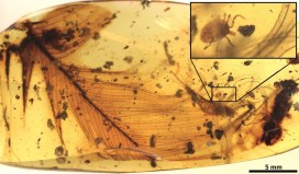
The paper that my colleagues and I have just published provides evidence that ticks fed from feathered dinosaurs about 100 million years ago, during the mid-Cretaceous period. It is based on evidence from amber fossils, including that of a hard tick grasping a dinosaur feather preserved in 99 million-year-old Burmese amber.
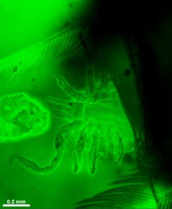
Fluorescence detail of the studied hard tick grasping a dinosaur feather. Extracted from the publication.

The probability of the tick and feather becoming so tightly associated and co-preserved in resin by chance is virtually zero, which means the discovery is the first direct evidence of a parasite-host relationship between ticks and feathered dinosaurs.

Fossils of parasitic, blood-feeding creatures directly associated with remains of their host are exceedingly scarce, and this new specimen is the oldest known to date. The tick is an immature specimen of Cornupalpatum burmanicum; look closely under the microscope and you can see tiny teeth in the mouthparts that are used to create a hole and fix to the host’s skin to suck its blood.

The structure of the feather inside the amber is similar to modern-day bird feathers, but it could not belong to a modern bird because, according to current evidence at least, they did not appear until 26 million years later than the age of the amber.

Feathers with the same characteristics were already present in multiple forms of theropod dinosaurs –  the lineage of dinosaurs leading to modern birds – from ground-runners without flying ability, to bird-like forms capable of powered flight. Unfortunately, this means it is not possible to determine exactly which kind of feathered dinosaur the amber feather belonged to.

But there is more evidence of the dinosaur-tick relationship in the scientific paper. We also describe a new group of extinct ticks, created from a species we have named Deinocroton draculi, or “Dracula’s terrible tick”. These novel ticks, in the family Deinocrotonidae, are distinguished from other ticks by the structure of their body surface, palps and legs, and the position of their head, among other characteristics.

Blood-engorged Deinocroton draculi tick (female). Extracted from the publication.

This new species was also found sealed inside Burmese amber, with one specimen remarkably engorged with blood, increasing its volume approximately eight times over non-engorged forms. Despite this, it has not been possible to directly determine its host animal:

Assessing the composition of the blood meal inside the bloated tick is not feasible because, unfortunately, the tick did not become fully immersed in resin and so its contents were altered by mineral deposition.
Dr Xavier Delclòs, an author of the study from the University of Barcelona and IRBio.

But there was indirect evidence of the likely host for these novel ticks in the form of hair-like structures called setae from the larvae of skin beetles, or dermestids, found attached to two Deinocroton ticks preserved together. Today, skin beetles feed in nests, consuming feathers, skin and hair from the nest’s occupants. But as no mammal hairs have yet been found in Cretaceous amber, the presence of skin beetle setae on the two Deinocroton draculi ticks suggests that their host was in fact a feathered dinosaur.

The hair-like structures, or setae, from skin beetles (dermestids) found attached to two Deinocroton ticks fossilised inside amber, in comparison with extant ones. Modified from the publication.

Together, these findings tell us a fascinating story about ancient tick behaviour. They reveal some of the ecological interactions taking place among early ticks and birds, showing that their parasite-host relationship has lasted for at least 99 million years: an enduring connection, bound by blood.

The paper “Ticks parasitised feathered dinosaurs as revealed by Cretaceous amber assemblages” is published as open access in Nature Communications. Direct link: http://dx.doi.org/10.1038/s41467-017-01550-z
Clean as a new pin

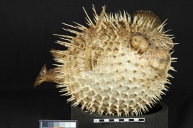
The spiky customer above has enjoyed a serious spruce-up from Stefani Cavazos, our current intern from UCL’s MSc in Conservation for Archaeology and Museums. Stefani tells us how she got this Spot-fin Porcupinefish looking shipshape, without receiving any serious injuries.

So far at the Museum I have been working on a range of specimens, from taxidermy and wet specimens to cleaning the whales, but my favourite project so far has been the conservation of this Spot-fin Porcupinefish from the displays. It is part of what is known as the Christ Church Collection, which came to the Museum in 1860. This makes the Porcupinefish at least 150 years old.

The specimen itself was covered in dust and all five of its fins were backed with deteriorating cardboard pieces. These were most likely attached to give some support during a previous restoration attempt. Unfortunately, cardboard is not a conservation grade material because over time it becomes acidic. Temperature and humidity changes in the Museum have caused it to bend forward, pulling the fins out of shape, so we felt it should be removed to prevent further damage.

The Spot-fin Porcupinefish (Diodon hystrix) before conservation treatment (left images), and close ups of the cardboard backings on the left pectoral fin (right images), where staining and bending are visible. The red arrow indicates where the paper has separated from the fin, and how thin the fins are.

The first step in the treatment was to clean the surface of the Porcupinefish using warm water and a cotton swab. This allowed me to get into the nooks and crannies of the body whilst (mostly) avoiding being poked by its spines. Next, the cardboard backings were softened with water vapor, causing them to break apart so they could be removed easily using tweezers and a scalpel blade.

Conservation can feel like detective work since we often uncover interesting information about specimens as we work on them. In this case, as we removed the cardboard pieces, we found writing on the underside. It appears to be from a shoe box! Though unexpected, it wasn’t entirely surprising. Preparators in the past used whatever materials were available to them at the time.

(Left) The cardboard was carefully removed from the caudal (tail) fin. (Right) The cardboard once removed from the fin, it appears to be from a shoe label.

After detaching the cardboard from all the fins, the remaining ink and adhesive residues were removed using a 50/50 alcohol and water mixture applied with a cotton swab. The edges of the fins were then coated with two thin layers of an acrylic adhesive to prevent any further breakage and to offer some support to the weakest areas. Cleaned, and free of damaging materials, this Porcupinefish is now ready to go back on display!

Photos of the Spot-fin Porcupinefish after treatment was completed. Without the cardboard backings, the fins are somewhat translucent.
Stefani takes her finished work out to one of our regular Spotlight Specimen sessions, giving visitors the chance to get a closer look and ask questions.

All that glitters…

The latest display in our changing Presenting… series showcases some of the incredible colours seen in many insects. Zoe Simmons, collections manager in our Life Collections, explains how such wonderful hues are created.

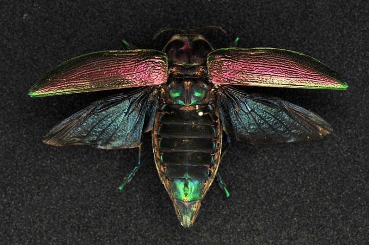
Reflected and refracted light creates the many bright and shining colours found in some insects. The dazzling natural display shown in the specimens here is formed through a combination of embedded pigments and sculpted surfaces on each insect’s external skeleton.

Some species can be variable in colour. Here a pair of Lamprima, a genus of Stag Beetles, shows off the range of colours present in the species.

Different pigment chemicals are responsible for different colours. Carotenoids produce yellow, orange and red hues, while bilins may be green, or blue if linked with proteins. They reflect and absorb different wavelengths of light, and the wavelengths that are reflected are the ones that we see as colour. Typically humans can see wavelengths of 390-700 nanometres, with the lower wavelengths perceived as blue, and the higher ones as red.

Many of the Leaf Beetles (Chrysomelidae) exhibit metallic colours.

Many insects also have multiple thin layers over their upper surfaces to help protect them and prevent dehydration. Variations in thickness and chemical composition of these layers can interfere with the transmission of light, refracting and scattering it back.

Some of the most striking metallic colours are found in the genus Chrysina, where species can be rose, silver or gold.

The shape of the surface layer can reflect light in a multitude of directions, with micro-folds, grooves, pits, hairs and scales all helping to produce complex colours and effects.

The formation and purpose of these colours is scientifically interesting, with research having applications in areas such as nanotechnology. But these insects are also simply beautiful examples of the spectacular diversity of the natural world.

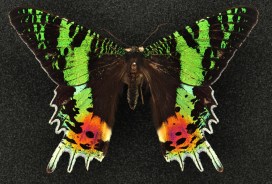
Sunset moths (Uraniidae) are so called because of the dazzling array of colours on their wings. As day-flying moths they are brightly coloured like many butterfly species.

Skeleton keys

by Chris Stimpson, visiting researcher from Queen’s University Belfast

Visitors to the museum will be familiar with the striking parade of mammal skeletons in the court, where they can get a close look at a polar bear’s jaws and peer up through the rib cages of Indian and African elephants, amongst many other things. But these mounted specimens are just a small sample of the animal skeletons that are looked after by the museum.

The main collection of skeletons is carefully stored in behind-the-scenes spaces such as the museum’s Tradescant Room. For researchers who work on animal bones found in archaeological sites, collections like these are not just important – they are essential.

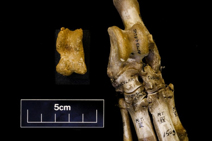
Comparison of an archaeological pig astragalus (ankle bone, left) with an articulated reference specimen from the museum collection (opposite leg, OUMNH.ZC.19948) of an Indonesian wild boar (Sus scrofa). Radiocarbon dating of charcoal indicates the archaeological specimen is over 17,000 years old.

Differences in size, shape, proportion, and the number and arrangement of bones and teeth are a great aid to identification. Teeth in particular often have features that help identify the animal they came from. Bones also have articulations and facets which can be helpful, though identification can be more challenging than with teeth.

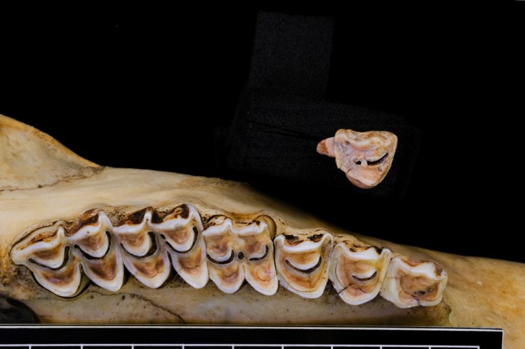
Comparison of an archaeological premolar (top), with the upper right tooth row of a goat-like animal called a serow (Capricornis sumatraensis) from the museum’s collection (OUMNH.ZC.21654). Radiocarbon dating of charcoal from the site indicates the archaeological specimen is over 5,000 years old.

These challenges are part of the work I am doing on the SUNDASIA Project which is undertaking archaeological and palaeoecological investigations in the Tràng An World Heritage Area, in Ninh Binh Province, Northern Vietnam. Working with Vietnamese colleagues, we are investigating climatic and landscape changes that have affected – and may affect – the limestone karst forest over thousands of years. In particular, we’re looking at the responses of human, animal and plant communities to these changes.

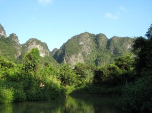
The limestone karst landscape of the Trang An World Heritage Area

During our cave excavations we have recovered bones from a variety of birds, mammals, reptiles, fish and amphibians. Radiocarbon dates from charcoal in the cave deposits suggest this material ranges from 30,000 to 5,000 years old. This is great, but what can these bones tell us of animal life and human hunters at different times in the past? What has changed and why? And what could it mean for the future of Tràng An?

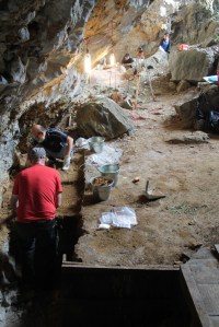
Excavations underway in Hang
Moi, a cave site in Trang An

Before we can begin to answer juicy research questions like these, we need to identify the bones. This is where collections like those held in the museum really come into play. Only with access to skeletons of known animals – where there is knowledge of family, genus or species classification – can we compare the excavated material and identify what we have found.

And while old bones and skeletons may smack rather of death, with a little patience and a good comparative collection like that in the museum, it is remarkable what a few specimens can tell you of life in different times and places that we otherwise know little about. Museum collections are a key to the past, present, and perhaps even to the future.

The ancient mariner

Helen J. Bullard is a PhD candidate at the University of Wisconsin–Madison whose research aims to tell the historical and cultural stories of the horseshoe crab. After visiting the museum, and reading the story of our Natural History After-School Club member’s horseshoe crab fossil find, Helen offered to write a guest post for the blog about these amazing, ancient mariners…

You’re reading this, so I’m guessing you like museums. But have you ever heard of living fossils? Animals such as sharks and crocodiles are often referred to as ‘living fossils’ because they appear pretty unchanged from their ancient fossilized relatives. Of course, by definition, you can’t be both alive and a fossil. But fossils allow us to become primary eyewitnesses to ancient life; we can literally see what life used to look like, how cool is that? They can also dole out some pretty valuable advice, if we just choose to listen.

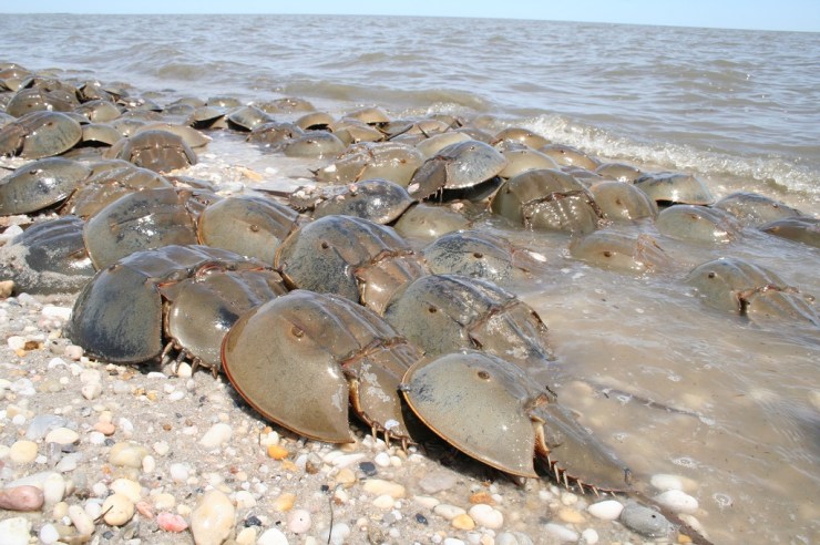
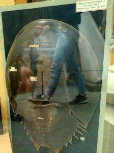
This summer during a visit to England, I spent some time at the Museum studying another so-called living fossil, the horseshoe ‘crab’. The horseshoe crab is not actually a crab, but is instead more closely related to spiders, scorpions and ticks. In fact, they are the closest living relatives of the extinct trilobites. But unlike their famous trilobite cousins, horseshoe crabs have survived all five of Earth’s major mass extinction events. Today, as a direct result of their ability to survive, the four remaining species of horseshoe crab play a vital role in global medical safety.

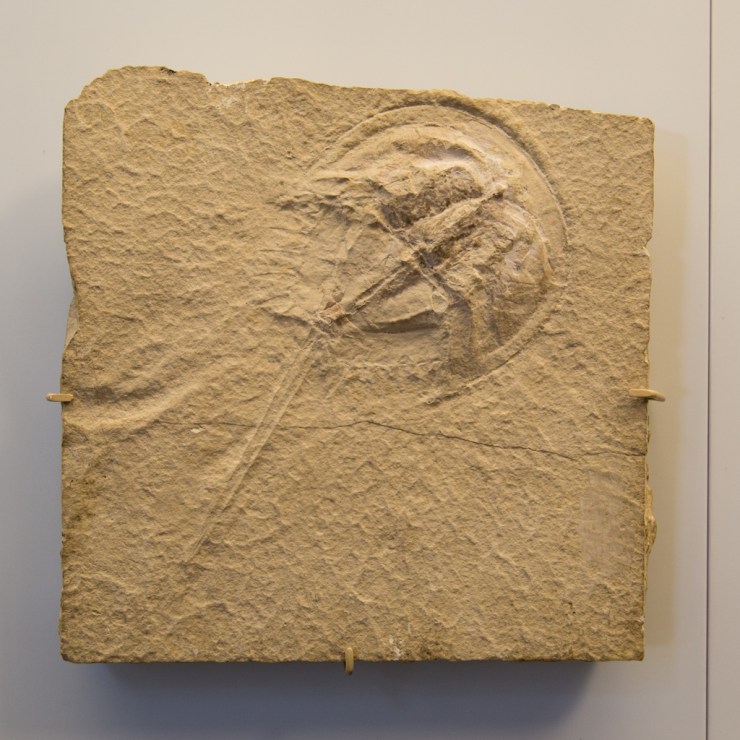
The Museum’s fossil specimen of Mesolimulus walchi, from the Upper Jurassic (163-145 million years ago), Solnhofen Germany, shows how little the form of the horseshoe crab has changed since

Not only do living horseshoe crabs look very similar to their early relations, they are also able to survive surprisingly severe injuries that often leave them missing body parts. Being able to see, through fossil evidence, how little their form has changed over time has helped to uncover the answer to this secret superpower. It lies in a very special life-saving trick that the crabs have kept for millions of years: a coagulating blood protein.

Horseshoe crabs on display in the Museum may provide food for thought for visitors

The blood of the horseshoe crab is able to clot quickly if bacteria are introduced, preventing infection, and saving the crab’s life. Since this discovery in the 1970s, this life-saving protein has been extracted from horseshoe crab blood and used in human medicine to test the safety of vaccines, medical laboratories, intravenous drugs, implants, and much, much more. The chances are that you owe a great deal of gratitude to the horseshoe crab.

But after all that surviving, horseshoe crabs, like many species, are now struggling for survival. They are losing their spawning grounds because of coastal development, industry, housing, marinas and coastal defense structures; they are collected and killed by the millions for bait, and bloodlet in their hundreds of thousands for medical use every year. It is likely that horseshoe crabs will not survive much longer.

But don’t despair. Museums are critical because they hold collections that can unlock knowledge about environmental change, and we can use that knowledge to protect life. Of course, horseshoe crabs are not alone in telling their stories through the fossils they leave – natural history museums are full of stories in stone, bones, pollen, and other traces. If you want to learn about and protect biodiversity, visit your local museum, or support organisations like Oxford’s Environmental Change Institute.

And to help the ancient horseshoe crab itself, join in with the efforts of the Ecological Research and Development Group – the crabs have saved us, so let’s return the favour.


Rehoming a dinosaur

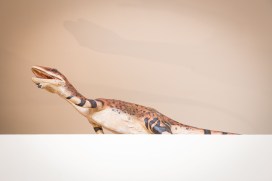
Last summer we ran an unusual competition: finding a new residence for our four metre-long model of Utahraptor ostrommaysorum. It had been hibernating in one of our off-site stores for a while, but following a reorganisation of collections we needed to find a new place for it to live. The competition to rehome the dinosaur was fierce, with 200 venues across the world vying to become the Utahraptor‘s new keeper…

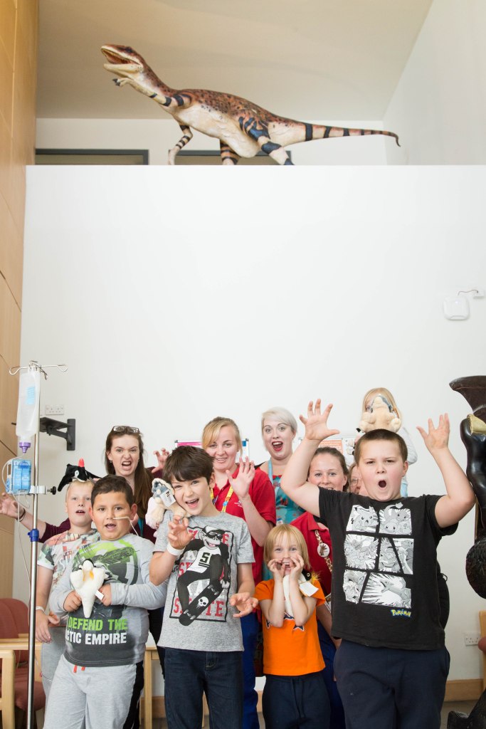
It’s taken some time, thanks to logistics and admin, but one year later we are really delighted to reveal that the Utahraptor has now been installed at the Children’s Hospital at the John Radcliffe Hospital in Oxford.

The bid to take in the Cretaceous creature came from Sarah Fletcher, who now works at the Churchill Hospital in Oxford. Sarah nominated the Children’s Hospital so that the dinosaur could amaze and inspire the young patients.

The idea of having a model Utahraptor in the hospital seemed like a lot of fun. Having been through the Children’s Hospital with my family, I knew that it would make such a difference to everyone who walks through those doors. But I never thought in a million years that we would win it – I am thrilled!
– Sarah Fletcher

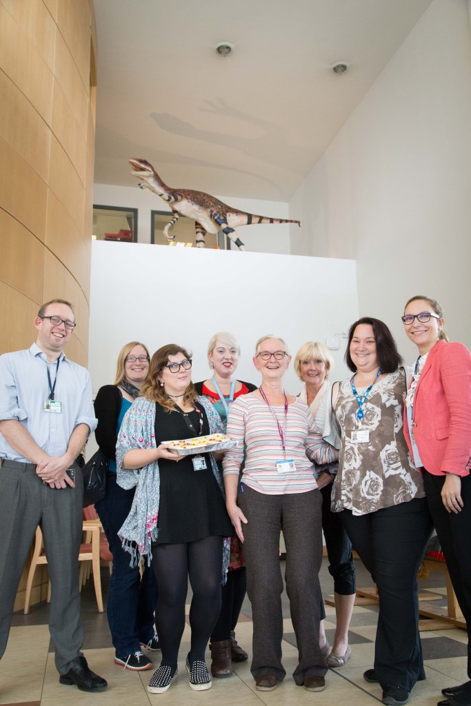
The Children’s Hospital team celebrate the arrival of their new ‘pet’

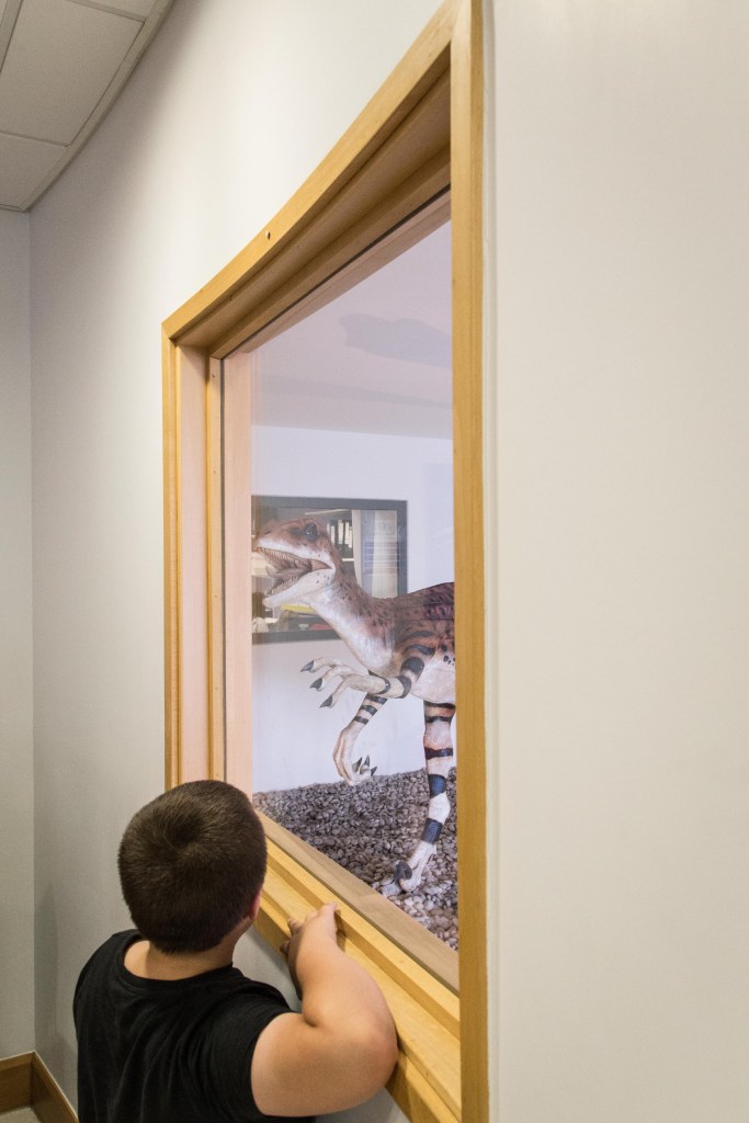
The model has been installed in the main entrance of the hospital, complete with new shadow-casting lighting, thanks to support from Oxford Radcliffe Hospitals Charitable Funds.

The team are now looking to develop new arts projects for young patients, themed around the dinosaur, including an all-important naming competition. We all hope it will bring pleasure to patients, provide a welcome distraction, and make their hospital visit a little more fun.

Patients, staff and visitors can peer at the dino on the way to the wards