by Steven Williams, Oxford Brookes University research student
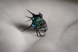
Described by A.G. Butler in 1873 as ‘the most gorgeously coloured spider in this genus’, Gasteracantha scintillans, with its metallic green iridescent abdomen, is the first of my Christmas Spiders.
Spiky and sparkly: Gasteracantha scintillans
The beautiful colour of the abdomen certainly has a very festive feel and it would not be out of place next to a bauble on a Christmas tree; at least not in my house. This species and the other closely-related metallic Thorn Spiders are currently only found on the Solomon Islands.
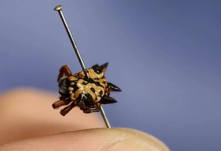
Christmas Island, in the Indian Ocean, is one of the locations where my second Christmas spider, Austracantha minax, can be found. Although not as striking as the metallic green of Gasteracantha scintillans, the layout of the abdominal spines on this spider almost give it the appearance of a star – perfect for the top of a Christmas tree, no?
Austracantha minax: starry, and found, amongst other places, on Christmas Island
The common name of ‘Christmas Spider’ is attributed to this species because in areas of Western Australia it is associated with the arrival of Christmas as the males reach maturity in mid-December and females in January.
Did you know that there is also an Eastern European folk tale of how tinsel came to be included in Christmas tree decorations? The legend tells of how spiders spun cobwebs on a poor family’s undecorated Christmas tree overnight. In the morning the webs turned to gold and silver and the family never lived in poverty again. So when you put the tinsel on the tree this year you could imagine you are a spider spinning a web!
With that spidery festive thought, have a very Merry Christmas from everyone at the Museum!
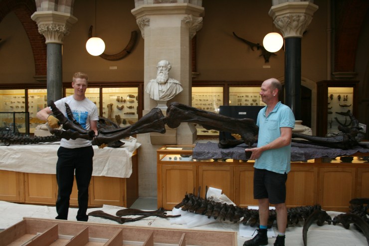
Last year we introduced you to Adam Fisk, our (then) new apprentice. He’s the slightly younger-looking one in the photo above; on the right is Pete Johnson, the Museum’s technician and Adam’s mentor and supervisor. Together, the two are a vital component that keeps the Museum machinery a-turning.
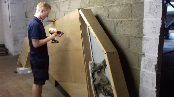
Over the past twelve months or so Adam has busied himself with many vital tasks: installing and de-installing our exhibitions, including Kurt Jackson’s Bees (and the odd wasp) in my Bonnet and Microsculpture; reconstructing an Edmontosaurus; and, more recently, sporting an excellent Star Wars Christmas jumper.
Packing an Attenborosaurus ready for transit
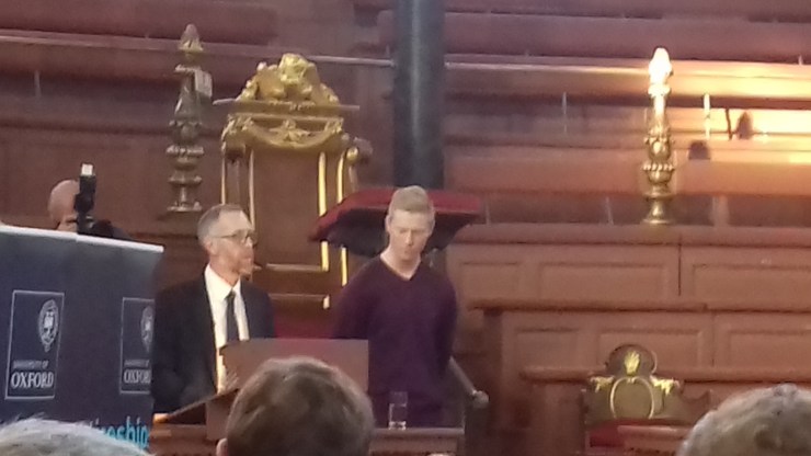
We’re therefore exceptionally pleased to announce that Adam has just been selected as a winner in the Annual University of Oxford Apprenticeships Awards. This calls for a small whoop, and a large ‘Well done Adam!’.
The awards, which celebrate the achievements of the University’s apprentices, supervisors and departments, were given out at a ceremony at the rather grand Sheldonian Theatre this afternoon.
Adam collecting his award at the Sheldonian Theatre in Oxford
When Adam joined the Museum last summer for his three-year apprenticeship he had just finished his GCSEs. Now he’s walking the stage at the Sheldonian… As Pete says:
Adam’s contribution to all aspects of work from maintenance and workshop demands to exhibition installation and Collections assistance has been outstanding. We have all benefitted greatly from his input, and he has helped us to achieve a high standard of public service.
Thanks for all the hard work Adam and we look forward to the next 18 months with you on the team.
Visitors to the Museum often comment on just how many things there are to see here, but in fact only a small percentage of the collection is on public display – less than 0.1%. The remaining 99.9% is held in storage for use in research, teaching and to loan to other museums and universities.
I receive hundreds of enquiries from researchers all over the world who are interested in many aspects of animal biology. In exchange for our information about the skeletons, skins, specimens preserved in fluid, nests, shells and taxidermy here, the Museum gets knowledge from world experts who are publishing new science.
Bramble Shark (Echinorhinus brucus). Image: Illustrations of South Africa / Wikimedia
One such recent enquiry, from researchers Dr Samuel Iglésias and Frederik Mollen, concerned the Bramble or Spinous Shark (Echinorhinus brucus). You may not have heard of the Bramble Shark – it’s an enigmatic bottom-of-the-sea dwelling species – but this is one of the reasons why the researchers are trying to track down all known specimens held in European institutions.
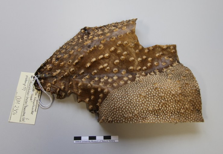
The characteristic brambles of the Spinous Shark, Echinorhinus brucus
Bramble Sharks are so called because of the large thorn-like structures, called dermal denticles, that cover the skin. These sharks are thought to be once common in European waters but are now virtually extinct.
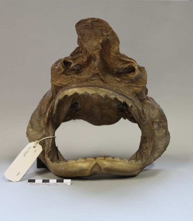
I checked the Museum databases and stores and found 14 Bramble Shark specimens in the collections: one taxidermy specimen; one set of jaws with skin; a section of skin; and 11 fluid-preserved dissections. Unfortunately, as is often the case in older natural history collections, the fluid specimens did not have data associated with them – information such as age, locality, who collected them, what the dissections were or who dissected them.
After passing images and this list of specimens to the researchers they sent through an 1875 paper titled The Brain and Cranial Nerves of Echinorhinosus spinosus with notes on the other viscera by Bruce Clarke and the excellently named Hatchett Jackson. This paper describes the dissection of two specimens at the University of Oxford, one of which was a female collected from Penzance on 15 February 1875.
Armed with this new information I was able to cross-reference this dissection with some of our archives to confirm that most of the dissected specimens actually came from this single female shark collected in 1875, which was dissected and the parts used in illustrations in a technical publication.
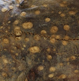
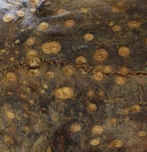
Section of skin previously identified as Bramble Shark but now re-identified as from a Porcupine Ray (Urogymnus asperrimus)
Another discovery came while photographing the skin specimen, and comparing it to the other dry specimens. It became clear that the thorns of the skin were very different to the typical Bramble Shark arrangement and it turned out that the skin was in fact from a Porcupine Ray (Urogymnus asperrimus).
The happy ending here is that the researchers now have better information for their publication and these specimens will be put ‘on the map’ in the technical literature for other researchers to access.
Although the Museum collections are effectively one Bramble Shark down (but up one Porcupine Ray skin), we do now have better information about the age, locality, relationship, identification, citation in the literature, and history of some these specimens. This makes them even more useful for research in the future.
Not all enquiries end with such a deeply satisfying result, but it is certainly nice when they do.
The latest display in our changing Presenting… case showcases some pioneers of photography and features a recently-discovered original print of Charles Darwin by Julia Margaret Cameron…
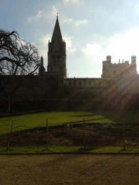
In the early days of photography, in the mid-19th century, a number of photographic innovators had close links to the Museum and its collections. Perhaps the most famous of these is Charles Lutwidge Dodgson (1832-1898), better known as Lewis Carroll. Dodgson was an author and mathematician at Christ Church College in Oxford, and also an accomplished early photographer.
Dodgson’s friends were often the subjects of his photos. A well-known example is Alice Liddell who, as well as sitting for portraiture, provided inspiration for Dodgson’s book Alice’s Adventures in Wonderland, as did the Dodo in the Museum’s collections.
The image at the top of the article is titled The Anatomy Lesson with Dr George Rolleston and was taken by Dodgson around 1857. It shows Dr Rolleston, Professor of Medicine at the University of Oxford at the time, pictured with a selection of specimens from the Museum’s collections.
Two of Dodgson’s subjects were accomplished photographers in their own right, and perhaps the most famous female photographers of their time: Julia Margaret Cameron (1815-1879) and Sarah Angelina Acland (1849-1930).
Cameron’s innovative and distinctive style of portraiture raised photography to a true art form. Her intentional use of shifted focus and experiments with process and finish gave an ethereal look to her works resembling the artistic style of the contemporary Pre-Raphaelites.
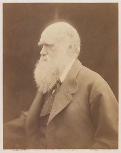
Many prominent scientists sat for Cameron, including naturalist Charles Darwin. She took the portrait above in 1868 at her home on the Isle of Wight. An original print of this photograph, made by Cameron, was recently discovered in the Museum Archive, uncatalogued. It features her signature on the back and the blind stamp from her printseller in London on the front.
‘Mr Ruskin & his old friend at Brantwood’ by Sarah Angelina Acland, 1893
Sarah Angelina Acland, inspired and taught by Cameron, is best known for her innovations in colour photography, but she also took many black and white photographs while learning the art form. The portrait above features her father Sir Henry Wentworth Acland and the artist John Ruskin at his home. Both Ruskin and Acland were instrumental in the founding of this Museum, and Sarah even helped to lay the founding stone in 1855.
In a 1904 Royal Photographic Society exhibition Acland was the first to exhibit work combining images taken with red, green and blue filters, three years before the launch of the colour photography process invented by the Lumière brothers.
These images, including the original print of Charles Darwin by Julia Margaret Cameron, are on display in our Presenting… case until 24 January.
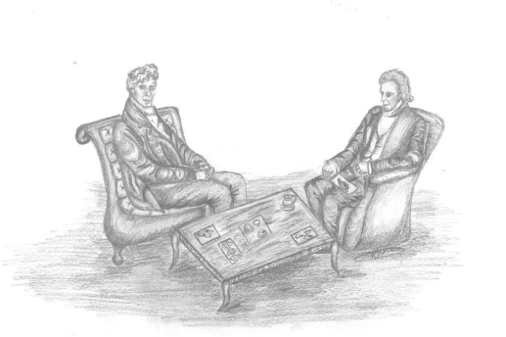
G.B. Sowerby and Charles Lyell discussing fossils. Drawn by Amy Jones.
Imagine this: Charles Lyell sitting with his friend George Brettingham Sowerby discussing and identifying fossils from Lyell’s last adventure whilst in a pair of plushy armchairs drinking tea. It is not known whether this is the case, but for me it is what immediately springs to mind when finding comments and identifications made by G.B. Sowerby which have been written on the back of display tablets. Some of these identifications seem to be in the handwriting of G.B. Sowerby himself. Whether this was G.B. Sowerby the elder or the younger remains to be discovered.
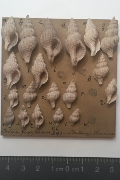
Reverse of specimen tablet with “Pollia fusiformis G.B.S.” as an example of G.B. Sowerby’s handwriting
Research on the Lyell Collection has revealed that G.B. Sowerby identified nearly 3,500 fossils for or with Charles Lyell, mainly between 1839 and 1841. These were mainly gastropods (snails) and bivalves (clams, mussels, and scallops). The majority were from France, but it seems Lyell called upon him to assist with identifications from many other localities.
Front of previous specimen tablet showing the fossil gastropods G.B. Sowerby identified
Now this is all well and good but if you are anything like me you will be wondering “well who were The Sowerbys?” Read on to find out what is interesting and extraordinary about them.
The Sowerbys contributed massively to the field of natural history in many different disciplines. 14 members of the Sowerby family wrote and or illustrated over 100 works on botany, mineralogy, palaeontology, and zoology. They worked with or for most of the great names of natural history in the 19th century, including Charles Darwin.
James Sowerby was the man who started it all. Father to 9 children including James de Carle and George Brettingham I. James Sowerby was a naturalist and an illustrator. He had black eyes and was told by his mother that all the girls would die for him. He couldn’t see how he would kill with his eyes or make girls hearts ache, and so decided that he was in fact ugly, concluding that all the talks of heartaches and killings were untrue. James recognised himself as a genius, titling his childhood reminiscences ‘Myself or the progress of a genious’ (his spelling). He produced not only his well-known work on plants (English Botany) but also works on mycology (the study of fungi), conchology (the study of mollusc shells), and mineralogy.
The “ugly” James Sowerby. Line engraving by Mrs D. Turner after T. Heap. From Wellcome Images. Accessed from here on 23 November 2016.
James de Carle Sowerby, the eldest son, was a mineralogist and illustrator. He studied experimental and analytical chemistry under Humphry Davy, and had the honour of assisting Davy with his experiments. James de Carle proposed classifying minerals according to their chemical composition, and by the age of 20 had named and arranged the collections of the Marchioness of Bath and other amateur collectors.
George Brettingham Sowerby I was a naturalist, illustrator, and conchologist. He became estranged from his family, with his name not appearing on family publications after 1822, and set up his own establishment working with natural history specimens. His son, George Brettingham II, was taken on by Charles Lyell in 1843 as his aid when in the USA. The name was passed down for 6 generations.
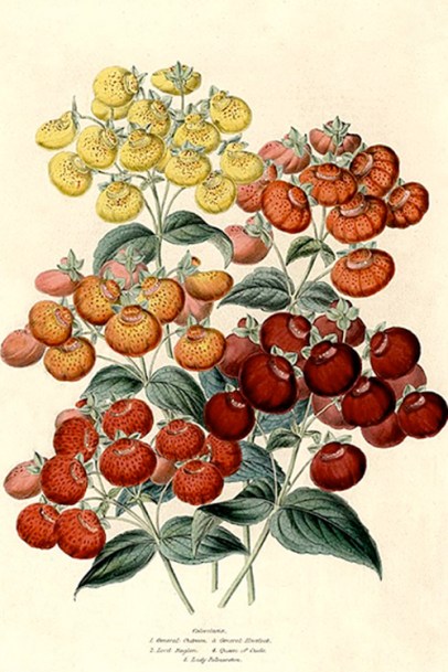
Charlotte Caroline Sowerby was the only daughter of George Brettingham I. She was a natural history illustrator with her high quality images present in The Illustrated Bouquet. Not only did she create botanical images she also illustrated a quartz crystal with asbestos inclusions, and volcanoes, proving it wasn’t just the men of the family who did the work!
Calceolaria Bouquet. Hand coloured zincography in The Illustrated Bouquet by Charlotte Caroline Sowerby. From The Illustrated Bouquet. Accessed from here on 23 November 2016
Hi, I am Lily, you may remember me from the Getting the Picture blog. I was the Museum Studies student working on the Lyell Project for 8 weeks and guess what – I’m back! I will be here for the next 6 months finishing off the project, so here’s a little bit of information about me.
Lily looking at Lyell fossils
I have been interested in palaeontology for as long as I can remember, since the first time I watched Jurassic Park. All I have wanted to do since then is work in a museum, giving information a new life by exploring the ways old stories can be used to engage people in unusual and exciting ways.
When I first visited OUMNH I fell in love. It was hands down the best museum that I had been to. All I wanted to do was work somewhere like that, and look at me now (follow your dreams, kids!). When I learnt earlier this year that I was able to undertake my work placement here I was ecstatic. The project looked amazing and I couldn’t wait to start. A few months later I arrived, and it was everything I hoped it would be and more. Now I am back I have the same feeling of elation, finishing a project that I loved working on. It felt like a dream when I was asked to return (for actual money this time) – I am still not quite sure it’s real. To all those out there looking at doing work placements one piece of advice: pick somewhere you can see yourself working and you will have an amazing experience. Trust me, I did. I can’t wait to see the end result of this project and I hope you guys are all with me on that. It’s going to be amazing.