Dr Ross Anderson climbs a steep, green slope surrounded by pine trees and dramatic mountain scenery.

Oxford team’s fieldwork revealing how complex life first evolved on Earth 

By Ross Anderson

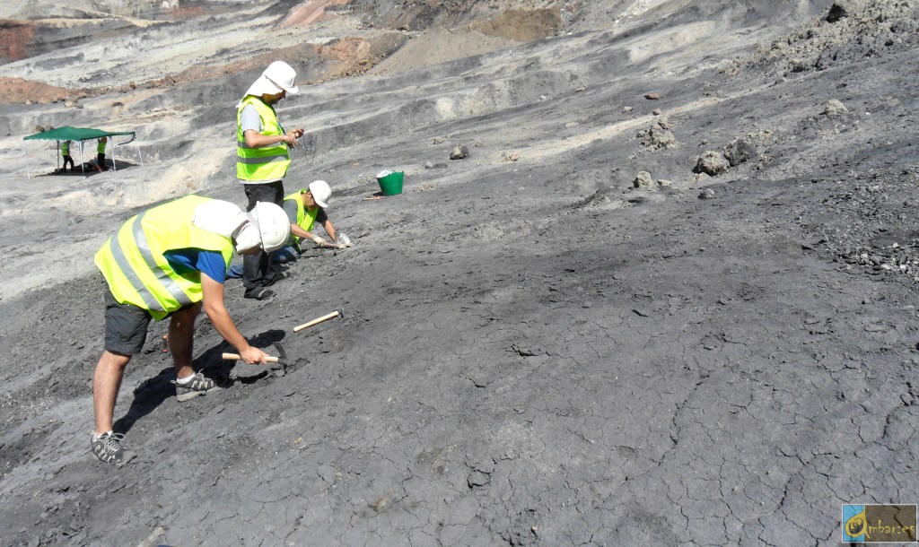
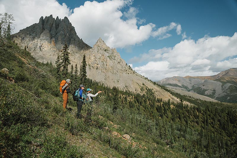
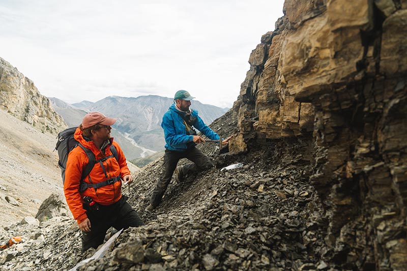
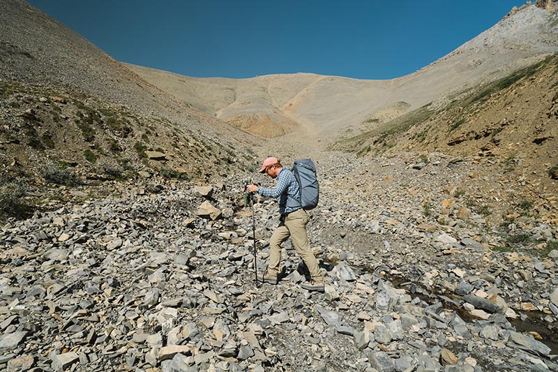
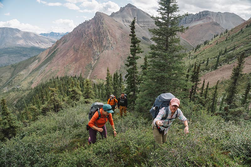
In Summer 2024, a team of palaeontologists and geologists from the University of Oxford, along with colleagues from Dartmouth College, the University of Washington, and Williams College in the USA, undertook an expedition to the Little Dal Group in the Mackenzie Mountains, Northwest Territories, Canada. Our purpose was to uncover some of the oldest fossil ecosystems that record complex life.
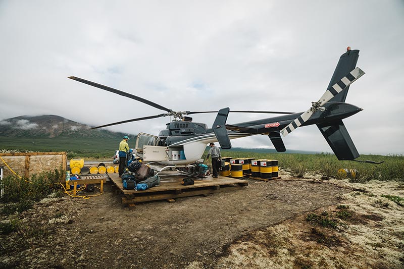
Complex life comprises all organisms whose DNA is enclosed in a cell nucleus. This includes animals and plants but excludes bacteria. Today, this complex life accounts for most of the Earth’s biomass, documented biodiversity, and oxygen production. Understanding when and how it first evolved remains one of the central unanswered questions in evolutionary biology.

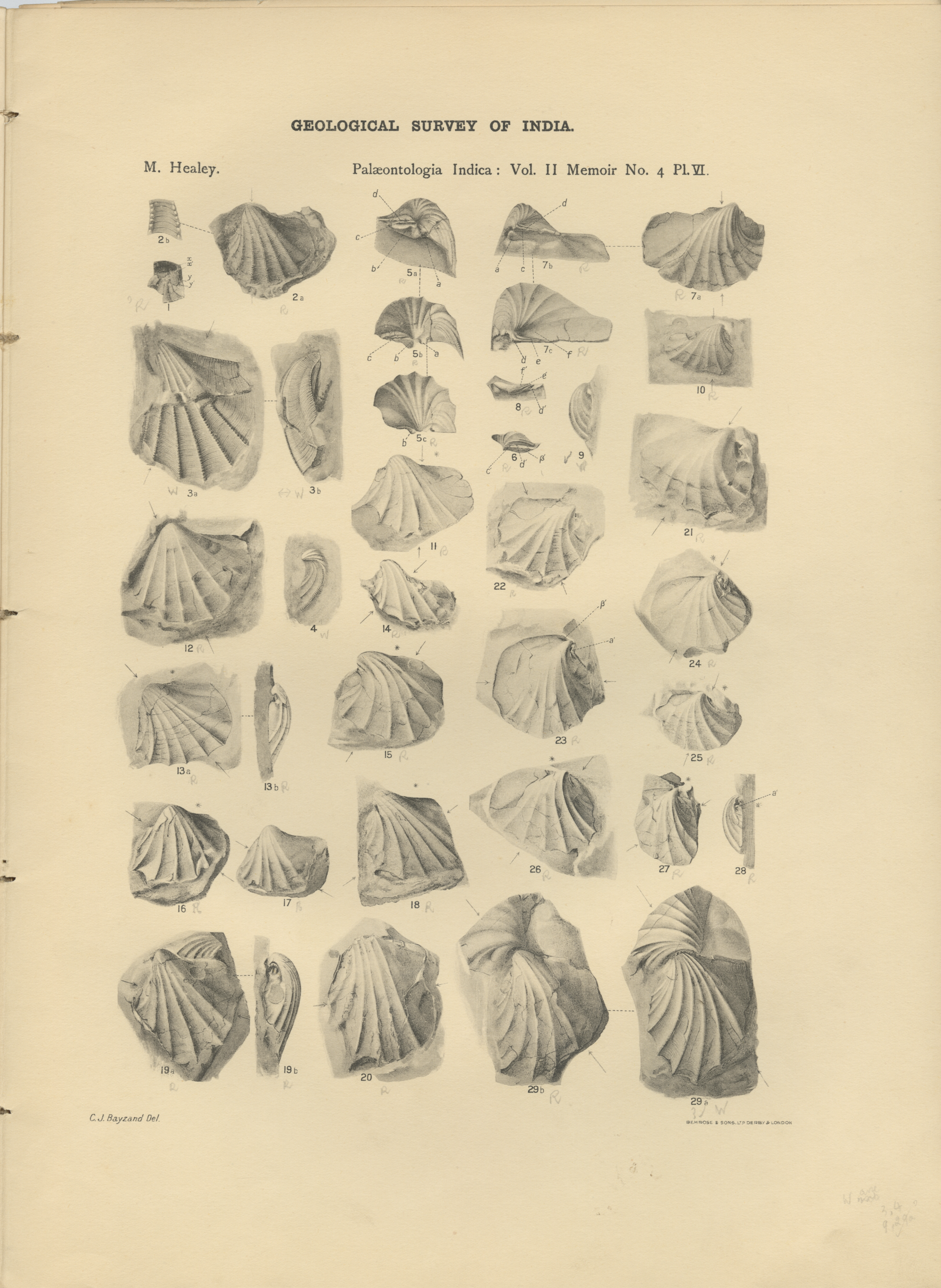
As palaeontologists, we normally use fossils to reveal the history of life. Fossils tend to preserve larger animals with hard shells or skeletons—creatures such as trilobites, ammonites, dinosaurs, and mammoths. However, the first complex organisms were microscopic and lacked such hard parts. As a result, their soft and fragile cells rarely fossilised. Put simply, we have found it a major challenge to trace the origins of complex life with fossils.

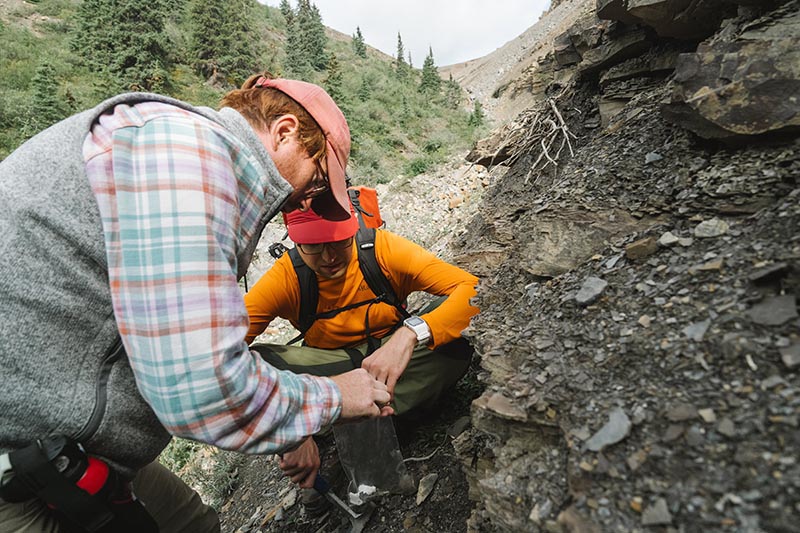
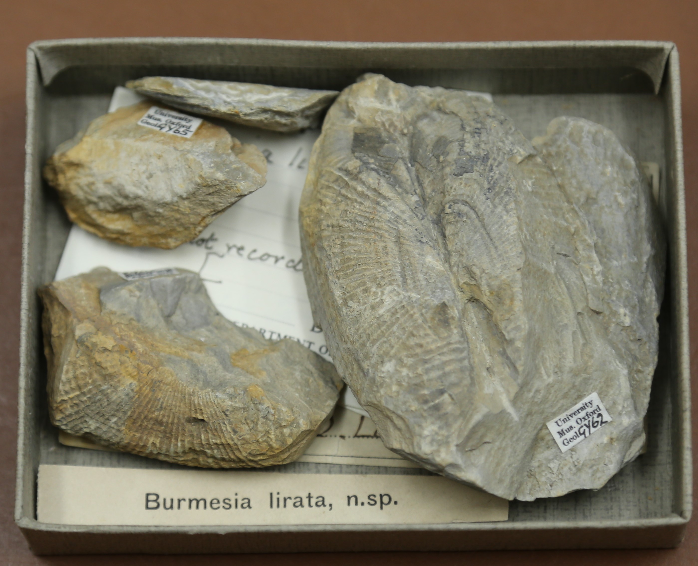
I have argued that finding rocks made up of antibacterial clay minerals holds the key. These minerals can slow the decay of organic cells long enough for them to survive as fossils. The Little Dal Group contains ~900-million-year-old rocks that are rich in just such clays, making it a prime target for new fossils that might help us unravel the origins of biological complexity.

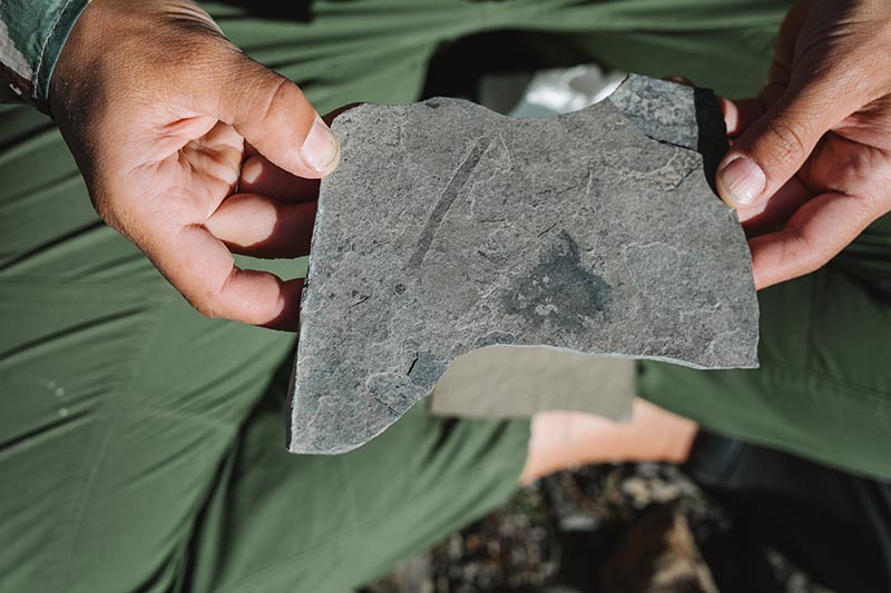
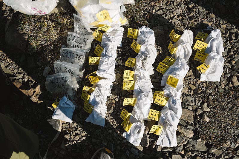
I was joined in Canada by my DPhil student, George Wedlake, from the Department of Earth Sciences. Together we spent two weeks collecting over 100 rock samples. The samples record an ancient tropical sea not unlike the Bahamas today, where early complex life likely flourished.

Back in Oxford, at the Museum of Natural History, George and I are now examining the samples; dissolving the rocks with hydrofluoric acid to extract and study the tiny fossils. We hope these new fossils will transform our understanding of how complex life first took hold on our planet.

Our fieldwork was funded by a Royal Society University Research Fellowship and by the Oxford NERC Environmental Science Doctoral Training Partnership. It was conducted under permit and with the support of the Sahtú Dene people.

A monochrome, three-dimensional rendering of a new, yet undescribed species of Achilidae (Hemiptera: Fulgoromorpha), preserved in Miocene Dominican amber (~15 million years old).

Amber Time Capsules and the Synchrotron’s X-Ray Vision

Imagine a drop of ancient resin. Inside is an insect, trapped for 53 million years, so well preserved it looks like it might twitch back into life. These amber fossils offer us a breathtaking glimpse into long vanished ecosystems. But there’s a catch: the most revealing details, like delicate mouthparts or microscopic genitalia, are sealed away under the resin’s glossy surface. Cutting into them would destroy what makes them precious.

Enter the SOLEIL synchrotron.

At SOLEIL, near Paris, a group of researchers led by OUMNH’s own Dr Corentin Jouault are preparing to shine one of the world’s brightest X-ray beams through over 100 blocks of Oise amber, each containing a fossilised insect no larger than a fingernail. The goal is to see inside without cracking them open. Using a technique called in-line phase-contrast synchrotron microtomography (a bit of a mouthful, so let’s call it “supercharged 3D X-rays”), the team hopes to reveal anatomy invisible to conventional CT scanners. Think of it as upgrading from grainy black-and-white TV to ultra-high-definition.

The impressive imaging setup of the ANATOMIX beamline at SOLEIL Synchrotron. In the foreground, a rotating platform holds the amber sample in the path of the X-ray beam, turning it a full 360° so that hundreds of 2D X-ray images can be captured from every angle. In the background are the scintillators, which transform invisible X-rays into visible light, and above them, the high-resolution camera that records these images. All the data are then processed by powerful computers to reconstruct a detailed 3D model of the fossil trapped in amber.

Dr Corentin Jouault carefully positioning a piece of Eocene Oise amber (approximately 53 million years old, from France), containing an undescribed extinct ant species, mounted on a scanning electron microscope stub, in front of the X-ray beam for a high-resolution scan (pixel size ≈ 1.3 μm).

Why does this matter? Well, these insects lived during the Early Eocene, around 53 million years ago, when flowering plants had taken over the world in what scientists call the Angiosperm Terrestrial Revolution (ATR). This upheaval transformed landscapes and diets alike, and insects, already an evolutionary success story, had to adapt. Some lineages thrived by exploiting new blooms, while others dwindled. By studying the fine details of insect mouthparts, researchers can track how feeding strategies shifted in tandem with the rise of flowers.

The plan is ambitious. Each piece of amber will be scanned in full at a resolution fine enough to spot features a few micrometres across (a micrometre is one-thousandth of a millimetre — about one-hundredth the width of a human hair). 20 chosen specimens will then be magnified further still for an even sharper look, down to less than half a micrometre per pixel. In other words, one pixel will cover an area 140 times smaller than the thickness of a human hair.

Three-dimensional renderings of a new, yet undescribed species of Achilidae (Hemiptera: Fulgoromorpha), preserved in Miocene Dominican amber (~15 million years old). The specimen was scanned at the SOLEIL Synchrotron (ANATOMIX beamline) using Synchrotron X-ray microtomography (SR-μCT). A. Habitus, ventral view. B. Habitus, dorsal view. (© Ancheng Peng & Corentin Jouault)

The resulting datasets will therefore be enormous, so the team has lined up banks of high-powered computers and even machine-learning tools to speed up the laborious task of reconstructing the fossils in 3D.

What emerges won’t just be pretty pictures. These models could rewrite parts of insect evolutionary history. For example, they may uncover the earliest records of certain insect families, refine the timeline of insect diversification, and provide the raw data needed to estimate how extinction and speciation rates shifted during the ATR. In short, these pieces of amber become not just a window into the past, but a testbed for some of the biggest questions in evolutionary biology.

And there’s a democratic twist: all the scans, reconstructions, and 3D models will be made freely available in open repositories. That means anyone, from entomologists to curious hobbyists, could spin, zoom, and explore these ancient insects in digital space.

So, next time you spot an insect hovering around a flower, think of its ancestors locked in amber, their secrets now teased out by beams of light brighter than the sun. The synchrotron doesn’t just illuminate fossils, it illuminates how deep the ties between bugs and blooms really go.

Disappearing Butterflies

HOW TO SOLVE A BIOLOGICAL MYSTERY USING MUSEUM COLLECTIONS AND DNA TECHNOLOGY


By Rebecca Whitla, PhD student at Oxford Brookes University


The Black-veined white butterfly (Aporia crataegi) was a large, charismatic butterfly with distinctive black venation on its wings. Once commonly found in the UK, the species unfortunately went extinct here in around 1925, with the last British specimens collected from Herne Bay in Kent. It isn’t fully understood why the species disappeared from the UK, but climate change, predation, parasites, and disease have all been suggested to have caused its disappearance — perhaps with several of these factors contributing to its decline. Central to solving the mystery of the disappearance of the Black-veined white will be the collections of butterflies that are stored in museums like OUMNH.

Butterflies tend to be well-represented in museum collections, and the Black-veined white is no exception. While the species has now been extinct in the UK for around 100 years, Lepidoptera enthusiasts from previous centuries often captured wild Black-veined white specimens for their personal collections. The abundance of Black-veined white butterflies in museum collections, like the collections at OUMNH, serve as a valuable repository for scientific research — including my own!

Black-veined white butterflies in the collections at OUMNH

Between June and December 2021, I undertook a research project using OUMNH’s Black-veined white butterflies. My task was to extract enough DNA from the butterflies to use for ‘whole genome sequencing’ — in other words, I was attempting to extract DNA from butterfly specimens to decode their complete DNA sequence. Getting DNA sequences from the historical specimens that are kept in Museums is no easy task, as DNA degrades over time. Nonetheless, animal specimens from natural history museums have successfully been used for whole genome sequencing and genetic analysis in the past, including species as diverse as longhorn beetles and least Weasels.

In order to work out how to extract DNA from the specimens, I had to try a variety of methods. This included experimenting to find out whether butterfly legs or abdomen fragments yielded more DNA, and whether non-destructive methods of DNA extraction were as effective as destructive methods. An example of a non-destructive method of DNA extraction would be a process like soaking a sample overnight and using the leftover liquid for DNA extraction, whereas a destructive method might involve mashing a whole leg or abdomen segment to use as a DNA source.

Preparing a DNA sample

Overall, I found that destructively sampling the legs of the butterflies gave the most reliable results, and also had the added benefit of not destroying the wings or abdomen of the specimens. Keeping the wings and abdomens of the butterflies intact will likely prove useful for conducting morphological studies in future.

Now that I have a reliable DNA extraction method, the next step in my research will be to analyse more Black-veined white specimens from a span of different time periods leading up to the species’ disappearance. I will then compare samples collected from each time period to calculate the genetic diversity of the species at each point in time, leading up to its disappearance. If I find a steady decline in the species’ genetic diversity over time, this may indicate a gradual extinction of the species. This is because we expect that, as numbers of a species decrease, inbreeding will become common, resulting in less diversity in the species’ DNA. However, if the populations of Black-veined white butterflies went extinct very suddenly, the decline in genetic diversity will probably be less pronounced. Learning more about the fate of the Black-veined White could not only help us unlock the historical mystery of the species’ decline in Britain, but will also help us understand more about the species’ decline in other parts of the world.


British Insect Collections: HOPE for the Future is an ambitious project to protect and share the Museum of Natural History’s unique and irreplaceable British insect collection. Containing over one million specimens – including dozens of iconic species now considered extinct in the UK – it offers us an extraordinary window into the natural world and the ways it has changed over the last 200 years. The HOPE for the Future project is funded by the National Lottery Heritage Fund, thanks to National Lottery players.

Sisters of Science

THE PIONEERING LEGACIES OF KATHLEEN LONSDALE AND DOROTHY CROWFOOT HODGKIN


By Leonie Biggenden, Volunteer


As Women’s History Month comes to a close, this blog post looks at two ‘sisters of science’, friends and contemporaries Dorothy Crowfoot Hodgkin (1910 – 1994) and Kathleen Lonsdale (1903-1971), and considers some links between these remarkable women.

Dorothy Crowfoot Hodgkin is the only female bust in the Oxford Museum of Natural History and is the only British woman to have been awarded the Nobel Prize for science.  When she was awarded the Prize in 1964 – for her ground-breaking discovery of the structures of vitamin B12 and penicillin – there was much scepticism about whether women belonged in the field of science. One newspaper commemorated her achievement with the headline “Nobel prize for a wife from Oxford”.

Hodgkin was assisted and supported in her endeavours by fellow scientist Kathleen Lonsdale, who worked in London while Hodgkin was based in Oxford.  Both women were pioneers who advanced the x-ray crystallography technique, in which x-rays are fired at crystals of molecules to determine their chemical structure. Lonsdale applied the technique to diamonds, benzene, and later kidney stones.  For her efforts, she had a type of diamond named after her: Lonsdaleite.  It was not just any diamond, but one formed in meteorites, as a result of the heat and pressure of impact into the Earth’s atmosphere.

Both had similar difficulties as girls wanting to study science.  Hodgkin was initially not allowed to take chemistry at her grammar school as it was considered a ‘boy’s subject’, but she thankfully managed to reverse the school’s decision, allowing her to pursue her scientific career.  Lonsdale had to transfer to a boys’ school to be able to study maths and science, as these were subjects not offered at her girls’ school. She later described how her love of maths was inspired by learning to count at school using yellow balls.

Both women were supported by strong male advocates and mentors, such as the scientist William Bragg. Bragg first met Lonsdale when he was assigned as one of her examiners, and subsequently asked her to join his research school at University College London (UCL).  Lonsdale would later follow Bragg when he moved his laboratory to the Royal Institution.  Bragg was also responsible for inspiring Hodgkin’s interest in the properties of atoms, giving her a copy of ‘Concerning the Nature of Things’ when she was 15 years old.

Lonsdale and Hodgkin worked hard to show that science was a viable option for girls.  Lonsdale’s essay, ‘Women in Science – why so few?’, argued that social expectations placed on women discouraged them from pursuing science [1]. In fact, she was so determined to encourage girls’ interest in the subject that, while ill in hospital, she received special permission to be able to leave to award prizes for science at a local girls’ school. Hodgkin advocated for female scientists and directly mentored several who went on to become important crystallographers in their own right.

Both women eventually became professors, and Lonsdale was one of the first two women elected as Fellows of the Royal Society (FRS) in 1945. In 1947, Hodgkin was one of the youngest people to be elected FRS. 

Both Hodgkin and Lonsdale were extremely concerned about the threat of nuclear war, and in 1976 Hodgkin became president of the Pugwash Conference which advocated for nuclear disarmament.  Lonsdale was also involved with Pugwash and was president of the Women’s International League of Peace and Freedom.  A lifelong pacifist, she went to Holloway prison in London for a month for failing to register for war service and not paying her £2 fine.  She became a dedicated advocate for prison reform after seeing the conditions of the women first-hand.

My favourite facts about both Lonsdale and Hodgkin are those that give us a glimpse of their ingenuity.  Lonsdale made her own hat to meet the Queen and have her Damehood conferred upon her.  It was constructed with lace, cardboard and 9d worth of ribbon.  Similarly, when awarded her first honorary degree, Lonsdale pinned a strip of beautiful material inside her gown as a substitute for buying a whole new dress.  Hodgkin was also very creative. As a child, she created her own personal laboratory in the attic and acquired acids from the local chemist to experiment with.

The two women held each other in great respect, as testified to by the fact that Hodgkin wrote a biographical memoir of Lonsdale.  She said of her friend: “There is a sense in which she appeared to own the whole of crystallography in her time.”  Let’s agree that both women can claim that crown. Looking back, we can remember these women for their remarkable stories, featuring precious gems, prisons, penicillin and peace. But, most importantly, we should remember Hodgkin and Lonsdale as pioneers who paved the way for future women scientists.


References

[1] Hodgkin D (1975) Kathleen Lonsdale 28 January–1 April 1971. Biogr Mems Fell R Soc 21:447–484

Reading Archival Silences

MAUD HEALEY AND HER GEOLOGICAL LEGACY


By Chloe Williams, History Finalist at Oxford University and Museum Volunteer

Email: chloegrace1000@gmail.com


“The professor regrets to have to record the loss of the invaluable services of Miss Healey, who as a result of overwork has been recommended to rest for an indefinite period. This will prove a serious check to the rate of progress which has for some time been maintained in the work of rearrangement, and it is hoped that her retirement may be only temporary.” So ends the Oxford University Museum of Natural History’s 1906 Annual Report, marking the near-complete departure of Maud Healey from the archival record.

Despite how little of her history has been preserved, it is clear that Maud Healey made significant contributions to the field of geology. After studying Natural Sciences at Lady Margaret Hall in 1900, Healey worked at the Museum as an assistant to Professor William Sollas from 1902–1906. Here, she catalogued thousands of specimens and produced three publications. These publications were at the centre of debates about standardising the geological nomenclature, and turning geology into a practical academic discipline that could sustain links across continents. However, Healey was continually marginalized on the basis of her gender. Closing the Geological Society of London’s discussion of one of her papers, “Prof. Sollas remarked that he had listened with great pleasure to the complimentary remarks on the work of the Authoress, and regretted that she was not present to defend before the Society her own position in the disputed matter of nomenclature.”[1] Predating the Society’s 1904 decision to admit women to meetings if introduced by fellows, Healey had been unable to attend the reading of her own paper.

Photo of the Geological Society of London centenary dinner in 1907, at which Maud Healey was present. Healey can be seen seated in the fourth row from the front, three chairs to the left. Of the 263 guests, 34 were women, 20 of whom were the wives or daughters of academics, and only 9, including Healey, were present ‘in their own right’. [2] Source: Burek, Cynthia V. “The first female Fellows and the status of women in the Geological Society of London.” Geological Society, London, Special Publications 317, no. 1 (2009): 373-407.

Healey later worked with specimens collected by Henry Digges La Touche in colonial Burma (now Myanmar). While Healey worked with the identification of species, acknowledged by La Touche himself as ‘a more difficult lot to work at’ than similar specimens assigned to her male contemporaries, the physical collection and therefore its name and record is attributed to a male geologist. [3] She continued her work identifying La Touche’s collection of Burmese fossils after retiring from the Museum in 1906 and published a report about them in 1908. What happened to her afterwards is unclear. Tantalizing snippets like a 1910 marriage record might suggest that she turned to a life of domesticity, but whether Healey continued to engage with geology as a hobby remains uncertain.

It is almost unbelievable that a professional of Healey’s calibre could abandon the work in which she excelled. However, Healey lacked any familial connections to geology, and apparently did not marry into money, which would have made it difficult for her to retain access to organizations like the Geological Society of London. The diagnosis of ‘overwork’ mentioned in the Annual Report makes it possible that a medical professional could have discouraged her from engaging further in academia. Unfortunately, any diaries or letters which might have provided us with further clues were not deemed worthy of preservation.

Maud Healey on a dig site (location unknown). Image from the Archives at Oxford University Museum of Natural History.

Tracing Maud Healey’s history to 1910, it might seem as though we hit a depressing dead end. Healey is one of many nineteenth-century female geologists who participated in an international community in a range of roles including collecting, preserving samples, and actively producing knowledge. However, like many of her colleagues, her contributions are largely absent from the historical record. My research doesn’t aim to simply ‘rediscover’ these exemplary women after previously being ‘hidden’ from history, but instead considers how history itself is constructed from a material archive created along lines of gender and class. A subjectivity which surfaces only rarely in appended discussions to academic papers, and in spidery cursive on ancient fossils, Maud Healey ultimately suggests the need for women’s history to read archival silences as their own stories.


Works cited

[1] Healey, M. ‘Notes on Upper Jurassic Ammonites, with Special Reference to Specimens in the University Museum, Oxford: No. I’, Quarterly Journal of the Geological Society of London 60, (1904), p.1-4.

[2] Burek, Cynthia V. “The first female Fellows and the status of women in the Geological Society of London.” Geological Society, London, Special Publications 317, no. 1 (2009): 373-407.

[3] La Touche, H.D. Letter to Anna La Touche, 1 August 1907. La Touche Collection. MSS.Eur.C.258/77. Asian and African Studies Archive, The British Library, London, UK.


Read more

Reconstructing the Cretaceous with Bones and Amber

A double window into the past

Post by Dr Ricardo Pérez-de la Fuente, Deputy Head of Research

Nature is wonderfully imperfect, and the data that we can gather from it is even further from perfection. Fossil localities, even those providing exceptionally well-preserved fossils, are inaccurate records of the past. Fossils can form from a variety of matter including organisms, their remains, or even traces of their activity. Yet not all of the material that can get fossilised at a particular site actually will. Among other factors, biases in the fossil record result from the nature of the materials responsible for fossilisation – usually sediments which are in the process of turning into rocks. In most cases, fossil localities offer us only a single ‘window of preservation’ – a skewed geological record of the ancient ecosystem that once existed there.


In 2012, a rich vertebrate bone bed was documented at the Ariño site in Teruel, Spain. Since then, researchers have unearthed more than 10,000 individual fossil bones, from which they have discovered new species of dinosaurs, crocodiles, and turtles. Plant fossils were also found, including pollen grains and amber, which is fossilised resin. Although amber was known to occur in this locality, this sort of material had remained unstudied… until recently.

Over the summer of 2019, I joined my colleagues to carry out amber excavations in the Ariño site – an open-pit coal mine that has an almost lunar appearance due to the dark carbonate-rich mudstone rocks and the total lack of vegetation. The scorching heat during a very hot summer was a bit maddening, but I did try to enjoy my yearly dose of sun before returning to the UK!


Resin pieces can be transported significant distances by runoff water before depositing on their final burial location, where they slowly transform into amber. However, we found amber pieces that had not moved from their original place of production. These large, round-shaped pieces preserved delicate surface patterns that would have been polished away even by the slightest transport. The resin that produced these amber pieces was formed by the roots of the resin-producing trees, and resembles sub-fossil resin my colleagues found in modern forests from New Zealand.

Large amber piece produced by roots (left) and assemblage of smaller amber pieces (right) from Ariño (Teurel, Spain).

The small amber pieces from Ariño contain an unusual abundance of fossils. These pieces come from resin produced by the branches and trunk of the resin-producing trees. From the almost one kilogram of amber we excavated, we identified a total of 166 fossils. These include diverse insects such as lacewings, beetles, or wasps, and arachnids such as spiders and mites. Even a mammal hair strand was found!1


We now know that the Ariño site provides two complementary windows of preservation — a bone bed preserving a rich variety of vertebrate animals, and amber with abundant inclusions. Aside from Ariño, only three localities that preserve both dinosaur bone beds and fossiliferous amber have been reported in Western France, Western Canada, and North Central United States. However, in these cases, either the bone bed or the amber have offered a much more modest abundance and diversity of fossils. Some of the fossils from these localities also show signs of significant transport, which means that the organisms could have inhabited different, distant areas even though they fossilised together. This makes Ariño unique because it offers two valuable ‘windows of preservation’ from the same ecosystem.

Thanks to all this evidence and other data, we have been able to reconstruct an ancient terrestrial ecosystem – a 110-million-year-old coastal swamp – with unprecedented detail and accuracy.2 The inherent incompleteness of the fossil record will always remain a headache for palaeontologists… but localities like Ariño make the data that we can recover from the past a bit more complete.

Reconstruction of the coastal swamp forest of Ariño, in the Iberian Peninsula, from 110 million years ago. Author: José Antonio Peñas. Source: Álvarez-Parra et al. 2021.

If you want to learn more about amber excavations, check out this post on Excavating Amber.


1Álvarez-Parra, Sergio, Ricardo Pérez-de la Fuente, Enrique Peñalver, Eduardo Barrón, Luis Alcalá, Jordi Pérez-Cano, Carles Martín-Closas et al. “Dinosaur bonebed amber from an original swamp forest soil.” Elife 10 (2021): e72477.

2Álvarez-Parra, Sergio, Xavier Delclòs, Mónica M. Solórzano-Kraemer, Luis Alcalá, and Enrique Peñalver. “Cretaceous amniote integuments recorded through a taphonomic process unique to resins.” Scientific reports 10, no. 1 (2020): 1-12.