Back to your roots

IMG_4714

If you have ever tried to trace your family tree and come to a dead end, the chances are that your missing ancestors were still living in the same place over a thousand years ago. A paper just published in Nature, and co-researched by the Museum’s environmental archaeologist Professor Mark Robinson, looked at the genotypes of more than 2,000 people and found some surprising results.

The People of the British Isles (POBI) survey selected people with grandparents who were born in shared rural locations, so as to remove the effects of recent population movements, and created the first fine-scale genetic map of any country in the world. It showed that the UK’s population could be divided into 17 genetically distinct groups, most with very little interbreeding for the last thousand years or more.

A genetic map of Britain created by the People of the British Isles study

The Romans, Danish Vikings and Normans, despite conquering Britain, seem to have made not much of a mark genetically. However, there is an Anglo-Saxon component to the population of south east, central and eastern England and, as might be expected, the inhabitants of Orkney are partly Norse (Norwegian). In both these areas, the earlier populations were not wiped out but merged with the invaders.

Amongst the surprising discoveries was the fact that many of the groups in north and west Britain seem to have been living in the same areas as their Celtic-speaking tribal ancestors since at least the 6th century. If you’re Welsh you may be more genetically similar to an Ice Age settler than you are to someone from Bristol or Liverpool. If you’re Cornish, you are most likely from a genetically different group to a Devonian.

And if you have ever thought of yourself as belonging to an ancient Celtic kingdom, you’d better decide which one as there was no single ‘Celtic’ genetic group. In fact, the parts of the UK in which the Celtic language survived longest (Scotland, Northern Ireland, Wales and Cornwall) are among the most different from each other genetically.

While our ancestral history is very interesting, it is not the primary purpose of the research study. Instead, the research group, led by Sir Walter Bodmer and Professor Peter Donnelly, is looking to decipher the genetic structure of the UK in order to track down genes associated with common human diseases.

Ancient arthropods

3. Aegirocassis benmoulae reconstruction low 
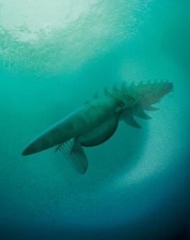
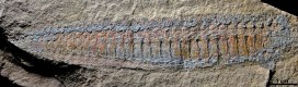
Meet Aegirocassis benmoulae – a 480 million year old, two-metre sea monster. This unlikely looking creature has been described, and imagined in this illustration, thanks to the work of one of the Museum’s research fellows, Dr Allison Daley.

Through collaboration with Dr Peter Van Roy and Professor Derek Briggs at Yale University, Allie has published a paper on Aegirocassis that is published in Nature this week. Here, Allie tells us a little bit more about it…

*

In December 2012, I met Peter Van Roy at the Palaeontological Association annual general meeting in Dublin. He told me about a new specimen that had just been unearthed in Morocco, and I almost couldn’t believe what I was hearing. Peter was working with professional fossil hunter Mohamed Ben Moula, discovering ancient Cambrian-type animal communities in the much younger rocks of the Ordovician period. What you can see above is a recreation of one of their finds, which was spectacularly preserved in three dimensions. Peter invited me to join him in studying this material, and I accepted with great excitement.

Reconstruction of the giant filter-feeding Aegirocassis benmoulae feeding on a plankton cloud in the sea approximately 480 million years ago. Aegirocassis grew to over 2 meters. Reconstruction by Marianne Collins, ArtofFact

Aegirocassis benmoulae belongs to a group of long extinct sea-dwelling animals called anomalocaridids. These were fearsome looking things: segmented bodies with wide swim flaps, a head bearing large eyes, a circular jaw with sharp teeth, and a pair of large claws. Anomalocaridids first appear in the fossil record during the Cambrian Explosion, a major evolutionary event that saw the rise of all animal life in a relatively rapid period of time.

They were early ancestors of the arthropods, the animal group that today includes spiders, insects, centipedes and lobsters. When they first evolved, in the Cambrian, anomalocaridids were apex predators and the biggest animals around, reaching up to about 50cm in size, but Aegirocassis benmoulae is a very different breed indeed.

Side view of a complete Aegirocassis benmoulae fossil, showing the pointed ‘flaps’ on the animal’s back. Photograph by Peter Van Roy, Yale University.

Most Cambrian anomalocaridids have one set of triangular swim flaps sticking out the side of the body, but the new Ordovician animal, Aegirocassis, shows us that the anomalocaridids actually had two pairs of body flaps. These two flaps correspond to the two branches of a limb that is characteristic of crustacea and represents an evolutionary stage before the two branches had fused. In other words, It allows us to trace the evolution of one of the key body features that made arthropods such a successful group of animals right through to the present day.

A side view of the fossilized spiny ‘net’ which Aegirocassis benmoulae used to filter its plankton food from sea water. Photograph by Peter Van Roy, Yale University.

As if that wasn’t enough, Aegirocassis also had a very different ecology from most anomalocaridids. While the Cambrian forms were mostly apex predators, this animal was a filter feeder – it used fine comb-like spines on its head appendages to filter plankton from the sea water. Only one Cambrian anomalocaridid also used filter feeding, but it remained a relatively modest size, while Aegirocassis was one of the largest arthropods ever to have existed.

This combination of gigantic size and filter feeding evolved from a previously predatory animal group is similar to the type of evolution seen later in whales. It makes Aegirocassis a very important animal for understanding both ecology and evolution in the oceans 480 million years ago.

Allison Daley holds up an Anomalocaridid fossil at the Burgess Shale in Canada. This area has yielded many previous Anomalocaridid fossils. Photograph by Parks Canada

Allison Daley – Research fellow You can also listen to an Oxford Sparks podcast with Allie, where she talks about the Cambrian Explosion, in the player here.

‘A thoroughly unhousewifely skill’

HodgkinBust etc 009

For International Women’s Day, the Museum of Natural History celebrates the life and career of Dorothy Hodgkin, one of its most eminent researchers. Hodgkin was awarded the Nobel Prize for Chemistry in 1964, and is still the only UK woman to have been awarded one of the science Nobels.

When the Museum of Natural History was designed in the 1850s, the building was intended not just to house a museum but also the burgeoning science departments of the University. The lettering above the doors facing the court continues to record these early affiliations: ‘Department of Medicine’, ‘Professor of Experimental Philosophy’, and so on.

Dorothy_Hodgkin_Nobel
Dorothy Mary Hodgkin (1910–1994) Image: Nobel Prize

As individual departments grew they moved into their own buildings across the science campus. One of the last research groups left in the Museum was the Department of Mineralogy & Crystallography, which, from the 1930s onwards, was the research home of the outstanding X-ray crystallographer Dorothy Hodgkin (1910-1994), winner of the Nobel Prize for Chemistry in 1964.

The Daily Mail famously celebrated her success with the headline ‘Oxford housewife wins Nobel’, but The Observer was no more enlightened, commenting that Hodgkin was ‘an affable looking housewife’ who had been awarded the Nobel Prize for ‘a thoroughly unhousewifely skill’.  That socially disruptive ability was an unparalleled proficiency with X-ray analysis, particularly in the elucidation of the structure of biological molecules.

Hodgkin undertook her first degree at Oxford from 1928 to 1932, initially combining chemistry and archaeology but later focusing on the emerging technique of X-ray crystallography. Her undergraduate research project was carried out using this technique in a Museum laboratory within what is now the Huxley Room, the scene of the 1860 Great Debate on evolution between Bishop Wilberforce and T. H. Huxley. She then journeyed across to Cambridge for her PhD before returning to Oxford in 1934 and resuming her association with the Museum.

Model_of_the_Structure_of_Penicillin,_by_Dorothy_Hodgkin,_Oxford,_c.1945
Model of the Structure of Penicillin, by Dorothy Hodgkin, Oxford, c.1945, in the Museum of the History of Science

Back in Oxford, Hodgkin started fundraising for X-ray apparatus to explore the molecular structure of biologically interesting molecules. One of the first to attract her attention was insulin, the structure of which took over 30 years to resolve – a project timescale unlikely to appeal to modern research funders. Other molecules proved more tractable, including the newly discovered penicillin, which Hodgkin began to work on during the Second World War, and vitamin B12.  It was for the determination of these structures that she was awarded the Nobel Prize.

Dorothy Hodgkin’s new X-ray laboratory was set up in a semi-basement room in the north-west corner of the Museum.  The room is now a vertebrate store but was once also the research home of Prince Fumihito of Japan, when he was based in the Museum for his ichthyological research (and It is still the only room in the Museum with bulletproof windows).

Initially, Hodgkin’s only office space consisted of a table in this room and a small mezzanine gallery above, which housed her microscopes for specimen preparation. Once prepared, she then had to descend a steep, rail-less ladder holding the delicate sample to the X-ray equipment below. Later, Hodgkin had a desk in the ‘calculating room’ (now housing the public engagement team) where three researchers and all of their students sat and undertook by hand the complex mathematics necessary after each analysis to determine the crystal structures of organic molecules.

Paul Smith – Director

If you would like to learn more about Dorothy Hodgkin and her work, then read Georgina Ferry’s excellent biography ‘Dorothy Hodgkin: A Life’ which has just been re-issued as an e-book and new, print-on-demand paperback by Bloomsbury Reader.

 This year’s Dorothy Hodgkin Memorial Lecture will be held in the Museum at 5 pm on Thursday 12 March, and is open to all. The lecture will be given by Dr Petra Fromme (Arizona State University) who is an international authority on the structure of membrane proteins.

The finest fossils

Collecting fossils%2c near Fuxian Lake%2c Chengjiangjpg 
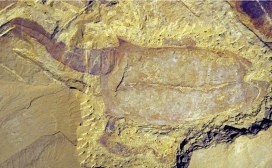
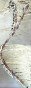
One of the most remarkable fossil sites in the world is located in Chengjiang in China, where exquisitely-preserved fossils record the early diversification of animal life. The 525 million year old mudstone deposits in the hills and lakes of Yunnan Province, South China are so fine that they have preserved not only the shells and carapaces of Cambrian animals, but also the detail of their soft tissue. In recognition, the site was added to the World Heritage list by UNESCO in 2012.

A  vetulicolian - a fossil which broadly speaking probably lies on the evolutionary lineage that leads to the vertebrates
A vetulicolian – a fossil which broadly speaking probably lies on the evolutionary lineage that leads to the vertebrates
The arthropod Synophalos showing numerous interlocking individuals in a chain

Professor Derek Siveter, a senior research fellow at the Museum, has been studying this material for a number of years, authoring a book – The fossils of Chengjiang, China: The flowering of early animal life – in 2004. But the rate of discovery of new fossils over the last decade has led to a wealth of new material to be documented.

So Derek recently headed back to the University of Yunnan for a two-week visit, where he began work on a revised edition of the book. Much of the documentation of these important fossils is currently in Chinese, so the new edition will bring the material to English-speaking researchers and fossils enthusiasts too. It introduces both the professional and the amateur palaeontologist – and all those fascinated by evolutionary biology – to the aesthetic and scientific quality of the Chengjiang fossils, many of which represent the origins of animal groups that have sustained global biodiversity to the present day.

Scott Billings –  Public engagement officer

Mystery critters

Cambrian muscle worm

Five hundred million years ago, in the Cambrian period, the oceans teemed with strange and unusual creatures that are now preserved as fossils. This period in Earth’s history is important because almost all known groups of animals appear very suddenly in the fossil record at this time. Many of them look just like their modern day counterparts, but several are much more weird and wonderful, with a very different appearance from anything alive today.

I was recently awarded the Whittington Award from the Palaeontological Association which includes a small research grant that will allow me to study one of these weird fossils. The creature in question is known as the “muscle worm”, or Myoscolex, and is particularly interesting because almost the whole fossil is made up of very well-preserved muscle fibres. It’s the oldest record of muscle tissue in the fossil record.

Me, taking a break from fossil hunting to cuddle an echidna

The material comes from the Emu Bay Shale fossil site, located on Kangaroo Island in South Australia. The original collection of fossils is nearly 35 years old now. These original fossils showed the muscle tissue of Myoscolex very clearly, but unfortunately we don’t know much about the rest of its body and scientists can’t even agree on what type of animal it is! Some people believe it is an annelid worm – a segmented creature – while others think it could be an early ancestor of either the arthropods, which includes animals like crabs, shrimp, spiders, centipedes, and insects, or the chordates, a group which includes any animal with a backbone, including ourselves.

Fortunately, many new fossils have been collected in the last few decades that will help us solve the mystery of the Cambrian muscle worm. Active collecting by the South Australian Museum and the University of New England has revealed hundreds of new specimens that show us more details about the anatomy of Myoscolex, including the head, legs, skeleton, and even its digestive system.

I have been working with researchers in Australia on Emu Bay Shale fossils for several years now, both on the fossil collections in the museum and in the field. The Whittington Award will allow me to travel to Australia to study the new muscle worm fossils. The research will involve taking photos, making drawings, and analyzing the nicest specimens under scanning electron microscopes. I will then come back to the Museum in Oxford and compare the fossil information with modern animals found in our collections here. All of this work should allow us to finally figure out what the Cambrian muscle worm really is.

Stay tuned as we try to solve this mystery….

Allison Daley – Museum Research Fellow