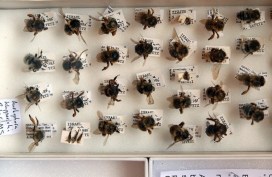
Plight of the bumblebee

nhm16

The ecological importance of bumblebees has become more widely appreciated in recent years, thanks to environmental campaigners and reports of species decline, and even some extinctions, in the UK.

To look at this issue, we have recently teamed up again with arts-science organisation Pale Blue Dot, which is launching a new research project to investigate why some species of bumblebee are declining and to raise awareness about the ultimate impact this has on people.

Here, Pale Blue Dot co-founder Jane King explains how the Bees & Weeds project brings together art students, public engagement, the Museum’s collections and a leading bumblebee scientist…

*

On 9 September we launched our latest project – Bees & Weeds – with the Museum of Natural History, building on our previous collaboration for the Lost & Found exhibition. We were joined by over 50 art students from Banbury & Bicester College to highlight the plight of the bumblebee, revealing how its decline is impacting everything from what we eat to where we live and work.

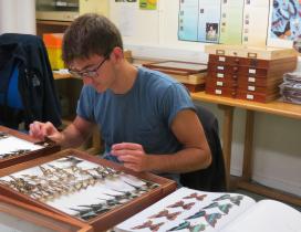
Amoret Spooner displays drawers from the collections

The students spent some time looking at methods of insect labelling and notation, before heading behind the scenes with entomologist Amoret Spooner to the Huxley Room, the location of the Great Debate on Darwin’s Theory of Evolution, which took place in 1860.

Amoret provided an insight into taxonomy – the science of species classification – as well as her work on the conservation of specimens. We visited the huge archive of bee specimens and learnt about some of the research that scientists are currently carrying out on UK bumblebee species to help prevent further decline.
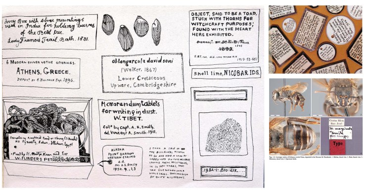
Student sketches of labelling and notation

During spring 2015, the art students from the Banbury & Bicester College, as well as students from Oxford Brookes University, will make and install hundreds of cycle seat covers on bikes in and around the Oxford city. The seat covers will carry messages about bumblebee decline in the UK countryside, showing how much we depend on their pollination services, which far outstrip those of the honeybee in their value to UK food production.

We are also working with Professor Dave Goulson from the University of Sussex, one of the world’s most important bumblebee scientists. He will present his research showing how well bumblebees are doing in gardens compared to the countryside, as well as the optimum range of flowering plants needed to help them thrive. Dave’s book, A Sting in the Tale, is already a best-seller, and the sequel – A Buzz in the Meadow – was published on 4 September.

Dave will also be speaking about his new book at the Museum on Thursday 9 October at 7pm. Book your tickets for that via Waterstones here.

Artwork from the Bees & Weeds project, together with cycle seat covers and bike paraphernalia, will be on show and for sale in the Old Fire Station in Oxford from next spring. If you cycle in Oxford, you may be lucky enough to receive one!

Pale Blue Dot is an arts-science organisation helping scientists to communicate their research to the public. It promotes an interdisciplinary approach to learning, living and working through exhibitions, publications and happenings.

Jane King – co-founder, Pale Blue Dot

Ready for your close-up?

IMG_3684
Image copyright Richard Kelsall

They say a picture is worth a thousand words, and at the Museum we make thousands of pictures: pictures to document, pictures to investigate, and pictures to wow. We use a lot of different imaging techniques too, from standard close-up photography to scanning electron microscopy, which reveals the most minute details.

To coincide with the final week of the Wildlife Photographer of the Year exhibition here, on Saturday 20 September we held a new adult workshop to give people some hands-on experience of some of these processes. Imaging Techniques in Modern Natural History gave participants the chance to get up close to some wonderful specimens and make their own images to take home.

I had planned to review the day here, but Rose Parkin, who took part in the workshops, very helpfully sent in her own write-up of the sessions. So here’s a special guest post from Rose, along with some pictures taken by people on the day.

*

By Rose Parkin

When I signed up for the digital imaging course I expected a fairly dry, tech-heavy day. Instead, the experience was really exciting. Not only did it provide hands-on experience of viewing and recording images with new technology, it also gave me a brief glimpse behind the scenes of my favourite museum.

Laser Scanning and Digital Modelling
For our first session our small group was led through a maze of corridors by Sarah Joomun, the Documentation Officer, to the laser scanning lab. It sounded a bit futuristic, and it turned out that it looks that way too. Sarah popped a fossil onto a mount, clicked a few buttons and red lasers appeared, scanning the fossil’s surface while it rotated. After ten minutes the first 3D image of the fossil was produced – a beautiful net of triangles, which looked like a teleporting object in a science fiction film.

Laser scanning Image copyright: Tom Nicholson-Lailey
Laser scanning
Image copyright Tom Nicholson-Lailey

Sarah turned the fossil and scanned it again. The challenge was then to fit  these two images together to make a complete 3D model. Amazingly, this technique enables other palaeontologists around the world to see and replicate, with the use of a 3D printer, the exact size and shape of a fossil without it ever leaving the museum.

Multi-plane Microscope Photography
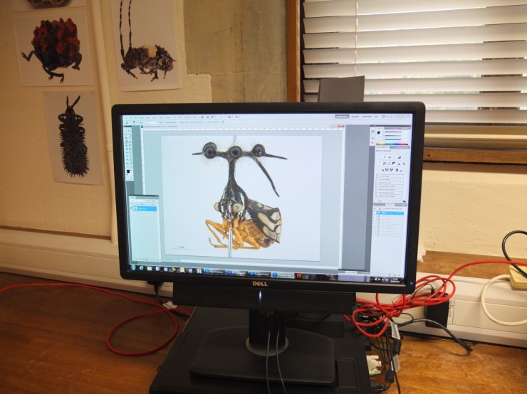
Our next session was upstairs, with artist-in-residence and photographer Katherine Child. Even though we were close to the main corridor of the museum it felt like a real working space, crammed full of equipment and insect specimens. Katherine had chosen the tiniest of insects for us to photograph with the multi-plane microscope. It looked like a small seed with some barely visible limb-like protrusions.

Multiplane photography. Image copyright Rose Parkin
Multiplane photography.
Image copyright Rose Parkin

But under the microscope a wonderfully strange insect became visible, with the most bizarre appendages and bright orange legs. While the microscope already showed a great deal of detail the multi-plane photography captured an incredibly crisp image. The microscope takes large numbers of photos of the specimen, using different focal planes each time, then the focussed elements are all stacked together to produce a crystal clear photograph.

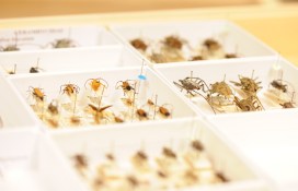
Once we’d chosen and photographed some other insects from the collection and poked around the room a bit (finding a disturbing collection of large pickled spiders), we were taken on a tour of the entomology department. Katherine led us through corridors of offices and labs, up to a stunning store room that felt almost church-like, with rows and rows of cabinets full of fascinating insects.

Scanning Electron Microscopy
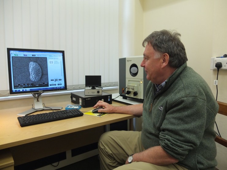
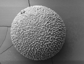
After lunch we had a laboratory session with museum director Paul Smith to look at sand under an electron microscope. Luckily, that was much more exciting than it sounds! The sand was taken from Dog’s Bay on the west coast of Ireland and is rich with a wide range of tiny fossilized organisms. Paul showed us how to carefully select individual microfossils from a tray using just a microscope and a paint brush.

PS and SEM
Professor Paul Smith demonstrates the scanning electron microscope. Image copyright Rose Parkin

We then viewed some of the microfossils using a scanning electron microscope. This allowed us to see an incredible level of detail. The microscope was so powerful that we could see hair holes in a fossil the size of a grain of sand.

DSLR Macrophotography
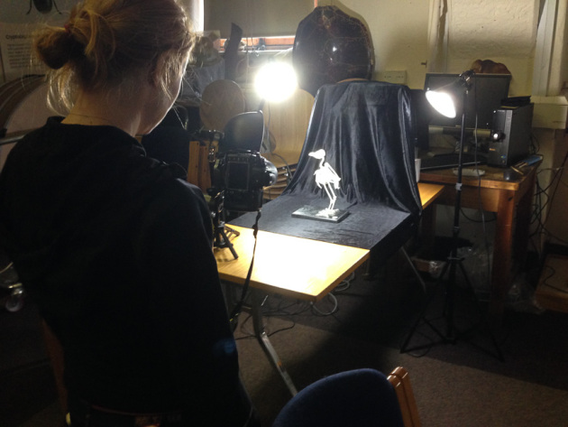
My final session was a crash course in macrophotography. Held in the seminar room, the low lighting and floor-to-ceiling collection of specimens lent an almost eerie feeling to the set-up.

Macrophotography. Image copyright Keith Barnes
Macrophotography.
Image copyright Keith Barnes
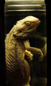
Bearded dragon. Image copyright: Rose Parkin
Bearded dragon.
Image copyright: Rose Parkin

Once prepped, we were let loose on four separate camera setups. Being able to choose and shoot at our own pace made this feel like a really creative experience. The help given by professional photographer Keith Barnes and public engagement officer Scott Billings was perfect – very hands on but not patronizing (despite my lack of DSLR experience).

With this digital imaging course the museum has created a really exciting snapshot of the work that goes on behind the scenes, reinforcing the fact that this impressive place is much more than just an ordinary museum.

A lesson from the past

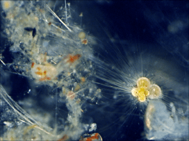
PETM Foram

If you’re a regular reader of this blog you might have heard of Dr Tracy Aze already, and may even recognise the strangely-shaped specimen above as an example of planktonic foraminifera, the single-celled marine organisms that Tracy has been researching. This morning we have issued a press release about Tracy’s research which offers a warning from history about carbon emissions and global warming.

Surprisingly enough, the study shows how the fossils of these creatures hold clues to the impact on our oceans of man-made global warming. Around 56 million years ago, in a period known as the Palaeocene-Eocene Thermal Maximum (PETM), a rapid rise in greenhouse gases caused sea surface temperatures to rise as high as 40°C, with significant impacts on marine life.

Worryingly, the PETM – which lasted for around 170,000 years – saw the release of roughly the same volume of CO2 as expected from modern fossil fuel consumption. Tracy explains:

The amount of CO2 that is predicted to be released from the Industrial Revolution to around 100 years from now is roughly equivalent to what happened in the PETM. But the big difference is the rate of release: today we are releasing greenhouse gases at a far faster rate than 56 million years ago.

Although the research was conducted by Tracy, the project was led by Professor Paul Pearson of Cardiff University and funded by the UK Ocean Acidification research programme.

Tracy and her team used newly-extracted planktonic foraminifera fossils from Tanzania, dating from the PETM period. The tiny shells of these organisms contain different proportions of oxygen isotopes and these proportions are largely determined by the sea temperatures at the time. So the fossil shells offer a glimpse of the way sea temperatures were rising alongside the release of greenhouse gases, as well as a record of the relative abundance of this planktonic life in the oceans.

The PETM shows us that rapid increases in CO2 in the atmosphere have significant impacts on global temperatures, with the new information from our study site showing that tropical sea surface temperatures may have exceeded 40°C with an associated local disappearance of marine life.

The research paper, Extreme warming of tropical waters during the Paleocene–Eocene Thermal Maximum, was published in the September issue of Geology and is available as open-access.

Scott Billings – Public engagement officer


It must be autumn – the interns have flown

James organising the South Sudanese butterflies
James Evry organising the South Sudanese butterflies

It seems very quiet this week, now the last of our undergraduate interns has left us. A week ago, third year Earth sciences student Keyron Hickman-Lewis identified and numbered his last tray of specimens from the 19th century Parker collection, photographed some of the finest Jurassic fish jaws, sharks’ teeth, and other beautifully preserved fossils, before heading home to enjoy a well-earned break.

We’ve been running paid undergraduate internships for several years now, supported by grants from the University’s E.P.A. Cephalosporin Fund. The students tackle curatorial projects – sorting, identifying, numbering and cataloguing specimens, or helping to organise and list archives. This enables us to get a lot of curatorial work done, and it gives the interns a chance to handle and learn about a wider range of specimens and materials than they would ever see on their degree courses, while learning new skills which will be useful in their future careers. This year we also had interns funded through Oxford University’s own internship scheme, all tackling projects with more of a research focus.

Naomi (l) and Branwen (r) numbering Freeman collection minerals

Second year biologist, Ellen Foley-Williams worked on the Long-horn Beetle collection, but she’s really interested in science communication, so we set her an extra challenge of running a blog where all the interns could share their experiences; have a look at More Than an Intern to discover more.

Naomi (r) showing off some Cumbrian iron ore from her home county, and a an iron meteorite from space in ‘Spotlight specimens’

Some of the interns rose to the challenge of joining our ‘Spotlight Specimens’ rota. Every weekday afternoon at 2.30, a member of staff takes some favourite specimens from behind-the-scenes, and talks about them to museum visitors. It may be a bit scary first time, but every one of the interns said it was really fun to do – if sometimes a little challenging with such a varied audience.

In total, we had eleven interns, each spending six weeks working on a specific project. So Branwen, Cecilia, Ellen, Emily G., Emily T., Grace, James, Keyron, Max, Naomi, and Steph, we’d like to thank you all for being hard-working and lots of fun to have around. We hope we’ll see lots more of you all in coming years.

Monica Price, Head of Earth Collections

Smaller than a dodo

Grace Manley, one of our interns, writes about her work on planktonic foraminifera…

morethananintern's avatarMore Than An Intern

The museum not only contains a huge array of specimens both on display and on the collections, but is home to active research undertaken by research fellows.

My internship involves the study of microfossils formed from organisms known as planktonic foraminifera: single celled DSCN1598 (1)organisms which create a shell up to the size of a few millimetres. As they die these shells fall to the seafloor and often become preserved in the deep sea muds, which may then be drilled up and prepared for study by washing over a sieve. Though a tray of foraminifera may look suspiciously like a tray of white dust, underneath a binocular microscope these small grains reveal a wide variety of shapes from which it is possible (though not always easy!) to identify different species. Study of foraminifera can therefore be carried out at species level over a long period of time; this is not possible…

View original post 241 more words

Not Just Dead Bugs

A glimpse from behind the scenes in our Life department to see what the interns are getting up to.

morethananintern's avatarMore Than An Intern

Longhorn4Not many people know that the Oxford University Museum of Natural History doesn’t just consist of the specimens on display – it also houses the Hope Entomological Collection: the second largest entomological collection in the UK. The space contains thousands of incredible pinned specimens as well as some live ones too (Geraldine the stick insect on the right). Entomology is the study of insects, and so the department is responsible for the curation of thousands of invertebrate specimens collected over hundreds of years by biologists such as Darwin and Wallace. We have all sorts in the collection – from beetles and flies (Coleoptera and Diptera) to bees and ants (Hymenoptera).

The collection consists of over five million specimens, which keeps the staff, volunteers and interns well occupied.  Each person working in the department has their own role and

Max6often their own speciality. Not only are the permanent staff experts, but the…

View original post 220 more words