

They say a picture is worth a thousand words, and at the Museum we make thousands of pictures: pictures to document, pictures to investigate, and pictures to wow. We use a lot of different imaging techniques too, from standard close-up photography to scanning electron microscopy, which reveals the most minute details.
To coincide with the final week of the Wildlife Photographer of the Year exhibition here, on Saturday 20 September we held a new adult workshop to give people some hands-on experience of some of these processes. Imaging Techniques in Modern Natural History gave participants the chance to get up close to some wonderful specimens and make their own images to take home.
I had planned to review the day here, but Rose Parkin, who took part in the workshops, very helpfully sent in her own write-up of the sessions. So here’s a special guest post from Rose, along with some pictures taken by people on the day.
*
By Rose Parkin
When I signed up for the digital imaging course I expected a fairly dry, tech-heavy day. Instead, the experience was really exciting. Not only did it provide hands-on experience of viewing and recording images with new technology, it also gave me a brief glimpse behind the scenes of my favourite museum.
Laser Scanning and Digital Modelling
For our first session our small group was led through a maze of corridors by Sarah Joomun, the Documentation Officer, to the laser scanning lab. It sounded a bit futuristic, and it turned out that it looks that way too. Sarah popped a fossil onto a mount, clicked a few buttons and red lasers appeared, scanning the fossil’s surface while it rotated. After ten minutes the first 3D image of the fossil was produced – a beautiful net of triangles, which looked like a teleporting object in a science fiction film.

Image copyright Tom Nicholson-Lailey
Sarah turned the fossil and scanned it again. The challenge was then to fit these two images together to make a complete 3D model. Amazingly, this technique enables other palaeontologists around the world to see and replicate, with the use of a 3D printer, the exact size and shape of a fossil without it ever leaving the museum.
Multi-plane Microscope Photography
Our next session was upstairs, with artist-in-residence and photographer Katherine Child. Even though we were close to the main corridor of the museum it felt like a real working space, crammed full of equipment and insect specimens. Katherine had chosen the tiniest of insects for us to photograph with the multi-plane microscope. It looked like a small seed with some barely visible limb-like protrusions.

Image copyright Rose Parkin
But under the microscope a wonderfully strange insect became visible, with the most bizarre appendages and bright orange legs. While the microscope already showed a great deal of detail the multi-plane photography captured an incredibly crisp image. The microscope takes large numbers of photos of the specimen, using different focal planes each time, then the focussed elements are all stacked together to produce a crystal clear photograph.
Once we’d chosen and photographed some other insects from the collection and poked around the room a bit (finding a disturbing collection of large pickled spiders), we were taken on a tour of the entomology department. Katherine led us through corridors of offices and labs, up to a stunning store room that felt almost church-like, with rows and rows of cabinets full of fascinating insects.
Scanning Electron Microscopy
After lunch we had a laboratory session with museum director Paul Smith to look at sand under an electron microscope. Luckily, that was much more exciting than it sounds! The sand was taken from Dog’s Bay on the west coast of Ireland and is rich with a wide range of tiny fossilized organisms. Paul showed us how to carefully select individual microfossils from a tray using just a microscope and a paint brush.

We then viewed some of the microfossils using a scanning electron microscope. This allowed us to see an incredible level of detail. The microscope was so powerful that we could see hair holes in a fossil the size of a grain of sand.
DSLR Macrophotography
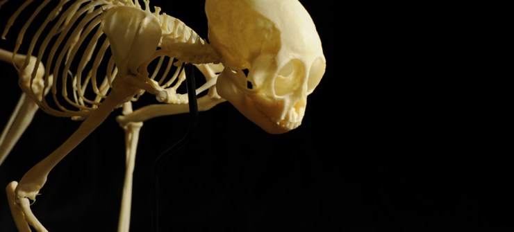
My final session was a crash course in macrophotography. Held in the seminar room, the low lighting and floor-to-ceiling collection of specimens lent an almost eerie feeling to the set-up.

Image copyright Keith Barnes

Image copyright: Rose Parkin
Once prepped, we were let loose on four separate camera setups. Being able to choose and shoot at our own pace made this feel like a really creative experience. The help given by professional photographer Keith Barnes and public engagement officer Scott Billings was perfect – very hands on but not patronizing (despite my lack of DSLR experience).
With this digital imaging course the museum has created a really exciting snapshot of the work that goes on behind the scenes, reinforcing the fact that this impressive place is much more than just an ordinary museum.

Published by