Lungfish, lithographs and libel


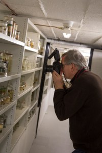
By Mark Carnall, Collections Manager


In addition to the many thousands of biological specimens that can be found at Oxford University Museum of Natural History, we also possess a variety of objects that originate from historical versions of the Museum’s displays. These include models, casts, and illustrations of various kinds, used to represent organisms that were otherwise difficult to preserve and display.

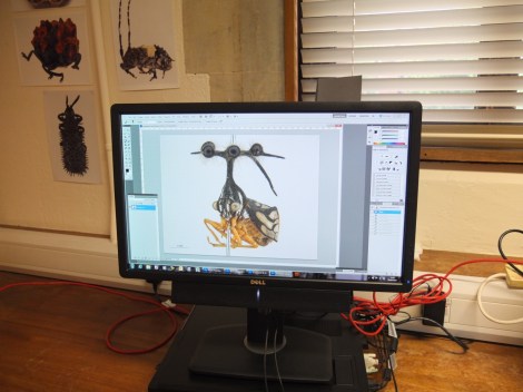
That any of these exhibition materials survive at all is down to pure happenstance and luck. At the time when they were removed from display, these artefacts would have just been seen as outdated ‘display furniture’ and all but destined to have been thrown away. One surviving piece of ex-display material, which catches my eye almost daily as it sits in my office, is a rather large pair of illustrations showing a South American and a West African lungfish mounted on a black backing board.

Mounted illustrations of West African lungfish, Protopterus annectens (top) and South American lungfish, Lepidosiren paradox (bottom). The board they are mounted on measures 93cm across.

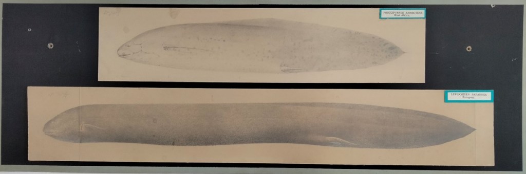
By pure coincidence, I recently came across lithograph reproductions of these illustrations in an 1895 publication by E. Ray Lankester. Had these fish not have been my office-mates, I might not have paid the lithographs in the paper much attention, nor recognised their significance. 

E. Ray Lankester was a noted Zoologist who studied at Oxford University and was the holder of the Linacre Chair. He was also heavily involved in adding to the collections and displays here at OUMNH. His 1895 paper – a smash hit I’m sure we all remember – was titled On the Lepidosiren of Paraguay, and on the external characters of Lepidosiren and Protopterus, and sought to add more reliable evidence on the appearances of lungfishes. 

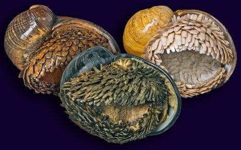
Lungfishes were of particular interest to scientists at the end of the nineteenth century. Though seemingly related, the different species of lungfish caused no small amount of head-scratching, given that they were found in freshwater ecosystems as far apart as Australia, Africa, and South America. As their name suggests, they are fish but also air-breathing, and the fact that they possess lungs also marked them for scientific interest at the time.

Comparison of Bayzand’s original drawing of Protopterus annectens (top) and screen-capture of the published figure (bottom). You’ll no doubt agree with Lankester that the changes to the scales are egregious and vexing. 

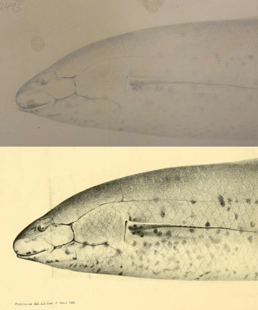
Interestingly (well, interesting to me!) is that Lankester adds an extensive note in the paper about the illustration of the specimens, explaining that he is unhappy with how Bayzand’s original drawings have been modified in the process of transforming them into lithographs for publication. According to Lankester, these modifications introduced inaccuracies. In particular, he complained that the lithographer had made it look like the lungfishes were covered in scales, and stresses that “[a]s a matter of fact, no scales at all[,] or parts of scales[,] are visible on the surface” of the lungfish. Instead, he makes clear that in real life (or, in this case, in preserved life) the scales of the fish are overlaid with soft tissue. Comparing the figure in the paper with the illustrations in my office confirms that the lithographer had, indeed, inaccurately reproduced the original drawings.

The happy coincidence of me finding Lankester’s paper led me to several important revelations. Firstly, we now know that Bayzand’s original drawings of the lungfish can still be found here at OUMNH. Secondly, we can surmise that, at some point in the past, these drawings were displayed in the Museum’s galleries. We can also corroborate that the original illustrations are different to the published versions, meaning that, if we are to believe Lancaster, they are also more accurate than those in the publication. Finally, we now know that two of the Museum’s specimens were cited with extra biographical information in Lankester’s paper.

Sadly, these exciting findings mean that my office mates will probably have to be relocated and take up residence in the Museum’s archives alongside their subject matter…

Shooting with Martin Parr

PAM2015011G00487

As you may have seen, the Museum was recently shortlisted as a finalist in this year’s Art Fund Prize for Museum of the Year – very exciting news for us. To add to the honour we hosted renowned Magnum Photos photographer Martin Parr, who spent a good few hours photographing the Museum, in the court and behind the scenes, as part of the Museum of the Year campaign.

The photograph above was captured by Martin during one of our primary school sessions on dinosaurs: the children have their mitts on a fossilised dinosaur egg – just one of the real specimens used during the session.

Martin Parr photographing some Darwin specimens in our collections

Having a Magnum photographer visit the Museum for a photoshoot isn’t something that happens every day, so it was a real privilege to take Martin Parr around the building and watch the types of things that caught his eye.

I am a keen photographer myself, with an interest in the history of photography as a technical process and as an art form, so it was especially exciting to not only meet Martin and watch him work, but also to photograph the process myself too. You can see a few of those shots here.

Martin Parr scrutinising our vertebrate spirit collections

Photo competition

Now it’s your chance. We’re inviting you to take photographs of the Museum and submit them to the Museum of the Year Photo Competition, with a chance to win a photography holiday in Berlin, photo gear and other prizes. Martin Parr will shortlist six photos, one for each of the six finalist museums, and the ultimate winner will be selected by a public vote.

So get snapping – with a posh camera or your phone; it doesn’t matter. Then either upload your pictures via the Art Fund website, or tweet or Instagram them using the #motyphoto hashtag and don’t forget to tag us in @morethanadodo.

Good luck!

Martin Parr photographing a primary school group for Art Fund Prize for Museum of the Year

Martin Parr, an Iguanodon and young visitors

Scott Billings – Public engagement officer


The Iron Snail

5

The Museum has recently received specimens of the enigmatic deep-sea vent snail, Chrysomallon squamiferum, the scaly-foot snail. In this post, Dr Chong Chen explains why this species is so extraordinary.

*

This is no ordinary snail. First of all, it lives in deep-sea hydrothermal vents in the Indian Ocean, more than 2,500 metres deep, just beside black smokers that are churning out superheated water exceeding 350°C. Second, it is the only known gastropod with a suit of scale armour. Thirdly, the scales as well as the shell are mineralised with iron sulfide. That’s right – these snails make a skeleton out of iron, and are the only animal so far known to do so.

A specimen of Chrysomallon squamiferum photographed live (Photo: David Shale)

Hydrothermal vents were first discovered in the Galápagos Rift as recently as 1977. This is just off the Galápagos Islands whose fauna famously inspired Charles Darwin in the development of his theory of natural selection. Vents are deep-sea ‘hot springs’ fuelled by geological activity; the hot erupting fluid is usually acidic and contains various metals, as well as hydrogen sulfide. This is what makes rotten eggs smell bad, and is toxic to most organisms. Some bacteria, however, are able to use it to produce energy in a process known as chemosynthesis.

Hydra, an active ‘black smoker’ vent chimney in Longqi field, Southwest Indian Ridge

Over geological timescales many remarkable organisms have adapted to live in these ‘toxic utopia’, and flourish by exploiting the energy produced by these bacteria. The scaly-foot snail has also harnessed the power of chemosynthesis, housing endosymbiotic bacteria – bacteria living inside another creature to mutual benefit – in an enlarged part of its gut. This produces the energy it needs. In another words – it has a food factory inside its body and doesn’t even need to feed! This is likely the reason it can grow to about 45mm in size, when most of its close relatives without endosymbionts are only 15mm or smaller.

Close-up of the scales, also showing the reduced operculum in middle

Scaly-foot snails were first discovered in 2001, at the Kairei vent field in the Indian Ocean. Its discovery came as a great surprise as even among those animals specialised for living at vents, it was very, very strange. And cool. Although the shell of a snail is well-known to be modified into a great variety of forms, this is not the case with hard parts on the foot, and apart from an operculum (the ‘trap-door’ serving as a lid when the animal retracts to its shell) no other gastropods have other mineralised structures on the foot. Yet C. squamiferum has thousands of scales!

The shell, although not particularly exciting in form, isn’t exactly ordinary either as the outermost layer is made of iron sulfide. And so are the scales. So this entire animal is covered in iron compound, mainly pyrite (FeS2, or ‘Fool’s gold’) and greigite (Fe3S4). As greigite is magnetic, the animal actually sticks to magnets. The function of the scales is postulated to be either protection or detoxification but their true use remains a mystery.

The three vent fields where Chrysomallon squamiferum is known from

So why blog about the ‘scaly-foot’ now, if it has already been known to science for more than a decade? Well, actually, despite numerous studies and publications on its strange biology this species has never been formally described and named, until now. A recent paper by Dr Chong Chen (Department of Zoology, University of Oxford) and colleagues finally gave it the scientific name you see here – Chrysomallon squamiferum.

The Museum received a set of five specimens as part of the description process, which will serve as key references for scientists who wish to study this extraordinary species in the future.

Here’s a video of the Longqi hydrothermal field featuring Chrysomallon squamiferum in their natural habitat:

Ready for your close-up?

IMG_3684
Image copyright Richard Kelsall

They say a picture is worth a thousand words, and at the Museum we make thousands of pictures: pictures to document, pictures to investigate, and pictures to wow. We use a lot of different imaging techniques too, from standard close-up photography to scanning electron microscopy, which reveals the most minute details.

To coincide with the final week of the Wildlife Photographer of the Year exhibition here, on Saturday 20 September we held a new adult workshop to give people some hands-on experience of some of these processes. Imaging Techniques in Modern Natural History gave participants the chance to get up close to some wonderful specimens and make their own images to take home.

I had planned to review the day here, but Rose Parkin, who took part in the workshops, very helpfully sent in her own write-up of the sessions. So here’s a special guest post from Rose, along with some pictures taken by people on the day.

*

By Rose Parkin

When I signed up for the digital imaging course I expected a fairly dry, tech-heavy day. Instead, the experience was really exciting. Not only did it provide hands-on experience of viewing and recording images with new technology, it also gave me a brief glimpse behind the scenes of my favourite museum.

Laser Scanning and Digital Modelling
For our first session our small group was led through a maze of corridors by Sarah Joomun, the Documentation Officer, to the laser scanning lab. It sounded a bit futuristic, and it turned out that it looks that way too. Sarah popped a fossil onto a mount, clicked a few buttons and red lasers appeared, scanning the fossil’s surface while it rotated. After ten minutes the first 3D image of the fossil was produced – a beautiful net of triangles, which looked like a teleporting object in a science fiction film.

Laser scanning Image copyright: Tom Nicholson-Lailey
Laser scanning
Image copyright Tom Nicholson-Lailey

Sarah turned the fossil and scanned it again. The challenge was then to fit  these two images together to make a complete 3D model. Amazingly, this technique enables other palaeontologists around the world to see and replicate, with the use of a 3D printer, the exact size and shape of a fossil without it ever leaving the museum.

Multi-plane Microscope Photography
Our next session was upstairs, with artist-in-residence and photographer Katherine Child. Even though we were close to the main corridor of the museum it felt like a real working space, crammed full of equipment and insect specimens. Katherine had chosen the tiniest of insects for us to photograph with the multi-plane microscope. It looked like a small seed with some barely visible limb-like protrusions.

Multiplane photography. Image copyright Rose Parkin
Multiplane photography.
Image copyright Rose Parkin

But under the microscope a wonderfully strange insect became visible, with the most bizarre appendages and bright orange legs. While the microscope already showed a great deal of detail the multi-plane photography captured an incredibly crisp image. The microscope takes large numbers of photos of the specimen, using different focal planes each time, then the focussed elements are all stacked together to produce a crystal clear photograph.

Once we’d chosen and photographed some other insects from the collection and poked around the room a bit (finding a disturbing collection of large pickled spiders), we were taken on a tour of the entomology department. Katherine led us through corridors of offices and labs, up to a stunning store room that felt almost church-like, with rows and rows of cabinets full of fascinating insects.

Scanning Electron Microscopy
After lunch we had a laboratory session with museum director Paul Smith to look at sand under an electron microscope. Luckily, that was much more exciting than it sounds! The sand was taken from Dog’s Bay on the west coast of Ireland and is rich with a wide range of tiny fossilized organisms. Paul showed us how to carefully select individual microfossils from a tray using just a microscope and a paint brush.

PS and SEM
Professor Paul Smith demonstrates the scanning electron microscope. Image copyright Rose Parkin

We then viewed some of the microfossils using a scanning electron microscope. This allowed us to see an incredible level of detail. The microscope was so powerful that we could see hair holes in a fossil the size of a grain of sand.

DSLR Macrophotography
My final session was a crash course in macrophotography. Held in the seminar room, the low lighting and floor-to-ceiling collection of specimens lent an almost eerie feeling to the set-up.

Macrophotography. Image copyright Keith Barnes
Macrophotography.
Image copyright Keith Barnes

Bearded dragon. Image copyright: Rose Parkin
Bearded dragon.
Image copyright: Rose Parkin

Once prepped, we were let loose on four separate camera setups. Being able to choose and shoot at our own pace made this feel like a really creative experience. The help given by professional photographer Keith Barnes and public engagement officer Scott Billings was perfect – very hands on but not patronizing (despite my lack of DSLR experience).

With this digital imaging course the museum has created a really exciting snapshot of the work that goes on behind the scenes, reinforcing the fact that this impressive place is much more than just an ordinary museum.

Seaside Minerals

Russell Society

Last week, members of the Southern Branch of the Russell Society came to visit the Museum. They especially wanted to see some of the minerals in our collection that came from the South Coast of England. The Russell Society, in case you are wondering, runs lectures, museum visits and fieldtrips for people who enjoy finding out about minerals. It is named after Sir Arthur Russell, one of Britain’s most gifted amateur mineralogists. I’m a member and so is one of our volunteers, Jane Randle.

Baryte from Babbacombe, Devon (80mm across)

Now, Jane and I know the Russell Society folk are a very knowledgeable lot, so we put our heads together a devised a challenge for them. We got out from the stores a whole lot of interesting minerals from quarries, mines, beaches and cliff exposures all along the coast from Kent round to south Devon, and laid them out in a random way. Then came the fun bit; we took away all the labels!

The task was to work as a team and organise the minerals according to where they were found, from west to east. Easy? Well, that depends on how good you are at recognising the different kinds of minerals and where they come from.

One of the fun things about minerals is that each kind  – calcite, quartz, gypsum, etc, – can look very different according to where it is found. Some are very easy to recognise but some are not. After much debate, looking at maps, and rearrangement of specimens came the moment of truth as Jane and I put the labels back with their specimens, and everyone could see whether or not they were right.

Fossil sponge preserved in chalcedony from Brighton beach (110mm across)

Our Russell Society folk certainly showed they know their local minerals!  They recognised the little round masses of baryte crystals from the Isle of Sheppey,  large transparent  gypsum crystals from Battle, powdery white masses of aluminite from Newhaven, rare blue vivianite crystals from near Southampton, sparkling pointed crystals of calcite from Charmouth, branching crystals of real gold from near Torquay, and many more. Others did elude them, and came from surprising places. Our beautiful polished slices of fossil sponges, for example, came from pebbles collected on Brighton beach in the 19th century!

Monica Price, Head of Earth Collections