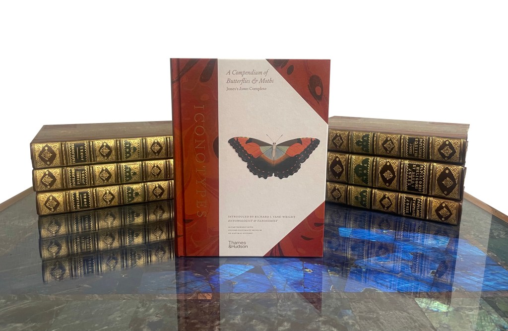
Iconotypes: A Compendium of Butterflies and Moths
By Danielle Czerkaszyn and Kate Diston
Today, the Museum is celebrating the publication of Iconotypes: A Compendium of Butterflies and Moths based on William Jones’ unpublished, six volume manuscript. Danielle Czerkaszyn, Librarian and Archivist, tells us more about the importance of Jones’ work…
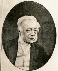
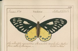
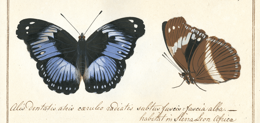
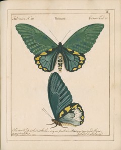
Since the 1920s the Museum has had in its care an original, unpublished manuscript containing 1,292 beautifully detailed and colourful paintings of butterflies and moths. Known as Jones’ Icones, this one-of-a-kind work was created in the late 18th century by retired London wine merchant, natural historian and Lepidopterist, William Jones (1745-1818).
In six volumes Icones depicts over 760 butterflies and moths from the collections of some of the most eminent naturalists in London at that time, including entomologist Dru Drury, explorer Sir Joseph Banks, the founder of the Linnean Society, Sir James E. Smith, and Jones’s own collection. A labour of love, Jones spent 30 years of his life – from 1780-1810 – using the finest materials to ensure Icones was both accurate and beautiful.
In addition to being a stunning work of art, Jones’ Icones is an extraordinarily important document in the history of entomology and insect collecting in Britain. At the time Jones was making these paintings, the British Empire was rapidly expanding. This was an exciting time to be an entomologist, and species from as far away as Africa, India and Australia were being described for the first time. Over such a long period of time, some of the butterfly specimens illustrated by Jones have been destroyed, lost or divided among private collectors, so Jones’s work represents a singular historical document of these early collections.
Jones’ Icones was even consulted by a student of Linnaeus, Johann Christian Fabricius – the man credited as the first to describe over 10,000 insects. Fabricius named 231 new species from the images in the Icones, citing Jones’ work in his publication Entomologica Systematica in 1791. The images from which new species are described are known as iconotypes. As the six volumes hold 231 iconotypes, Icones constitutes part of the foundations of butterfly taxonomy and systematics making it one of the most scientifically important items in the Museum’s archive.
Icones also provides early documentation of global butterfly fauna in a pre-industrial world which carries important messages for today’s conservation biologists. Studies show that global insect abundance has declined by as much as 45% in half a century and several of species illustrated in the manuscript are now in decline or locally extinct.
In spite of Jones Icones huge importance to the history of entomology in Britain, the manuscript was not made available beyond the reading room of the Museum’s archive until recently. Several attempts to publish Icones for a wider audience failed or were abandoned. However, as a part of a 2013-14 National Heritage Lottery Fund project, Flying Icons, all 6 volumes were digitised and keen amateurs and specialist entomologists were invited to identify all the species represented in Jones’s Icones.
Expanding on this momentum, Oxford University Museum of Natural History’s newest publication, Iconotypes: A compendium of butterflies and moths, publishes Jones’s seminal work for the very first time. This enhanced facsimile is accompanied by expert commentary, contextual essays and annotated maps with modern taxonomic names and historical references clarified. Moreover, with over 1,600 colour illustrations, Iconotypes is visually stunning. This book represents an exciting step in the long history of trying to make William Jones’s masterpiece more accessible and we could not be more excited to share it with you all.