Image: (c) Mark Garrett

All hail the swift

Image: Maciej Szymański

By Chris Jarvis, Education Officer

This week is Swift Awareness Week and that means it’s time to celebrate our screaming summer visitors – the avian ones, that is.

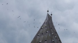
Here at the Museum we eagerly await the return of these long distance migrants each May. This is not only because for many of us they herald the start of summer, but also because the swifts that nest each year in the Museum tower are part of the longest-running continuous study of any bird species in the world.

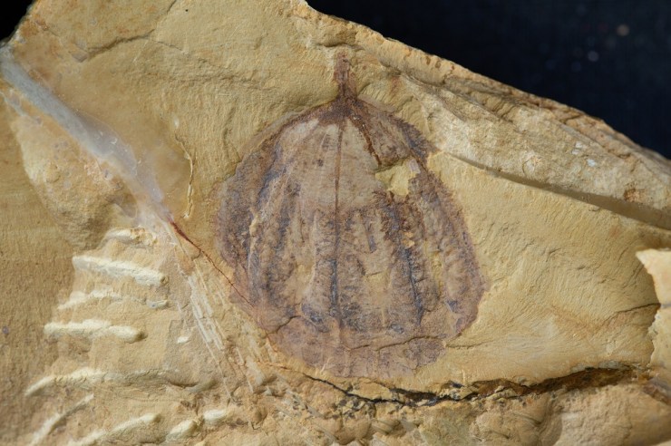
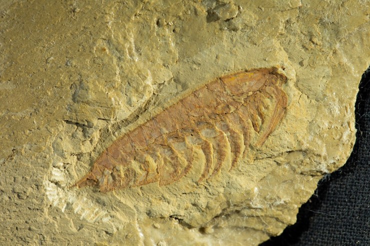
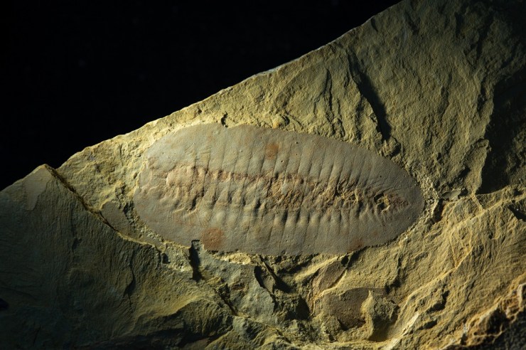
Taking the long view of these amazing birds we know that, like all birds, they evolved from a particular group of dinosaurs. Birds, in effect, are living dinosaurs. The earliest fossil swift, the ‘Scania Swift’, is around 49 million years old and shows us that by this time they had already evolved in forms that are virtually indistinguishable from today’s birds. Today, they have diversified into around 100 different species including our Common Swift (Apus apus).

Swifts in the tower nests as seen on webcam
Swift chicks in a nestbox in the Museum tower, shown on the webcam feed

Swifts have taken life on the wing to the extreme. Not only are they the fastest recorded bird in level powered flight, reaching speeds of nearly 70mph, but once launching themselves from the nest that they hatched in they may not land for the next two years of their lives!

They are so adapted to life in the air that they are capable of eating, mating and even sleeping on the wing. During sleep, it is thought that the two hemispheres of the brain take it in turns to nap as the swift slowly circles at heights of up to 30,000 feet. They do not even land to collect nesting material, instead relying on whatever feathers or pieces of plant material are floating in the air to build their nests.

During this two-year flight they will follow their food – the seasonal blooms of flying insects that appear after summer rains – on a 14,000 mile annual migration to southern Africa and back, living in perpetual summer.

Whilst for a long time scientists thought swifts were closely related to similar looking birds, swallows and martins, DNA analysis has revealed that they are the product of another amazing type of evolution – called convergent evolution – where organisms with similar lifestyles independently evolve similar traits. It turns out that whilst swifts may look like swallows, they are actually more closely related to hummingbirds; swallows, on the other hand, are more closely related to kingfishers than to swifts.

Swifts flying around the Museum tower
Swifts circle the entrances to the nest areas in the Museum’s tower. Image: Gordon Bowdery

Studies show that the population of breeding swifts in the UK has roughly halved between 1995 and 2016. The causes of this decline are debated: Lack of nest sites, lack of food, and changes to global weather patterns have all been implicated. The truth is that a bird which lands only once a year is extremely difficult to study.

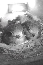
We hope for a successful breeding season here in the tower, but if you would like to observe them yourself you can watch the swifts on our nest cam and compare the ups and downs of their populations over the years on our website.


Exceptional Chinese fossils come to Oxford in new partnership

by Imran Rahman, Deputy Head of Research

China is world-famous for its unique and exceptionally preserved fossils, which range from some of the oldest animals on Earth, to spectacular feathered dinosaurs. We are therefore very excited to announce that the Museum, along with other institutions from across Europe, is a partner in a major new venture with Yunnan University in China: the International Joint Laboratory for Palaeobiology and Palaeoenvironment.

Collaboration between this Museum and Yunnan University dates back to the 1990s, driven by the work of Professor Derek Siveter – a former Senior Research Fellow and current Honorary Research Associate at the Museum. He collaborated with Professor Hou Xianguang, director of the International Joint Laboratory for Palaeobiology and Palaeoenvironment, to study fossils from the internationally renowned Chengjiang biota, which was discovered by Hou Xianguang in 1984.

Museum researchers Duncan Murdock, Jack Matthews and Derek Siveter (l-r) visit the Precambrian-Cambrian Section

The Chengjiang fossil site is important and exciting because it preserves both the soft and hard parts of a range of early animals. This fossil record captures the rapid diversification of life about 520 million years old – in an event referred to as the Cambrian explosion. Derek Siveter was instrumental in a successful bid to have the Chengjiang biota designated a UNESCO World Heritage site in 2012, preserving it for future generations.

In December 2018, Museum researchers Duncan Murdock, Imran Rahman and Jack Matthews travelled with Derek to Kunming, China, for the first meeting of the International Joint Laboratory for Palaeobiology and Palaeoenvironment. The lucky researchers spent three days on field trips to the region’s most spectacular fossil sites, including Lufeng World Dinosaur Valley and the Chengjiang biota itself, followed by two full days of scientific talks and discussions.

The International Joint Laboratory is funded by the Ministry for Education of China and includes the University of Leicester, the Natural History Museum, London, the University of Munich, and the Bavarian State Collection of Zoology, along with Oxford University Museum of Natural History and Yunnan University.

The arthropod Haikoucaris ercaiensis. Sometimes referred to as a ‘short-great-appendage’ arthropod, Haikoucaris had a pair of prominent grasping appendages adjacent to the head (right-hand side of this image). Credit: Scott Billings
The arthropod Leanchoilia illecebrosa. Sometimes referred to as a ‘short-great-appendage’ arthropod, Leanchoilia illecebrosa had a pair of prominent grasping appendages adjacent to the head (right-hand side of this image). Credit: Scott Billings

A significant first outcome of this new partnership will be the loan of iconic Chengjiang fossil specimens from Kunming to Oxford for our First Animals exhibition which opens on 12 July and runs until February 2020. Most of these fossils have never been outside of China before, and some have never been seen by the public before. We invite you to visit First Animals to see these exceptional fossils first hand!

The arthropod Saperion glumaceum. Saperion had a flattened, segmented body and jointed appendages (not visible in this specimen). Credit: Scott Billings.

Top image: The comb jelly Galeactena hemispherica. Unlike modern comb jellies, which are soft-bodied animals, Galeactena and its relatives had hardened ‘spokes’ on the sides of the body (appearing as dark bands in this photograph). Credit: Scott Billings.

Bacteria that changed the world: Leuconostoc

In our Bacterial World exhibition we offer a selection of ten bacteria that have changed the world, some in profound ways. In this series of short fact-file posts we present one of the ten each week. This week’s bacteria are…

Leuconostoc
– the food-fermenters

Where they live
Vats of bubbling syrup in sugar factories first yielded samples of Leuconostoc. In 1878, a scientist called Philippe van Tieghem found and studied the bacteria, which people use to make fermented food all over the world.

Why they are important
Leuconostoc bacteria play a part in creating traditional dishes in many countries, including sauerkraut, kimchi, kefir and sourdough bread.

How they are named
Van Tieghem named Leuconostoc after another bacterium that he thought it resembled. Today, however, new bacteria are named according to rules that are governed by the International Committee on Systematics of Prokaryotes.

How they work
In a pickled food dish like sauerkraut or kimchi, Leuconostoc converts the sugars in vegetables into lactic acid, preserving them and leading to a characteristic sour taste. A similar process takes place in the starter culture for making milk into kefir, and for giving sourdough bread its flavour.

Top image: Coloured scanning electron micrograph (SEM) of Leuconostoc citreum. Copyright: Science Photo Library

Bacteria that changed the world: Lactobacillus acidophilus

In our Bacterial World exhibition we offer a selection of ten bacteria that have changed the world, some in profound ways. In this series of short fact-file posts we present one of the ten each week. This week’s bacteria are…

Lactobacillus acidophilus
– the gut-guzzlers

Where they live
Lactobacillus acidophilus is one of the hundreds of species of bacteria that live in your gut. This particular species is found all through the gut from your mouth to your anus.

Why they are important
In your gut, this species digests lactose in milk, splitting it into the simpler sugars glucose and galactose. People suffering from diseases such as HIV and cancer tend to have abnormal levels of Lactobacillus in their gut – either too many bacteria, or too few.

How they are named
Lacto is Latin for milk and bacillus refers to the rod shape of these bacteria. Acidophilus means ‘acid-loving’ in Latin – this species makes sure that its home remains slightly acidic by releasing its own acid, which helps to keep other bacteria at bay.

How they work
Not only does Lactobacillus acidophilus produce sugar from milk, but it may also produce tryptophan – an essential nutrient that we cannot produce ourselves.

Top image: Coloured transmission electron micrograph of the Gram-positive rod-shaped bacteria Lactobacillus acidophilus. Copyright: Science Photo Library

Bacteria that changed the world: Wolbachia

In our Bacterial World exhibition we offer a selection of ten bacteria that have changed the world, some in profound ways. In this series of short fact-file posts we present one of the ten each week. This week’s bacteria are…

Wolbachia
– the man-killers

Where they live
Up to 60 percent of insect species are infected with the bacterium Wolbachia, as are other species such as nematode worms.

Why they are important
Wolbachia selectively kills off males in many species of insect and alters the sex ratio of the population to its own advantage. However, some species of insect rely on it for protection against other threats.

How they are named
The bacteria take their name from Simeon Burt Wolbach, who along with Marshall Hertig co-discovered Wolbachia in 1924 in a mosquito.

How they work
Infected female insects pass the Wolbachia to their offspring – so the bacteria do everything they can to ensure females survive. Their strategies include killing male larvae, making males infertile, and rendering females able to reproduce without males.

Top image copyright: Joshua Blight (University of Oxford) & Steven Sinkins (University of Glasgow)

How the sea cucumber lost its armour

By Imran Rahman, Research Fellow

You have probably heard of sea cucumbers. If you’re lucky, you might have seen one, if not in the wild, then perhaps in a nature documentary like Blue Planet or the children’s cartoon Octonauts. If you’re less lucky, you might have eaten one – they are most commonly described as slippery and bland in taste!

Despite their appearance, sea cucumbers are actually marine animals most closely related to sea urchins, rather than to worms or slugs. Over the past century palaeontologists have uncovered a range of ancient fossil relatives of modern sea cucumbers that allow us to piece together the story of how they evolved from armoured ‘tanks’ into the naked slug-like forms we see today. One such fossil is described in a new paper by my colleagues and I, just published in the journal Proceedings of the Royal Society B.

The fossil in question is 430-million-years-old, and it comes from a site of exceptionally-preserved fossils in England called the Herefordshire Lagerstätte. Herefordshire has produced many exciting discoveries over the years, from prehistoric parasites to an ancient ‘kite runner’. The new fossil is the first of its kind from this deposit.

Like all fossils from Herefordshire, the specimen was preserved in an egg-shaped nodule of rock. Because the rock has the same chemical composition as the fossil, it could not be studied with modern imaging methods such as CT scanning. Instead, it had to be studied by painstakingly grinding away the fossil, a few hundredths of a millimetre as a time, with photographs taken of each exposed surface using a digital camera. This allowed us to build up a dataset of hundreds of slice images through the fossil, which were digitally reconstructed as a 3-D ‘virtual fossil’ on a computer.

The 3D computer reconstruction revealed a very peculiar animal, about 3 cm wide, with 45 tentacle-like ‘tube feet’ and a large mouth surrounded by five teeth. The animal had a skeleton made up of numerous hard plates, which were composed of the mineral calcite. After studying this fossil and comparing it to other similar ones from the same time period, we were able to identify it as a species new to science. We named the species Sollasina cthulhu, for its resemblance to monsters from the Cthulhu universe created by author H.P. Lovecraft.

One of the most useful things about our 3D computer reconstruction was that it enabled us to study the inner features of the fossil, as well as the parts visible on the outer surface. This revealed internal soft parts that had never previously been described in this group of fossils. In particular, it allowed us to see an internal ring-like structure within the main body cavity.

3D reconstruction of Sollasina cthulhu. Left-hand image shows part of lower surface. Right-hand image shows same view with outer surface partly transparent to reveal inner ring (in red). Credit: Imran Rahman, Oxford University Museum of Natural History

We interpreted this inner ring as part of the water vascular system – the system of fluid-filled canals used for feeding and movement in modern sea cucumbers and their relatives, such as sea urchins and starfish. In life, the ring was connected to the large tube feet, which were filled with seawater. Most of these tube feet were used for crawling over the seafloor, with those nearest the mouth used for capturing food. The teeth could cut and crush food items, which were then eaten by the animal.

Life reconstruction of Sollasina cthulhu. Credit: Elissa Martin, Yale Peabody Museum of Natural History

To work out the evolutionary relationships of Sollasina cthulhu, we assembled a list of characteristics for various fossil and modern sea cucumbers and sea urchins. We analysed this matrix using several computational methods to determine how these different animals were related to one another. The results confirmed that Sollasina cthulhu and closely-related forms were ancient relatives of modern sea cucumbers. This allowed us to reconstruct the early evolution of sea cucumbers, back to their shared common ancestor with sea urchins, over 450 million years ago. Our study demonstrates this was a story of loss, with fossil sea cucumbers becoming progressively less armoured as they evolved into modern forms.

This discovery has greatly improved our understanding of sea cucumber evolution, but several questions remain. One intriguing question is when and how did sea cucumbers lose their teeth, and did these evolve into any features seen in living sea cucumbers? Future study of existing and new fossil sea cucumbers and sea urchins will help to answer this and other intriguing questions.