Wasp Faces: Power Struggles and Royal Drama
By Kristian Suszczenia, Intern
The second you step into the Museum of Natural History you will notice a remarkable thing about nature: its diversity. Instantly, you see huge whale bones, a distant elephant skull, birds, fish, dinosaurs, and a mounted kaleidoscope of colourful insects. This variation is what many of us love most about biology.
But diversity occurs at a much finer scale than the magnitudes of difference that exist between species. Often there is ‘Individual Variation’ — differences that occur within species. Person to person, specimen to specimen, each organism is unique, just like us humans!

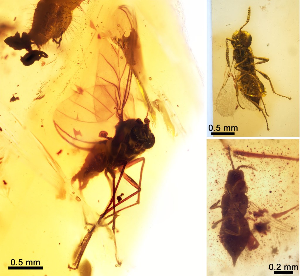
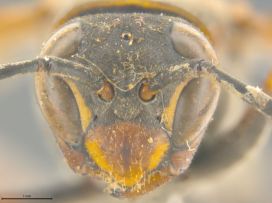
Buried under the 5 million other insects of the HOPE collection is a drawer that houses a species of the genus Polistes, a social paper wasp collected from Brazil. A close look at their faces reveals staggering individual variation. Before getting confirmation from specialists, the Museum staff found it hard to believe that these four faces could even belong to the same species. The question is, why are they so different? Is there an evolutionary benefit to all these wasps having their own style of eyeliner?

It is well-known that individual variation can give certain members of a species an edge over others, especially when it comes to dating! From guppies to fruit flies, females often prefer mates that stand out with unique colours and patterns, perhaps because they are simply more noticeable.
Yet dating is not a sufficient explanation for our fashionable paper wasps. They get a very different benefit from their unique looks — not so much standing out, but being memorable. For them, a memorable identity is a way to remember who goes where in a critical pecking order.
Thanks to a lot of elegant work by Polistes specialists, we understand that bespoke face markings tend to evolve in species that have multiple queens in a linear hierarchy. A Polistes queen can start a hive alone but often benefits from forming a group of queens that can all cooperate together in a single hive. In order to cooperate, the queens must decide on a dominance hierarchy amongst themselves. To do this they take part in brutal one-on-one battles as they assess each other’s prowess.
After they establish an initial order, each wasp will constantly test the adjacent ranks (their closest match) with darts and lunges as they try to climb the ladder for extra reward and simultaneously defend their place. Four punishes Three for transgressions and plots against Five’s downfall. It’s a royal reality show.

This part-insect, part-spartan society is certainly fascinating, but what does it have to do with the wasps’ faces?
In order for queens to defend their rank from their adjacent competitors, they need to know exactly who’s who. Unique faces are more recognisable and more memorable. Being able to recognise and recall every individual wasp allows queens to track their rivals based on their faces and avoid a lot of violent misidentification. Imagine if a queen were to look forgettable; every other queen in the hive would see her as a potential challenge to their power. She wouldn’t last long. But having a memorable face allows individuals to avoid unnecessary scraps and make for a more efficient hive overall.
After learning the story of the wasps, it seems plausible that humans may have evolved our fantastically recognisable faces for societal advantages too — perhaps to avoid getting mistaken for an enemy, perhaps so we can trade favours, or maybe just to avoid general confusion. It would certainly make life difficult if all your co-workers had the exact same face. Remembering names is hard enough already!