Diving into deep time

Our current First Animals exhibition is extending its run until 1 September, and to mark the extension our Research Fellow Imran Rahman takes a look at how animal life in the ancient oceans was brought to life in our Cambrian Diver interactive installation.
One of the biggest challenges in developing the First Animals exhibition lay in visualising rare fossil specimens as ‘living’ organisms, transforming them from two-dimensional imprints in the rock into three-dimensional animated computer models.
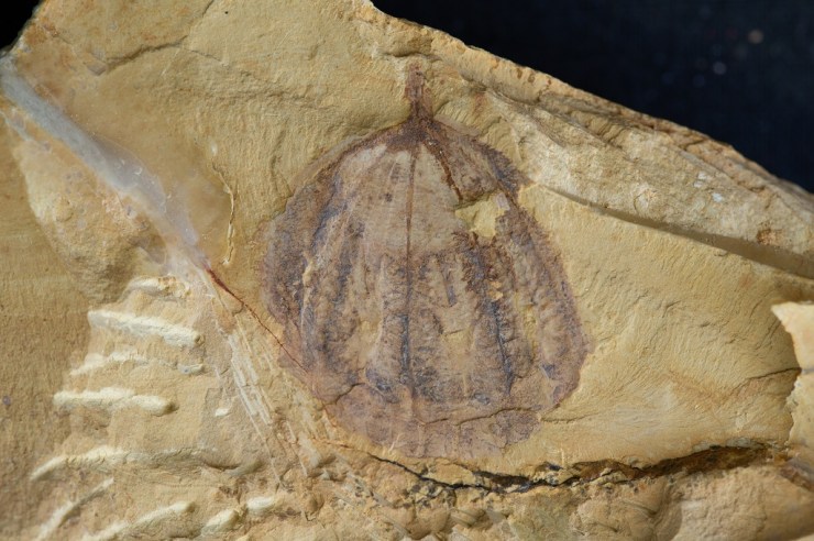
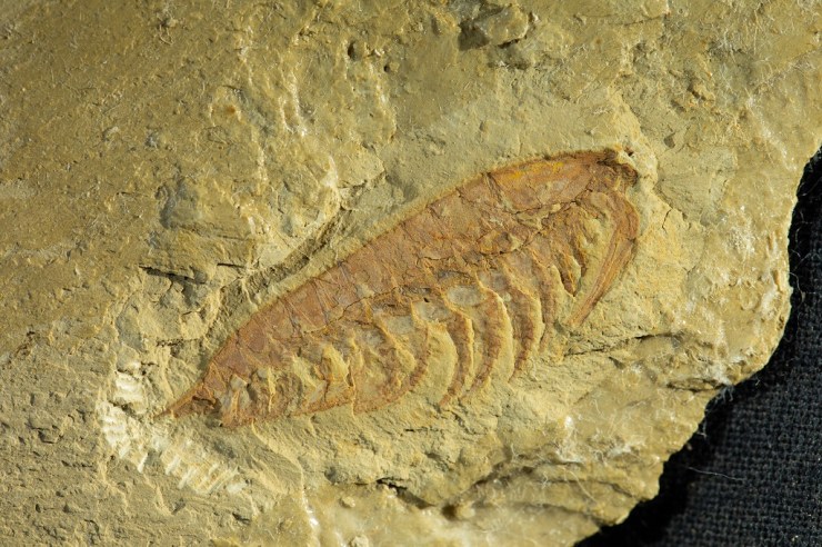
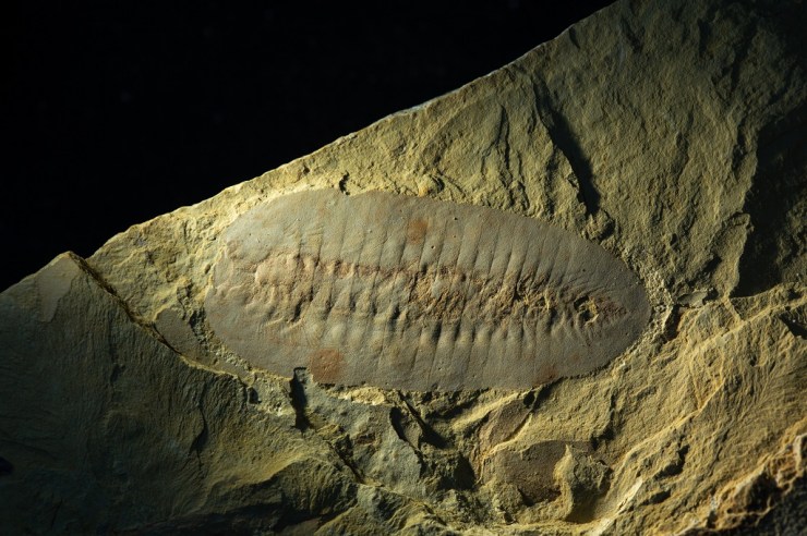
Many of the specimens on display in First Animals were collected from sites of exceptionally well-preserved fossils called Lagerstätten. These deposits preserve the remains of soft-bodied organisms that are almost never seen in the fossil record; things such as comb jellies and worms, as well as soft tissues such as eyes, gills and muscles. Even so, most of these fossils are flattened and two-dimensional, which makes it very difficult to reconstruct what they looked like in life.

To help exhibition visitors visualise the animals in a living environment we worked closely with Martin Lisec and his team at Mighty Fossils to create a set of detailed computer models of a key set of animals. We have worked with Martin before on the video of a Jurassic sea inhabited by plesiosaurs and other marine animals for our Out of the Deep display. That was very successful, but our idea for First Animals was even more ambitious: to create a unique interactive installation called the Cambrian Diver.
The material focused on the Chengjiang animals from the Cambrian of Yunnan province, China, which provides the most complete record of an early Cambrian marine community, from approximately 518 million years ago. Using fossil evidence of the organisms thought to have lived at the time we selected 12 species that were representative of the diversity of the Chengjiang biota.
The first phase was collecting as many materials as possible to be able to create 3D models. As usual, we started with rough models, where we set basic dimensions, shapes and proportions of body parts. Once approved, we moved to very detailed models for the animations, artworks and textures for less detailed models to be used within the interactive application. – Martin Lisec, Mighty Fossils

To provide two-dimensional templates for Mighty Fossils to work from we scoured the scientific literature for the most recent accurate reconstructions available for each of the species.
The predatory arthropod Amplectobelua symbrachiata is a good example. We drew heavily upon a 2017 paper by Dr Peiyun Cong and colleagues, which included a very detailed reconstruction of the head region. This reconstruction shows that the underside of the head of Amplectobelua consisted of a rod-shaped plate, a mouth made up of two rows of plates, and three pairs of flaps with spiny appendages, all details that are included in our 3D model.

Colour and texture were another consideration. To inform these we looked at living species that are thought to have similar modes of life today. For Amplectobelua, a free-swimming predator, we examined the colouration of modern marine predators such as sharks. Many sharks have countershading, with a darker upper side of the body and a lighter underside, which acts as camouflage, hiding them from potential prey.
We reconstructed our Amplectobelua model with similar countershading camouflage, with blue and red colouration inspired by the peacock mantis shrimp, a brightly coloured predatory arthropod that lives in the Indian and Pacific oceans.

The next vital step was establishing how the animals moved and interacted with one another. This is a major challenge because in many cases there are no modern equivalents for these extinct early animals. For Amplectobelua we inferred that the flaps on the sides of the body were used for swimming, with the tail fan helping to stabilize the animal as it moved through the water. This agrees with previous interpretations of swimming in closely related animals such as Anomalocaris.
The models were built and textured by Mighty Fossils using the 3D gaming engine Unity. The video below is an accelerated sequence showing how the elements of the model are layered together.
The finished, animated and annotated Amplectobelua model is shown below, and can be zoomed and rotated. All the models generated by Mighty Fossils for the First Animals exhibition are gathered in a collection on our Sketchfab page.
Once animated models of all 12 species were created we placed them in a realistic marine environment. Study of the rocks preserving the Chengjiang fossils suggests these animals lived in a relatively shallow, well-lit sea, perhaps 50 metres deep and characterised by a flat, muddy seafloor. A continuous shower of organic particles is thought to have filled the water column, as in modern oceans.

Based on present-day marine ecosystems, we infer that the number of immobile suspension feeders would have been much greater than the number of predators. As a result, we included multiple individuals of the suspension feeders Cotyledion, Saetaspongia and Xianguangia, which were tightly grouped together, but only a small number of the active predators Amplectobelua and Onychodictyon.

The final step involved setting up a camera and user interface to allow visitors to discover the various animals in our interactive environment. For this we worked with creative digital consultancy Fish in a Bottle to identify eight locations, each focused on a different animal.
As the video above shows, users can navigate between locations by touching an icon on the screen, and when the Cambrian Diver sub arrives at a location information about the animal, its mode of life and its closest living relatives is presented on-screen. A physical joystick allows users a 360-degree rotation to look around the scene, and explore the ancient watery world.
This project was significantly bigger than the Out of the Deep work we had done previously with the Museum, mainly because of the complicated approval procedure needed for 20 individual 3D models. Along with three large illustrations, two animations and the interactive application this was a big workload! Fortunately, we managed to finish the whole project on time for the opening of the exhibition. – Martin Lisec