A shark’s tale

Following the completion of our Life, As We Know It redisplay project earlier this year, we are taking a look at some of the exceptionally skilled people who helped to bring it all together. In this article, model maker Jeremy Hunt and our Vertebrate Palaeontology Collections Manager Emma Nicholls take a deep dive into what we believe is the world’s first 3D reconstruction of the extinct shark Ptychodus…

In six of our newest displays, fossil material found in and around Oxfordshire is used to reconstruct ecosystems from slices of geological time. One of these recreates a marine environment of the Cretaceous Period, between 94 and 84 million years ago, when most of northern Europe was submerged under a warm shallow sea. In these seas swam a formidable 8-metre long shark called Ptychodus.

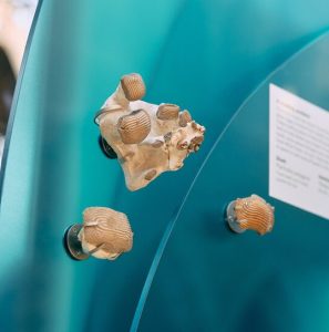
Fossils of Ptychodus teeth in the new display

Until recently, Ptychodus remained something of a mystery: teeth and vertebrae were the only remains scientists could use to work out what it looked like. Its robust, low-domed teeth are covered in deep ridges of enamel that created a mosaic in each jaw. They were capable of crushing shelled prey like a giant nutcracker. Similar teeth in modern sharks are found only in slow-moving species that prowl the seabed, so it was presumed that Ptychodus behaved in the same way.
But then in 2024 an incredible fossil with much of the animal’s soft tissue preserved was published by Dr Romain Vullo and co-authors. This new fossil revealed clues to the shapes of the fins and their position on the body. The study concluded that Ptychodus was in fact not a bottom-dwelling animal at all, but much more likely to be a fast, agile predator living higher in the water column, and perhaps related to the Great White Shark.

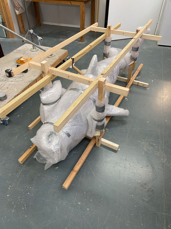
Armed with this recent discovery, we decided to create a three-dimensional model of the reimagined Ptychodus for our Cretaceous display. It had to be a quarter of life-size to fit it in the showcase, but would still be a huge two metres long.
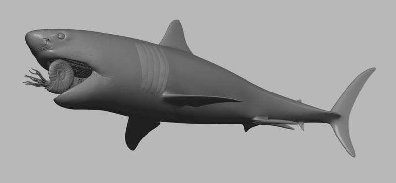
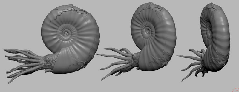
Digital 3D model of a shark, shown in grey, without any skin colouring or texture. The mouth is open and inside is an ammonite being crushed by the shark's plate-like teeth.
An early computer model of the shark and ammonite

“Working with Dr Vullo and others we began to decipher the shark’s possible appearance,” says Emma.  “Aspects to sink our teeth into included the skull shape, how it would have fed on hard-shelled free-swimming prey such as ammonites, how the mosaic of teeth sat in the mouth, as well as where and what shape the fins would have been.”

The next stage was to turn these ideas into a realistic model, and for this we turned to specialist model maker Jeremy Hunt. “When I was asked to make the reconstruction of Ptychodus I thought it was a great opportunity to incorporate modern digital techniques and processes that would give great accuracy, but also and flexibility so that adjustments could be made at later stages. This would be really difficult using a traditional clay sculpting approach,” he explains.

Together, Emma and Jeremy began to translate the fossil research into draft models on the computer, digitally sculpting versions on-screen. As well as being scaled down to quarter size, the position and pose of the model also needed to work inside the showcase structure and the designs of the rest of the display. Combining computer models helped us to make sure that Ptychodus would fit perfectly and that its method of support could be worked out well ahead of installation.
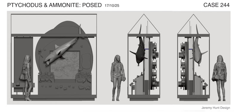
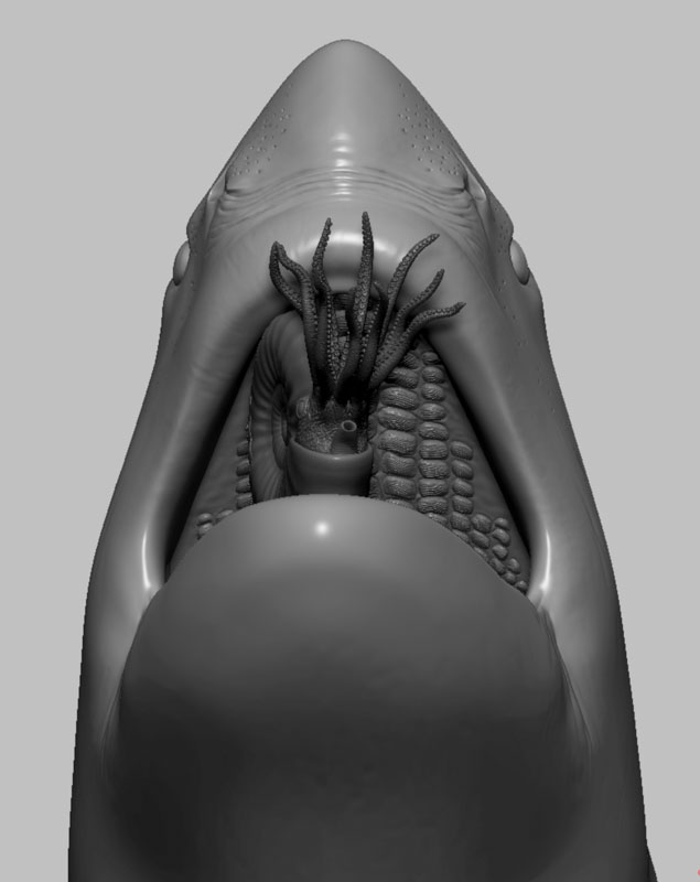
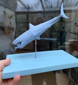
To give us a sense of how the final model would actually look, Jeremy 3D-printed little maquettes just a few centimetres long. On these we could check that the anatomy and pose were just what we needed. The maquettes also showed how the shark would be suspended in a diving angle, chomping down on an ammonite which Jeremy was modelling separately.

A hand holds out a wooden plinth, painted blue, onto which a 3D printed shark model is mounted on a vertical metal rod. The 3D print is also blue and is approximately 15cm  long.
3D-printed maquettes of the model helped us to check its form and pose

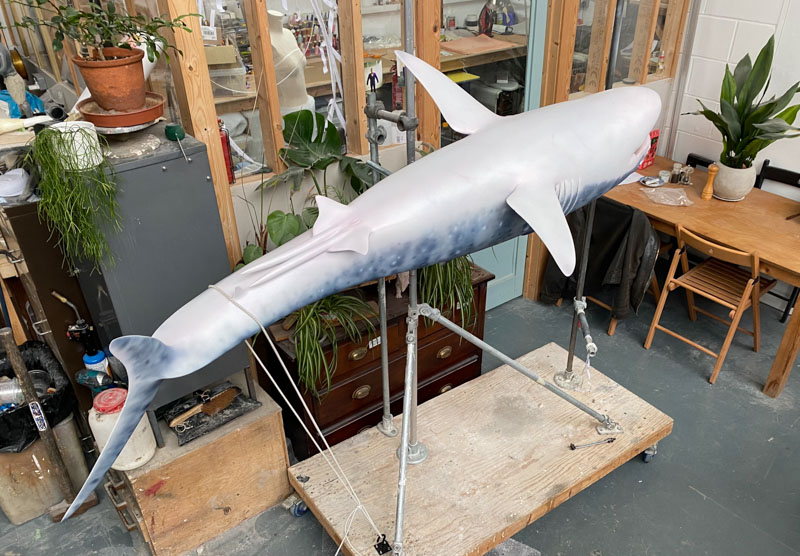
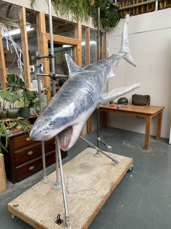
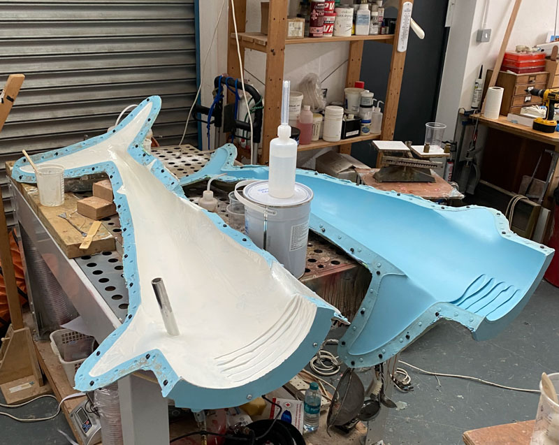
Once ready, the model was 3D-printed at its display size of two metres long, allowing Jeremy to create a silicone rubber mould to produce a fibreglass cast of the final structure. Fibreglass is both strong and light, perfect for the display. But the model really came to life when Jeremy hand-painted and airbrushed its colourings and markings based on a research by Emma and a concept drawing by palaeo-artist Julius Csotonyi.
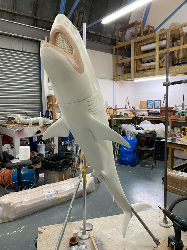
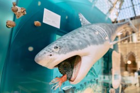
Once Ptychodus and the rest of the display were installed, the ammonite was placed in the mouth. It is held safely there by hidden strong magnets, caught permanently between those crushing plates, about to become shark food.
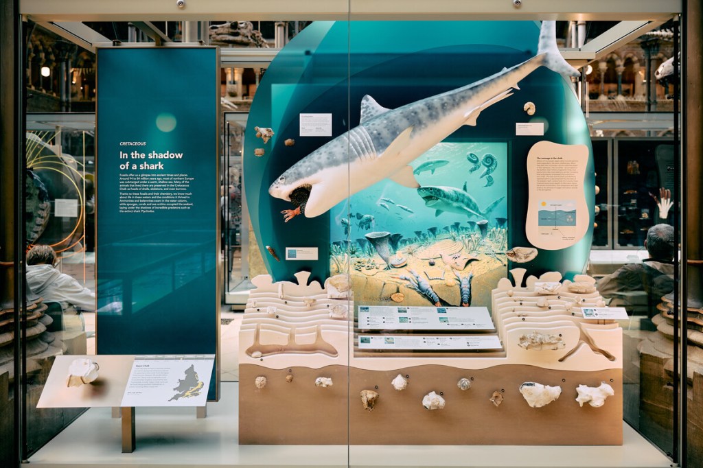
Museum exhibit showing a 2 metre long shark model against a backdrop artwork of an ocean containing drawings of other sharks, fish, ammonites and belemnites in the sea, along with corals, shellfish and lobsters on and burrowing into the seabed. Below this scene are a selection of chalk fossils. On the left the title panel is printed on blue acrylic and shows the display's name as 'In the shadow of a shark'.
The Ptychodus model in the final display at the Museum

“This was a great project to be involved with, and I hope the new displays will be enjoyed by all who visit the Museum,” says Jeremy.

Jeremy Hunt, model-maker

*

See more of Jeremy’s work at: www.instagram.com/memsculpt

A grasp of the past

by Ricardo Perez-De-La Fuente, research fellow

Few creatures look weirder – or are cooler, in my opinion – than mantidflies. There are around 400 species of these small predatory insects known worldwide – a scarce diversity by insect standards.

Like praying mantises, mantidflies have long ‘necks’ and forelegs armed with powerful spines and other structures used to hunt their prey with a sudden lethal grasp. The unfortunate victims become immobilised until they are meticulously eaten alive – not the best way to spend your last minutes on Earth!

Mantidflies belong to the Neuroptera order of insects and so aren’t actually related to praying mantises, but to insects such as lacewings and antlions.

A new paper that a colleague and I have published presents a new fossil mantidfly from Spanish amber that is important in understanding the evolution of their gripping – or raptorial – forelegs. The finding is presented in the open access journal Scientific Reports today.

Although the discovery has just been published, we excavated the new fossil during the scorching summer of 2010 in Teruel, northeastern Spain.

Amber excavations are very romantic – while they take place we carefully store the amber, piece by piece, into muddy plastic bags, remaining oblivious of what creatures are being unearthed because the amber surfaces have become opaque during fossilisation. Later, in the laboratory, the surfaces of the amber pieces are polished and screened for inclusions. Then a first glimpse is gained into what has remained frozen in time for millions of years.

It is only when the amber inclusions are carefully examined and studied that the implications of the specimens that were dug up years earlier start to be revealed. In this case, a specimen that was preserved in fragments, nothing spectacular at first look, ended up being truly exceptional.

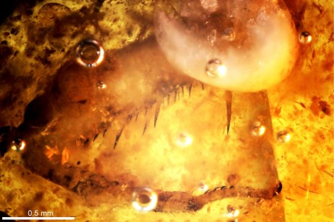
Foreleg of Aragomantispa lacerata, showing powerful spines and other structures adapted to strike and hold prey.

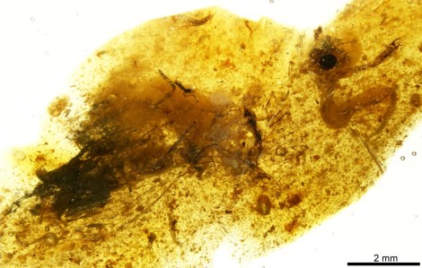
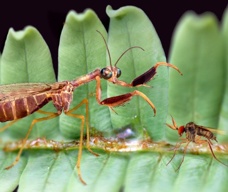
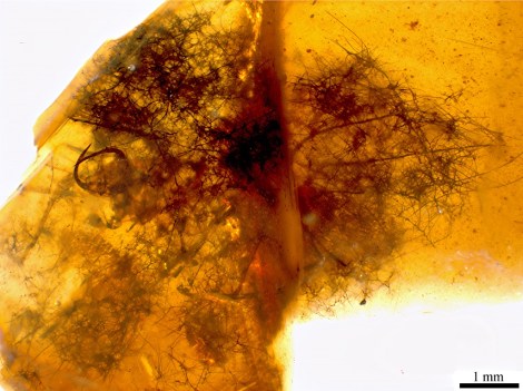
Extinct true mantidflies, particularly those preserved in amber, are extremely rare. Our new fossil, pictured above at the top of the article, is 105 million years old, from the Cretaceous period. It currently stands as the oldest true mantidfly known in amber. The new extinct species, named Aragomantispa lacerata, has allowed us to compare the structures of the raptorial forelegs between extinct and extant mantidflies with an unprecedented detail.

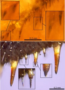
Comparison between the foreleg spine-like structures of the new fossil mantidfly (up), with those from a close modern species (bottom).

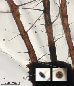
Present-day mantidflies have forelegs with spines that bear minute cones at their tip. These cones are sensory organs that elicit the striking reflex and feel the prey’s movements once captured and restrained by the mantidfly’s tight embrace.

The forelegs of Aragomantispa lack these cones at the spines’ tip, instead having larger, icicle-shaped tips. We do not know how sensitive the mantidfly forelegs were in the Cretaceous, but the spines of at least some of these insects seem to be not as specialised as those from their present-day relatives.

Some mantidflies have smaller, reclined hair-like structures forming an edge on the leg’s surface opposing the spines. These reinforced edges create a scissor effect that stuns prey when the forelegs strike. Although Aragomatispa has these structures on the forelegs, they are also different in shape to those found on extant mantidflies.

Reconstruction of Aragomantispa lacerata striking at a hypothetical prey on a fern in the Cretaceous Spanish forest.

The fossil record offers the only direct means to assess when and how the traits characteristic of a given animal group originated in time. However, this kind of fossil evidence appears very occasionally. Our discovery shows that the foreleg spine-like structures of recent mantidflies were not fully developed in at least some of their Cretaceous ancestors.

The most exciting part is to think that this story and literally thousands more lie waiting to be discovered – or otherwise forgotten forever – buried underground.

Crafty camouflage

Last week we brought you snails that attach all manner of pebbles, fossils, corals and shark teeth to their shells. Today we give you a newly-discovered fossil green lacewing larva that attached pieces of soil to its body as an act of camouflage. Our research fellow Ricardo Pérez-de la Fuente, lead author of the new paper, explains…

Visual camouflage is one of the most successful survival strategies in nature. Camouflaging is usually defensive, allowing animals to be left unnoticed by their predators, but it can also be used aggressively by predators themselves to approach their prey undetected.

Some camouflaging animals can actively change their colouring to match that of the background ‒ a technique called crypsis. Others can make their bodies resemble elements of the environment, such as leaves or twigs, which is called mimicry.

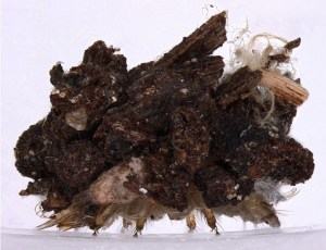
Italochrysa italica, an extant green lacewing larva carrying a dense debris packet made of soil fragments. Taken from the open access publication Tauber & Winterton, 2014.

Yet another approach to camouflage involves collecting diverse materials from the environment and incorporating them on the animals’ bodies in order to better blend with the surroundings. This is known as debris-carrying, trash-carrying, or decoration, and it can be found across a wide variety of animals including sea urchins, gastropods, and arthropods, such as decorating crabs, or sand- and mud-covering spiders.

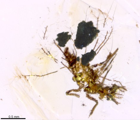
My colleagues and I have just published the discovery of a fossil green lacewing larva, pictured at the top of the article, that has been preserved carrying bits of soil that it used for camouflage and physical protection. It’s a new larval species just 1.5 mm in length, and is preserved in Early Cretaceous Lebanese amber. We have named it Tyruschrysa melqart after the Phoenician city of Tyre and its tutelary god Milk-Qart (if you want to learn the reasons behind this name check out our open access paper!).

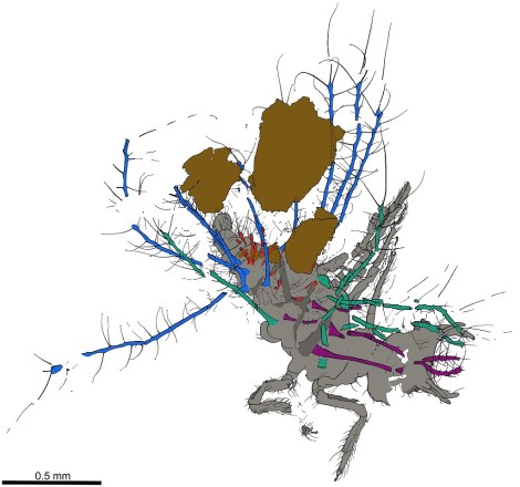
Interpretative drawing of Tyruschrysa melqart: body in grey, ‘tubes’ with setae coloured according to which body part they are attached to, and soil debris in brown.

Green lacewing larvae are active predators that eat other insects such as aphids, using sickle-shaped ‘jaws’ to pierce their prey, suck out their fluids and liquefy their tissues; eating is easier when there is no need to chew! Some green lacewing larvae are debris carriers, entangling all kinds of debris among their velcro-like ‘hairs’ called setae, which extend from relatively short ‘bumps’ on their backs. This debris is carefully selected and gathered with meticulous head and body movements to form a so-called debris packet on the back of the insect.

‘Tubes’ bearing setae of Tyruschrysa melqart, with detail of their mushroom-shaped endings (bottom), used for anchoring bits of soil.

The new fossil and similar ones described from younger Cretaceous ambers differ from modern relatives because instead of short ‘bumps’ with setae on their backs they have relatively long ‘tubes’, giving them a bizarre appearance.

These tubes have setae with mushroom-shaped endings of a kind never seen before in extinct or living green lacewing larva species. The mushroom-shaped ending is a special adaptation to anchor debris, which in the case of Tyruschrysa melqart are fragments of soil.

Hallucinochrysa diogenesi, another Cretaceous green lacewing larva bearing long ‘tubes’ with setae on its back, but carrying a debris packet made of plant hairs (trichomes). Preserved in Spanish amber (105 million years old).

It was already known that Cretaceous green lacewing larvae like Tyruschrysa had long tubes on their backs and that they collected plant hairs and other plant material to construct their packet of debris. But thanks to the new discovery we now know that these immature insects also used bits of soil, and that in the deep past debris packets were probably as diverse as those we see today.

Green lacewing larvae have been gathering debris to camouflage and protect themselves for about 130 million years, giving rise to the different body adaptations we see amongst these fascinating tiny collectors.

‘A soil-carrying lacewing larva in Early Cretaceous Lebanese amber’ Ricardo Pérez-de la Fuente, Enrique Peñalver, Dany Azar and Michael S. Engel is published as open access in Scientific Reports this month.

Dinosaur WLTM friendly new carers

DSC_8255 small

It’s not often that one of our residents flees the roost to take up home elsewhere: usually once you’re in the Museum that’s it, accessioned for life (or, more accurately, death). However, one of our former dinosaur aisle characters is now looking for a new home…

The four-metre long Utahraptor model has been with the Museum since 2000, during which time it escaped to take up temporary residence in Blackwell’s book shop in Oxford city centre as part of the award-winning Goes to Town project. This time, however, the Utahraptor will be leaving us for the last time as part of a reorganisation of the Museum’s offsite store, where the model is currently residing.

The Utahraptor model in Blackwell's bookshop as part of the Goes to Town project. Photo: Mike Peckett
The Utahraptor model in Blackwell’s bookshop as part of the Goes to Town project. Photo: Mike Peckett

But rather than just ditch this Cretaceous creature unceremoniously we’d like to offer it out to new keepers, ideally somewhere with a public space where the model can be enjoyed by others. So, fancy yourself as a dinosaur owner? If so, check out our selection form for details of what it takes to keep such a pet.

We’re asking people to make a case for the Utahraptor to move to their venue and we will donate the model free of charge to whoever is selected. The closing date for submissions is 5 August and the selected venue will be announced by 12 August. We need to deliver the model to its new home on or before 23 September.

Get me outta this place!
“Get me outta this place!” – the Utahraptor is currently in the Museum’s offsite storage

At this point you probably want some Utahraptor facts to help with your decision, right? Well, here you go:

Utahraptor means ‘Utah’s predator’ and the animal is known from fragmentary fossils found only in Utah in the United States. There is just one known species, Utahraptor ostrommaysorum, which was alive in the early Cretaceous period, around 125 million years ago.

It is thought that like most dinosaurs of its type (dromaeosaurids) the Utahraptor was feathered, although no direct evidence has yet been found. The Museum’s model, made by Crawley Creatures, does not represent a feathered example of this species. It’s likely the beast was not bright orange too, but who knows?

We looking forward to reading your submissions…

www.bit.ly/utahraptor

For more information email communications@oum.ox.ac.uk.