A shark’s tale

Following the completion of our Life, As We Know It redisplay project earlier this year, we are taking a look at some of the exceptionally skilled people who helped to bring it all together. In this article, model maker Jeremy Hunt and our Vertebrate Palaeontology Collections Manager Emma Nicholls take a deep dive into what we believe is the world’s first 3D reconstruction of the extinct shark Ptychodus…

In six of our newest displays, fossil material found in and around Oxfordshire is used to reconstruct ecosystems from slices of geological time. One of these recreates a marine environment of the Cretaceous Period, between 94 and 84 million years ago, when most of northern Europe was submerged under a warm shallow sea. In these seas swam a formidable 8-metre long shark called Ptychodus.

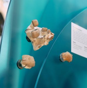
Fossils of Ptychodus teeth in the new display

Until recently, Ptychodus remained something of a mystery: teeth and vertebrae were the only remains scientists could use to work out what it looked like. Its robust, low-domed teeth are covered in deep ridges of enamel that created a mosaic in each jaw. They were capable of crushing shelled prey like a giant nutcracker. Similar teeth in modern sharks are found only in slow-moving species that prowl the seabed, so it was presumed that Ptychodus behaved in the same way.
But then in 2024 an incredible fossil with much of the animal’s soft tissue preserved was published by Dr Romain Vullo and co-authors. This new fossil revealed clues to the shapes of the fins and their position on the body. The study concluded that Ptychodus was in fact not a bottom-dwelling animal at all, but much more likely to be a fast, agile predator living higher in the water column, and perhaps related to the Great White Shark.

Armed with this recent discovery, we decided to create a three-dimensional model of the reimagined Ptychodus for our Cretaceous display. It had to be a quarter of life-size to fit it in the showcase, but would still be a huge two metres long.
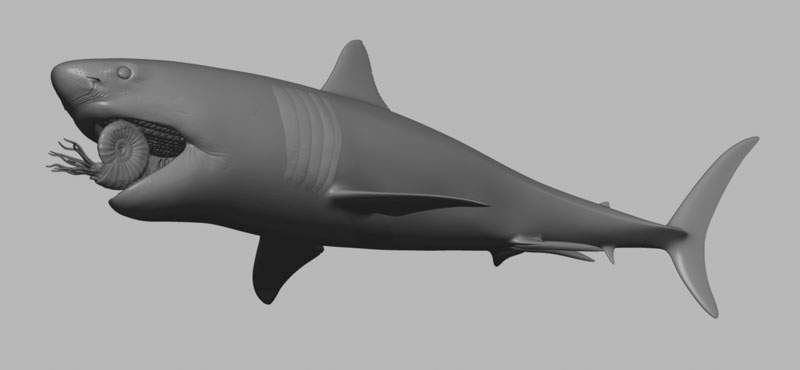
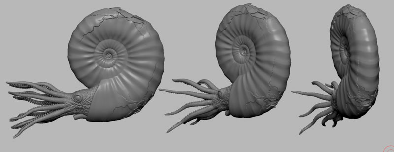
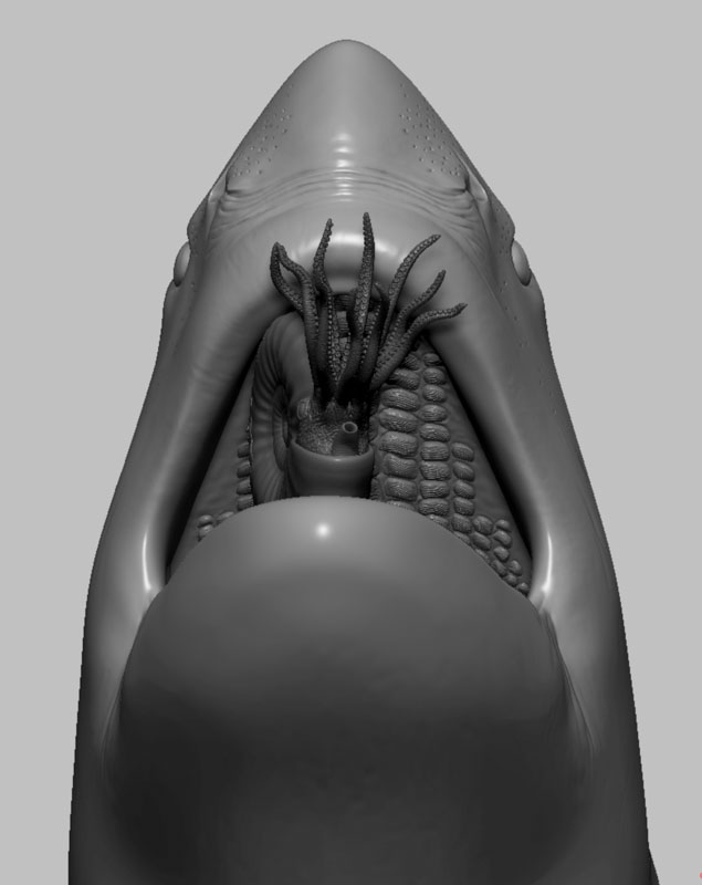
Digital 3D model of a shark, shown in grey, without any skin colouring or texture. The mouth is open and inside is an ammonite being crushed by the shark's plate-like teeth.
An early computer model of the shark and ammonite

“Working with Dr Vullo and others we began to decipher the shark’s possible appearance,” says Emma.  “Aspects to sink our teeth into included the skull shape, how it would have fed on hard-shelled free-swimming prey such as ammonites, how the mosaic of teeth sat in the mouth, as well as where and what shape the fins would have been.”

The next stage was to turn these ideas into a realistic model, and for this we turned to specialist model maker Jeremy Hunt. “When I was asked to make the reconstruction of Ptychodus I thought it was a great opportunity to incorporate modern digital techniques and processes that would give great accuracy, but also and flexibility so that adjustments could be made at later stages. This would be really difficult using a traditional clay sculpting approach,” he explains.

Together, Emma and Jeremy began to translate the fossil research into draft models on the computer, digitally sculpting versions on-screen. As well as being scaled down to quarter size, the position and pose of the model also needed to work inside the showcase structure and the designs of the rest of the display. Combining computer models helped us to make sure that Ptychodus would fit perfectly and that its method of support could be worked out well ahead of installation.
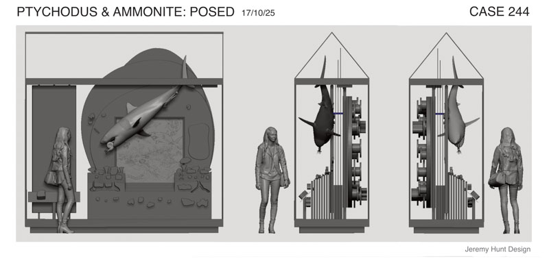
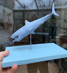
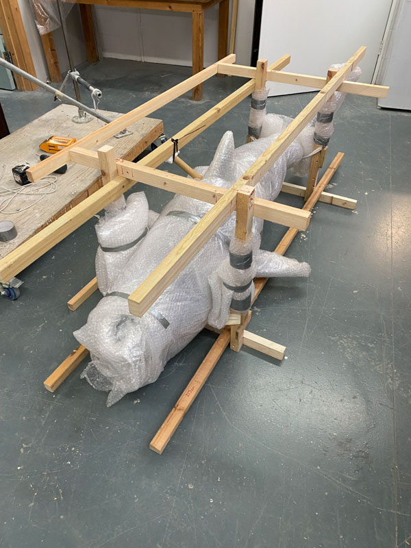
To give us a sense of how the final model would actually look, Jeremy 3D-printed little maquettes just a few centimetres long. On these we could check that the anatomy and pose were just what we needed. The maquettes also showed how the shark would be suspended in a diving angle, chomping down on an ammonite which Jeremy was modelling separately.

A hand holds out a wooden plinth, painted blue, onto which a 3D printed shark model is mounted on a vertical metal rod. The 3D print is also blue and is approximately 15cm  long.
3D-printed maquettes of the model helped us to check its form and pose

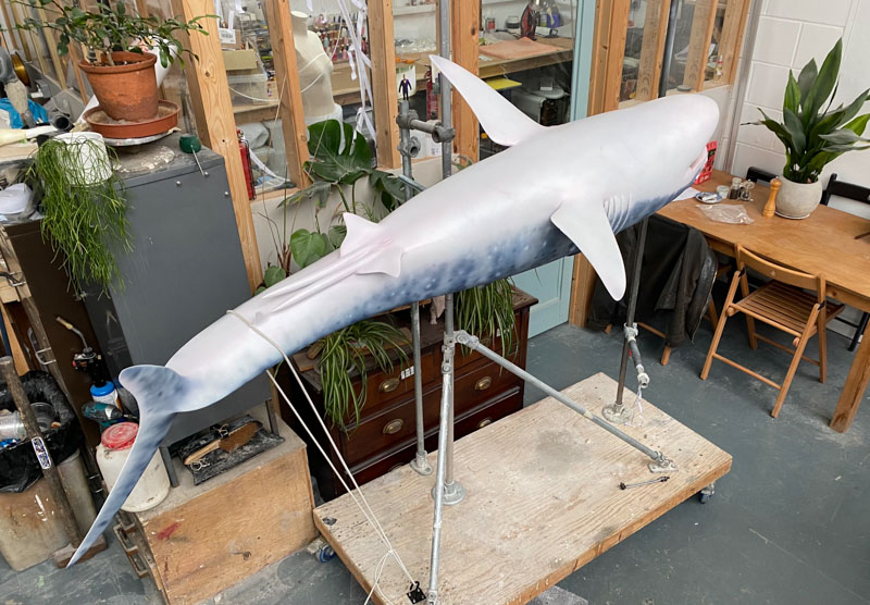
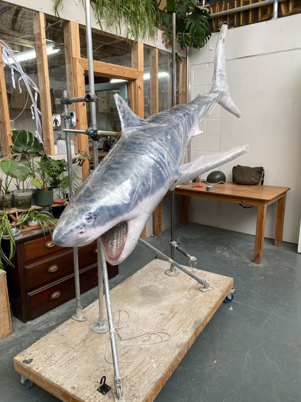
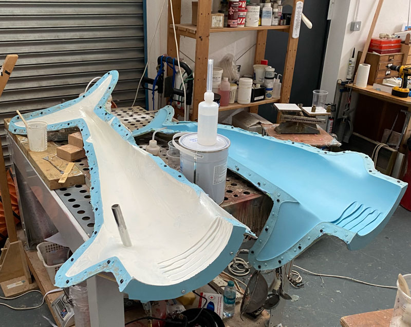
Once ready, the model was 3D-printed at its display size of two metres long, allowing Jeremy to create a silicone rubber mould to produce a fibreglass cast of the final structure. Fibreglass is both strong and light, perfect for the display. But the model really came to life when Jeremy hand-painted and airbrushed its colourings and markings based on a research by Emma and a concept drawing by palaeo-artist Julius Csotonyi.
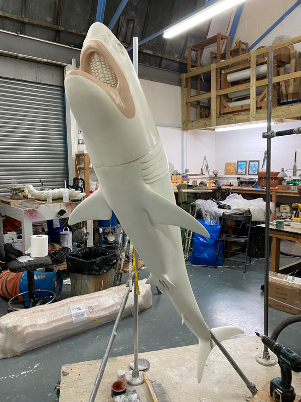
Once Ptychodus and the rest of the display were installed, the ammonite was placed in the mouth. It is held safely there by hidden strong magnets, caught permanently between those crushing plates, about to become shark food.
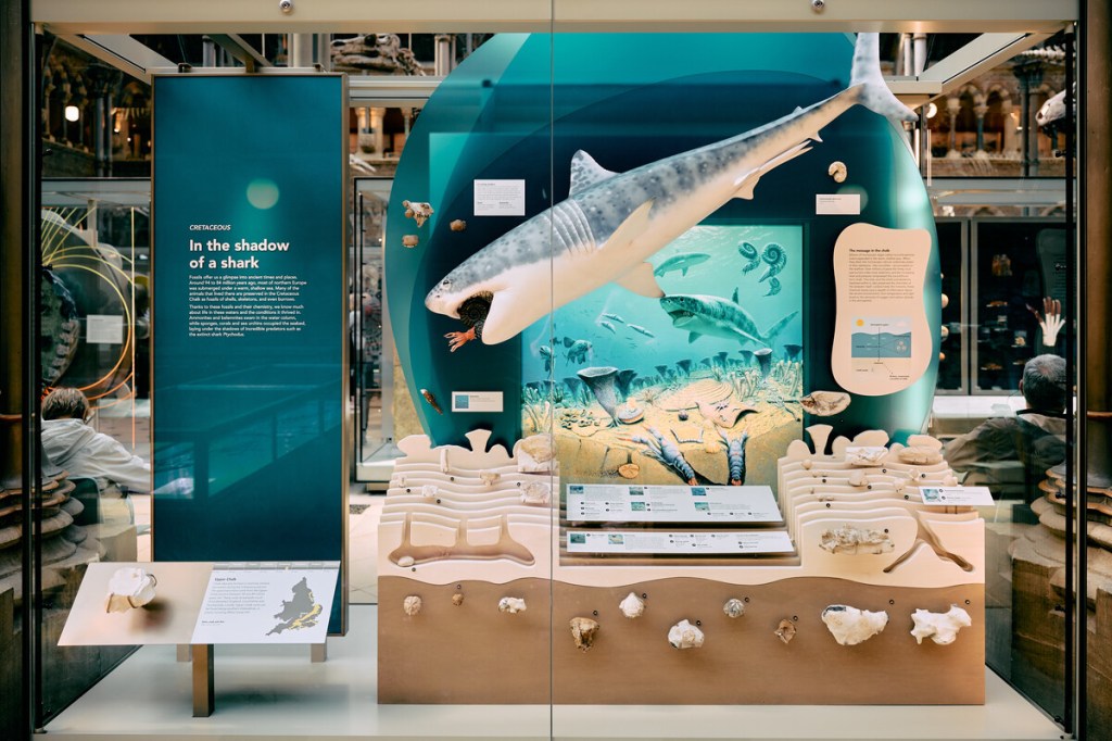
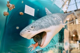
Museum exhibit showing a 2 metre long shark model against a backdrop artwork of an ocean containing drawings of other sharks, fish, ammonites and belemnites in the sea, along with corals, shellfish and lobsters on and burrowing into the seabed. Below this scene are a selection of chalk fossils. On the left the title panel is printed on blue acrylic and shows the display's name as 'In the shadow of a shark'.
The Ptychodus model in the final display at the Museum

“This was a great project to be involved with, and I hope the new displays will be enjoyed by all who visit the Museum,” says Jeremy.

Jeremy Hunt, model-maker

*

See more of Jeremy’s work at: www.instagram.com/memsculpt
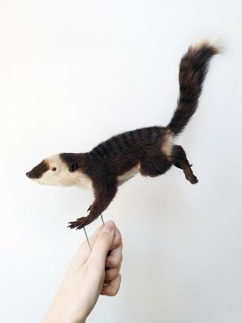
From Fossil To Flesh: Reconstructing a Phascolotherium

Following the completion of our Life, As We Know It redisplay project earlier this year, we are taking a look at some of the exceptionally skilled people who helped to bring it all together. In this article, taxidermist Alexandra Rose Cherrett tells us about the ‘unusual proposal’ that she received one day…

I was sitting at my studio desk ready to start the day with some admin. I often receive emails enquiring about commissions ranging from peoples’ pets to wild animals that have been found by the public, asking for them to be preserved after death through taxidermy. At the top of my inbox was an email with an “unusual proposal” from the Museum of Natural History in Oxford.

As a taxidermist I have been lucky enough to work on a wide range of animals over my short career, from kingfishers to vultures, hedgehogs to lemurs and many more in-between. Safe to say I was not expecting the request to taxidermy an animal that hasn’t walked the earth for 165 million years.

I was then introduced to Phascolotherium; a small, furry mammal that lived in the Mid Jurassic alongside Megalosaurus, the first dinosaur ever named and discovered, right here in Oxford, UK. A proverbial stone’s throw from the museum itself.

Now, I have always been rather obsessed with dinosaurs. My obsession and adoration for dinosaurs and wildlife has been evenly matched for as long as I can remember. Suddenly I found my Jurassic Park dreams were coming to life. An impossible task? I thought, “Life, uh…finds a way”.

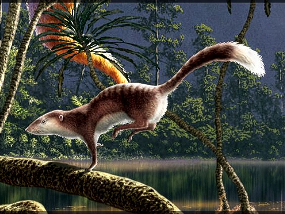
The first challenge was that only the jawbone and teeth of Phascolotherium had ever been discovered. I was given a reference image by Paleo artist, Julius Csotonyi, from which I primarily  worked.

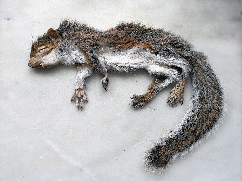
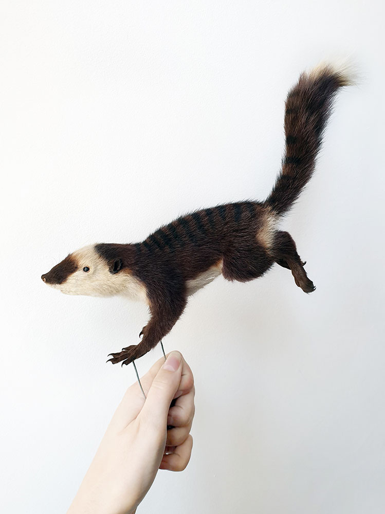
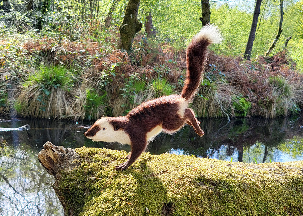
It was rodent-like in nature, with an elongated head, a lithe body and a long, plumed tail. My first thought was to use a squirrel as the base as the limb structure and body shape were so similar. I knew I had a pair of juvenile grey squirrels in my freezer that could be the perfect size, one was donated as a cat kill and the other perished at a wildlife rescue.

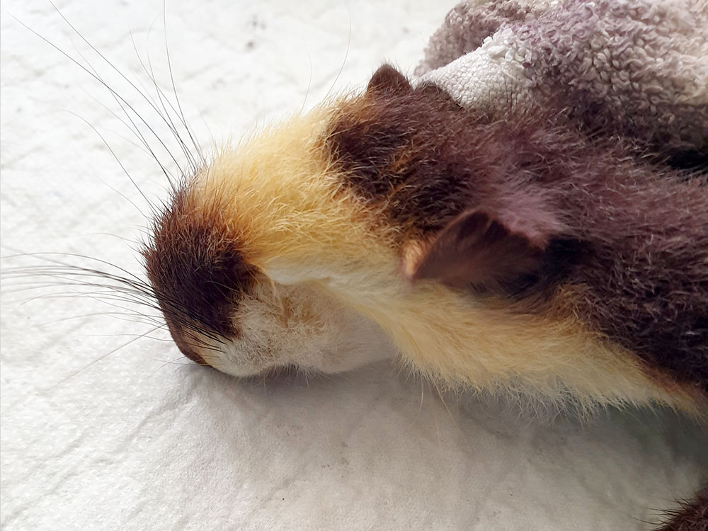
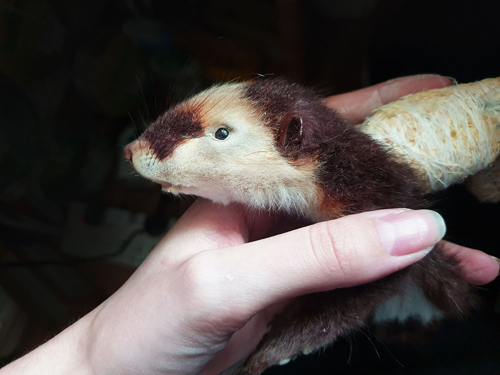
The most obvious visual changes were the colouration. The artworks showed a body of a dark reddish hue, with patches of white on the face, stomach and tip of the tail and stripes stemming down the spine. My next step of course, was Boots. A few boxes of Garnier hair dyes and peroxide bleach later and I had a completely different animal skin laid out on my desk.

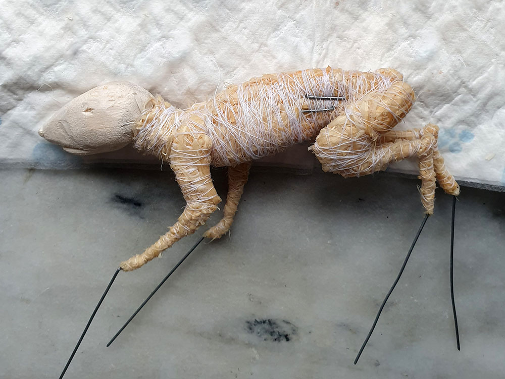
My next process was to create the body form over which the skin would be fitted. For mammal taxidermy, I usually carve or cast a head out of balsa wood or PU foam and make a bind up copy of the body and limb shape from woodwool.

With the head essentially being a non-existent shape, I copied the paleo-art and carved a small balsa block to a similar shape as a large shrew. The body was very similar to the young squirrel already so that was a simple workaround.

With the nature of its surroundings and the looming Megalosaurus, I imagined this creature was indeed, always on the run. So, an action pose was created, mid run, which highlighted its slight build and balancing tail very nicely.

When dressing the skin, the biggest alteration was the eye and ear placement. The young squirrel had eyes around 8mm, whereas Phascolotherium measured in at 4mm. Not to mention they were set further back and lower, as were the ears at two thirds their original size and shape. I made an incision across the eyelid and manipulated the skin to fit its new size and position, copying the same methodology for the ears. Almost instantly, a face that hadn’t been seen for 165 million years was right there looking back at me.

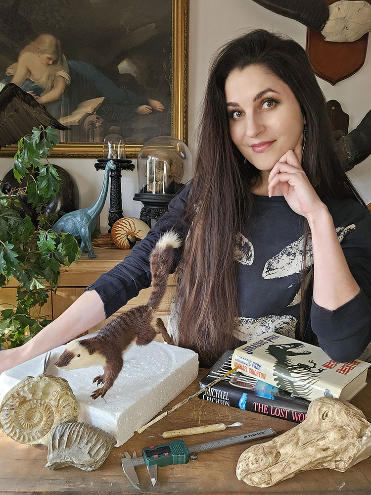
Me with the completed Phascolotherium

I have been fortunate enough to work with the Museum of Natural History on a few projects now and it’s safe to say this project has been one of my biggest challenges and greatest pleasures to date.

www.alexandrarosetaxidermy.co.uk

Instagram: @alexandrarosetaxidermy

Facebook: @alexandrarosetaxidermy

Presenting… Christmas Island

By Eileen Westwig, Collections Manager in the Museum’s Life Collections.

About 320 km south of Java in the Indian Ocean lies Christmas Island. Although discovered and named on Christmas Day in 1643, the island remained unexplored until its first settlement in 1888, a development which had dire consequences for some of its native species.

Christmas Island is home to a variety of endemic animals such as rats, land crabs, butterflies and many birds. The accumulation of bird droppings over thousands of years made the island rich in phosphate, and the commercial potential of these deposits brought many expeditions to the island. With the ships’ cargo came black rats.

Two species of endemic rats, Maclear’s Rat (Rattus macleari) and the Bulldog Rat (Rattus nativitatis) went extinct within 20 years of settlement, despite having been previously very numerous on the island.

One of the skins of Maclear’s Rat (Rattus macleari) collected by H.E. Durham, and now held in the Life Collections of the Oxford University Museum of Natural History.

Maclear’s Rat, seen at the top of the page in an illustration from an 1887 publication, was described as chestnut brown above, with a partly white, long tail. It was once the most numerous mammal on the island ‘occurring in swarms’. The Bulldog Rat had a much shorter tail and a layer of subcutaneous fat up to 2 centimetres thick, the function of which is unknown to this day.

The likely cause of their extinction was the introduction of diseases by the ship rats, to which the Christmas Island rodents had no immunity. The disappearance of the native rats also had a knock-on effect: the parasitic Christmas Island Flea (Xenopsylla nesiotes) depended on the rats as hosts, and so the fleas became extinct with the rats’ demise.

In 1901 Dr. Herbert E. Durham, a British parasitologist investigating the origins of beriberi disease, led an expedition to Christmas Island. During his visit he collected several specimens of Maclear’s Rat, but was unable to find any Bulldog Rats, despite a lengthy search and the offer of a reward. Two of the nine Maclear’s Rats Durham obtained showed abundant parasites, trypanosomes, in their blood.

Christmas Island possesses quite a number of peculiar species in its fauna, and it is regrettable that observations were not made before animals had been imported to this isolated station, as well as that my own notes are so incomplete.

Dr. Herbert E. Durham

Durham also found blood parasites in the native fruit bats (Pteropus melanotus) but noted that these were unlikely to have been introduced, instead were “an old standing native occurrence.” These bats still inhabit various islands in the Indian Ocean, including Christmas Island, where they are critically endangered.

Original letter by H.E. Durham offering his Christmas Island specimens to the Museum in 1938.

The Museum holds a range of material from Christmas Island, including six skins and three skulls of Rattus macleari, which were collected by H. E. Durham in 1901-02, and donated in 1938.

Visit the Museum’s Presenting… case between now and 6 March to see Christmas Island specimens from the collections.

Deal or no deal

Melinopterus

by Darren Mann, Head of Life Collections

In a previous article on this blog I reported the discovery, in an insect collection, of the 21st British specimen of the ‘Regionally Extinct dung beetle Melinopterus punctatosulcatus. And since then, I’ve been on the hunt for more…

Heading out to numerous other museum collections I discovered more specimens, all collected in the same locality – Deal in Kent. In Ipswich Museum there are six, collected by C. Morley in 1896; there are two in the Natural History Museum, London, collected by G.C. Champion; and in the Museum of Zoology, Cambridge there are two collected by G.C. Hall in 1883.

Ipswich Museum
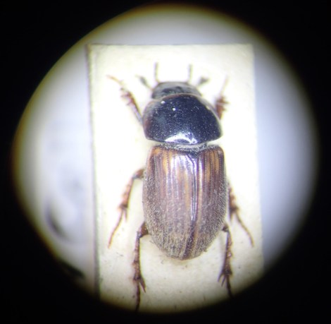
A view through the microscope of Melinopterus punctatosulcatus held in the collections of Ipswich Museum, collected by C. Morley in 1896

But the earliest and most recent finds are both in the National Museum of Scotland – one from May 1871, in the G.R. Waterhouse collection, and one from 1923, in the T. Hudson-Beare collection. So now we know of 42 specimens of this beetle with data and we know that the species occurred at Deal for about 50 years. But why are there no records after this time?

The Deal sandhills in Kent were famous for their insects, but even as long ago as 1900 entomologists* were discussing the negative impact of “summer camping-out stations and the modern craze for the ‘Royal and Ancient Game of Golf'” on beetles and butterflies in the area.

Today, most of the sandhills are gone and there are no grazing animals other than a few rabbits. Most of the surrounding land is either developed as a golf course or under agricultural management. So, is the possible local extinction of this dung beetle due to habitat loss and a lack of dung?

Deal
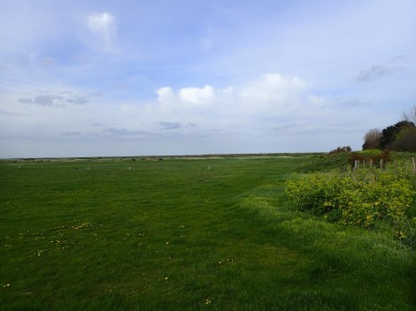
Deal, Kent: the original locality for Melinopterus punctatosulcatus, with remnants of the sandhills in the distance

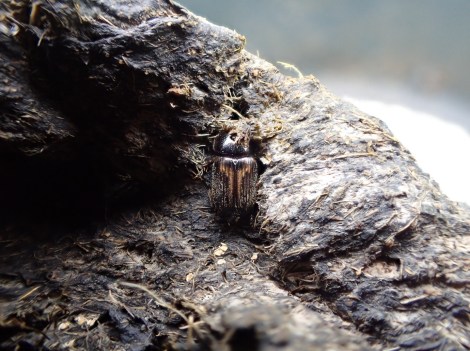
To try and answer this question, naturally I went looking for poop in Deal. In a field in Sandwich Bay I could hear sheep bleating in the distance, although poo was scarce. Eventually I found a few old plops and inside were ten Calamosternus granarius, a small dung beetle. This was good, but my main target was Melinopterus punctatosulcatus.

Melinopterus punctatosulcatus edit
A specimen from the Museum of Melinopterus punctatosulcatus, previously listed as ‘Regionally Extinct’ in Britain, but now rediscovered in Deal, Kent

I probed the poop further. To my delight, crowded in the remaining squishy bit were four other species. On close inspection, one of these was hairy, so a male, and much darker than its close relatives. It fitted perfectly with my expectations for Melinopterus punctatosulcatus after seeing so many examples in museum collections. Success! This beetle, misidentified in museum collections for so long, and not seen since the 1920s in Deal, is indeed hanging on in Kent.

Disappointingly, after a further few days of searching, only a handful more specimens were seen. This suggests that either the species exists at low population levels, or that it was it was not the peak emergence period when I was there. Nonetheless, a species not recorded anywhere in the UK for over 70 years is actually still here.

Now hopefully we can encourage local land owners to help conserve this all-important dung fauna and flora.

* Walker, J.J. 1900. The Coleoptera and Hemiptera of the Deal Sandhill. Entomologist Monthly Magazine 36: 94-101.