Doctoral researcher Elaine Charwat is exploring the value and meaning of models and casts in the Museum’s collections as part of her PhD. She has recently been studying some fabulous models that help to visualise and understand some of the very, very smallest of specimens…
By Elaine Charwat
The first time I encountered a Radiolarian was in a book – Ernst Haeckel’s (1834-1919) weird and wonderful Kunstformen der Natur (Art Forms in Nature, 1899-1904). It took comparative morphology – comparing the shapes of organisms – to new giddy heights, scientifically, philosophically and artistically. I felt that giddiness when looking at page after page crammed with crustaceans, orchids, hummingbirds, moths and even bat faces, all exquisitely arranged to celebrate their symmetries, the evolution and kinship of their shapes and forms. It also made visible organisms that are normally all but invisible.
Illustration of Cyrtoidea (table 31) from Kunsterformen der Natur (1899 – 1904) by Ernst Haeckel. By permission of the Linnean Society of London.
Foraminifera and Radiolarians are microscopic sea-dwelling organisms. Species may be found as fossils dating from Cambrian times, ca. 500 million years ago, right up to living specimens today.
To Haeckel, they were living proof of Darwin’s theory of evolution, and for his own belief that morphology was the key to understand the actual processes of evolution, catching it in the act. However, these organisms had two big disadvantages – their unwieldy taxonomy, or the way they are classified, and their minute size: they were difficult to examine and display.
Illustrations of Radiolarians, (table 28). from Die Radiolarien (1862) by Ernst Haeckel. By permission of the Linnean Society of London.
Through his illustrations, Haeckel widely popularized them – triggering a Victorian craze for microscopes and microorganisms, as well as influencing art nouveau art and architecture. But there were limits to what an illustration could communicate. Models stepped in, representing these organisms in ways illustrations could not.
Detail from Haeckel’s Kunstformen der Natur (1899 – 1904)
One defining feature of Radiolarians and Foraminifera is their shells – called “tests”. Variations in shapes of the tests not only indicate that they are different species, but also, excitingly, provide clues about space and time. The tests of Neogloboquadrina pachyderma, for instance, record ocean temperature over geological timescales – their shells coil to the left when water temperatures are relatively cold, and to the right when it is warmer. The potential for research into climate change is obvious. Foraminifera are also important “signature fossils”, helping geologists to determine geological strata.
You really need to see them in glorious 3D to appreciate these tests across geological time, to understand their complex, beautiful shapes. And I felt a similar twang of excitement to my first encounter with them through Haeckel when discovering these extraordinary models here in the Museum as part of my PhD research.
Václav Frič (1839-1916) was a natural history dealer based in Prague. He developed a series of 100 plaster of Paris models of Foraminifera (1861), as well as the stunning papier-maché models of Radiolaria (listed in his catalogue of 1878). He worked closely with Ernst Haeckel.
A selection of Frič’s models in the Museum’s stores
The Frič models oscillate between visible and invisible, illustration and model, art and science, philosophy and theory. They bear witness to a key period in the history of science when they were used to give tangible shape and proof to Charles Darwin’s poignant phrase: “[…] from so simple a beginning endless forms most beautiful and most wonderful have been, and are being, evolved.”
Through the models we can “grasp” microorganisms that have been around for over 500 million years; organisms that truly have stood the tests of time.
Real or fake? Do replicas have a value of their own? Elaine Charwat is exploring this in her PhD, using the Museum’s large collection of natural history models and casts to research their role in science. Here she tells the story of the fascinating fish that caught her imagination…
By Elaine Charwat
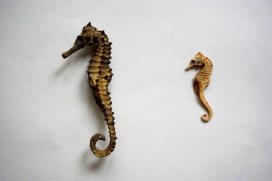
It all started with a seahorse. Last year, I walked into a little seaside shop, and I spotted a seahorse. I instantly flipped back to the happy day I bought my first dried seahorse as a child, the beginning of a life-long passion for the natural world. The man behind the counter smiled: “It’s a fake.” Really? “3D printed.” It looked absolutely perfect. Tracing its lines with my fingers, I said, “It’s a model”.
Ever since I became interested in models and replications, I have encountered this perception of them as “fakes”. Quite recently, I heard the curator of a natural history museum call the cast of a dinosaur skeleton a “fake”. Models in natural history – and in this I include casts and reproductions – are what the Germans call “Wissensdinge”, objects that contain, distribute and generate knowledge. In this aspect, the real specimen and the model meet. Models are made from a vast array of materials with often astonishing skill and technologies. They represent what we know about a particular organism at a certain point in time. They have a history, a context.
Long live the replica! Most of our most beloved dinosaur skeletons in museums are partly or fully casts of bones, like Stan’s here at the Museum. Almost complete skeletons like Stan’s are extremely rare, and casts allow us to share and preserve them. Accompanying models give the bones “flesh and blood” – and provide a snapshot of what was known about the dinosaur when the model was made.
But they are also ambassadors, and this is something I realised when I held the “fake” 3D-printed seahorse in my hand. While it becomes ethically problematic to buy specimens of organisms like seahorses, something of it is captured, and communicated, in a reproduction. I can still trace its exoskeleton, and marvel at its strange symmetry. This symmetry, incidentally, is being analysed for its potential in robotics. Seahorses have unusual tails – instead of the cylindrical trail structure found in most animals, theirs have a square cross-sectional architecture, resulting in a unique combination of toughness and flexibility. In fact, when studying the unique abilities of the seahorse’s tail, researchers have actually used 3D-printed specimens.
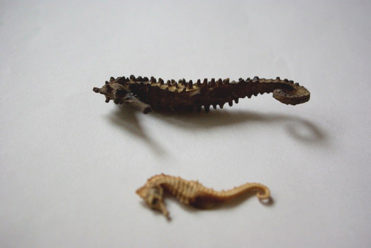
Seahorse from the Museum’s collection. Even in Victorian times, long before 3D printers, there seems to have been a desire to emphasise that souvenir seahorses were “natural” – i.e. not man-made. Was it because seahorses are easily preserved and so attractive when dead and dried?
The Oxford University Museum of Natural History has a largely unexplored wealth of models and casts. Many of them date to the second half of the 19th Century, the heyday of their production. Made from glass, wax, metal, wood, plaster, papier-mâché or, indeed, actual bone and feathers, they were modelled, cast, sculpted, glued, painted and mounted to enhance and preserve our understanding and appreciation of nature. But they also tell of scientific discoveries and controversies, research and teaching, rivalries and collaboration, politics and society, ideas and identities.
Spot the replica – both the specimen and the 3D printed seahorse are “Wissensdinge”, they have a context and provide valuable information.
I will trace these complex relationships in a collaborative and interdisciplinary PhD project called “Nature of Replication”. This is funded by the AHRC and jointly supervised by the Institute of Archaeology, University College London, and the Oxford University Museum of Natural History.
The 3D-printed seahorse now lives alongside my real seahorse. So I like to think of my project as a journey that started with one seahorse, and continues with another.
When working on the dissertation for my MSc in Archaeological Science last year, I explored the medieval craftsmanship of sealing wax. I was interested in the way the medieval wax seals had flaked, as the beeswax dried out. Drawing on my previous education in conservation techniques, I began a close investigation of the prestigious material, beeswax.
Medieval craftsmen used a range of dangerous materials to make sealing wax. The red pigment cinnabar, a mercury (II) sulphide, and red lead, are now known to be extremely poisonous.
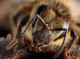
Although some of the ingredients of sealing wax are very hazardous, there is nothing dangerous in beeswax… except the bees! Produced by honey bees, Apismellifera, honey and beeswax were important commodities in the Middle Ages. Beekeeping was a skilful profession, housing colonies in woven hives, known as skeps. Colonies were carefully selected to overwinter for the next season.
Manuscript illuminations provide detailed information on the types and construction of beehives in the Middle Ages.England, 13th century. British Library Royal 12 C XIX f. 45.
Beeswax was also important in the Middle Ages for lighting, and beeswax candles were preferred for their pleasant smell. After the Protestant Reformation in the 16th and 17th centuries, the religious use of candles decreased, so demand for beeswax declined.
Even today, the Catholic and Orthodox Churches still require the candles they use to contain a proportion of beeswax.
On my quest to understand the degradation of beeswax in sealing wax and write my disseration, I was very lucky to use some samples from the entomological collections from the Oxford University Museum of Natural History. After some early mornings spent amongst the Westwood collection, I found the perfect specimens of natural honeycombs, from the 19th century. The old hand-written labels were also a lovely encounter when exploring the historical collections.
I compared the samples to modern beeswax and medieval seal samples, and learned that the degradation of beeswax is caused by multiple factors, triggered also by storage conditions. The composition of beeswax is very complex, and there are differences caused by the age of the bee in addition to geographical provenance.
A selection of bee specimens from the Museum’s collection.
The recent catastrophic decline of bee populations has drawn focus to save the bees, and in my PhD research (University of Copenhagen and University of Cambridge) I will explore the recovery of ancient DNA and proteins of bees from beeswax, to cast light on the health of bee populations over time.
In our Bacterial World Science Short event series, researchers present their latest findings related to themes in the exhibition. At a recent Science Short, Hannah Behrens, a University of Oxford PhD student, explained how bacteria become resistant to antibiotics and how the species-specific antibiotics she studies might reduce the worrying rise in antimicrobial resistance.
Bacteria that are resistant to antibiotics present a huge problem. I work on developing new antibiotics that will slow the development of bacterial resistance.
But let’s not get ahead of ourselves. Your body is full of bacteria. In fact, there are more bacteria than human cells in your body. Most of these bacteria are good for you; they help you digest food and protect you from diseases.
But once in a while a harmful bacterium causes an infection. This could be a lung, wound, or bladder infection, or something with a fancy name like, Black Death, tuberculosis, leprosy, syphilis or chlamydia. The doctor will then prescribe you antibiotics to kill the offending bacteria.
Hannah Behrens delivers her Science Short talk at the Museum
The development of antibiotics in the 20th century was a major breakthrough. For the first time bacterial infections could be effectively and rapidly treated. Since 1942, when antibiotics first became available, we have discovered many new antibiotics which have saved millions of lives.
However, in the last 30 years we have not managed to develop any new antibiotics. During the same time, many bacteria have adapted to become resistant to the antibiotics we do have. In 2017, a woman in the US died because she had an infection with bacteria that were resistant to all available antibiotics. It is estimated that already 700,000 people in Europe alone die because of resistant bacteria per year. What is happening?
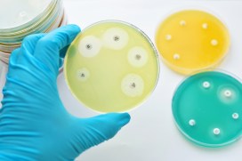
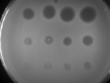
Bacteria are forming a lawn on this plate (light areas); where an antibiotic has been spotted on the bacteria they die and leave the surface blank (dark areas).
Every time we treat bacteria with antibiotics, most die, yet a few resistant bacterial cells survive. Like Rudolph the red nosed reindeer, the resistant bacteria are usually at a disadvantage until a special situation arises (a foggy night for Rudolph; treatment with antibiotics for resistant bacteria).
Under usual circumstances, producing a resistance mechanism is a disadvantage: it wastes energy and slows down growth, so very few bacteria are resistant. Only when all the non-resistant bacteria are killed by antibiotics do the resistant ones thrive. They have no more competition, and have all the resources, such as food and space, to themselves.
The more we use antibiotics, the more resistant bacteria we get. It is essential not to use antibiotics carelessly.
More antibiotics are used in animal farming than on humans. If we eat less meat, and so reduce the farming of livestock for food, we may reduce the growth of resistance bacteria. Another approach is to only take antibiotics when the doctor prescribes them. Antibiotics do not help against viral infections like colds. In many low and middle income countries, antibiotics are available in supermarkets and it is no coincidence that these countries have higher levels of resistant bacteria.
The precision antibiotics research group in the Department of Biochemistry at the University of Oxford
Apart from avoiding the unnecessary use of antibiotics, scientists – including me – are trying to develop better therapies against bacteria. I study precision antibiotics: drugs that specifically kill one species of bacteria. The advantage of this is that all good bacteria remain unharmed and only the disease-causing species is targeted. This also means that only resistant bacteria from this one species get an advantage to thrive.
I am interested in species-specific antibiotics against Pseudomonas aeruginosa. This bacterial species causes lung and wound infections and, according to the World Health Organization, is one of the three bacteria for which we most urgently need new antibiotics. Colleagues of mine tested different precision antibiotics against Pseudomonas and found one that is better than the others, called Pyocin S5.
Hannah’s painting of how researchers think pyocin antibiotics kill bacteria. The pink bacterium produces pyocins (pink balls), which enter the susceptible blue bacteria through pores (blue). The blue bacteria mistake the antibiotic for a nutrient and open the pore to let it in. Once inside the bacterium it forms a pore in the inner membrane which causes leakage of the cell contents and kills the cell.
I am now investigating how stable this antibiotic is, how it recognises this specific species of bacteria and how it enters the bacterial cells. This knowledge is important to decide on how to store, transport and administer the drug. I also hope that understanding why Pyocin S5 is more effective than the other antibiotics will allow us to design more effective, targeted antibiotics in the future.
My hope is that one day we will treat all bacterial infections with precision antibiotics and that antibiotic resistance will become a problem of the past.
Since we posted about ten-year-old Sarah’s amazing beetle discovery, we’ve had lots of queries as to why the insect needed to be caught and pinned. It’s a question we’re often asked, so here’s Darren Mann, Head of Life Collections at the Museum, to explain the value of ‘voucher specimens’.
The Museum’s collection houses over five million insect specimens, amassed over the past 300 years. This collection is, in effect, a biodiversity database, but unlike virtual databases, each data point has an associated ‘voucher specimen’ that was caught, pinned and labelled.
Although technical advances in digital macro-photography do reduce the need for some collecting, it is impossible to dissect an image to confirm an identification. So for many groups, even the best photograph in the world is inadequate for identification purposes.
Shingle CrawlerD18 (Psammoporus insularis Pittino, 2006) one of our few endemic insects.
Unlike plants and birds, many insects can only be identified with the aid of a microscope, to study tiny features that distinguish closely-related species. Some groups even require the dissection of minuscule genitalia to really tell them apart.
Entomologists take voucher specimens to enable this correct identification and these are later deposited in museum collections, making them available for further study in years to come. From an entomologist’s point of view, we believe we need to know what a species is, where it occurs and as much about it as possible, so we can inform biodiversity conservation.
The conservation assessment of UK insects by Natural England in their Species Status Reviews has only been possible with the data provided by entomologists, generated from collecting and identifying voucher specimens.
Entomologists follow a Code of Conduct for responsible collecting, which ensures they don’t remove too many species or damage the environment during their work .
There are numerous examples of the value and use of insect collections in contemporary science, including the discovery of previously unknown species in the UK and population genetics for butterfly conservation. Recently a species believed extinct in the UK was rediscovered. This was only made possible by checking the identification of several thousand museum specimens.
Museum collections also contain numerous examples of species now considered extinct in the UK. Without voucher specimens much of this research would be impossible and our understanding of insect distribution patterns, ecology and conservation would be significantly diminished.
Large Tortoiseshell butterflies, now considered to be extinct in the UK. The voucher specimens act as record in time of its occurrence in the UK.
What is rare? Sarah’s False Darkling Beetle (Anisoxya fuscula) has been described as ‘rare’, but what does that mean in reality? For most invertebrates when we talk about a rare species we are not talking about a tiny number of individuals. This conservation status is based on their known distribution and the level of threat they face. A species can be rare if it is only found at one or two locations, but at those locations there may be many thousands of individuals.
The greatest threats to biodiversity are well known and include habitat loss, fragmentation and degradation and pollution, such as pesticides and light. Taking a small number of voucher specimens to confirm the identification of species has negligible impact on its population. But if we don’t know it’s there because we couldn’t identify it, then a housing development destroys its entire habitat… well you get the picture!
This article is taken from European research magazine Horizon as part of our partnership to share natural environment science stories with readers of More than a Dodo.
Unravelling one of the most elaborate forms of non-human communication – the honeybee’s waggle dance – could help researchers better understand insect brains and make farming more environmentally friendly.
It’s part of a field of work looking at insect neurology which is helping to unravel the complexity of their brains.
Bees have evolved a unique, and ingenious, way to communicate with each other – the waggle dance. By shaking their abdomens in a particular way, a bee can tell others in its hive the specific direction and distance of a food source or a new site for a nest.
‘If nectar or pollen is in the direction of the sun, a bee will run a figure of eight that is orientated towards the top of the hive. If pollen is found 90 degrees from the sun they will point that way instead,’ explained Dr Elli Leadbeater, a bee expert from the School of Biological Sciences at the University of London, in the UK.
The longer the bees spend dancing corresponds to the better quality of a food source, while the more time spent on each figure eight represents the distance from the pollen or nectar.
Researchers now believe that decoding this information-packed dance further could reveal a link between bees’ brains and how the surrounding environment affects them. In a project called BeeDanceGap, Dr Leadbeater is working to identify the exact genes in the bee brain that play a role in helping the insects understand this waggle dance.
To do this, researchers must first identify the best dancing bees in a test hive and watch them as they reveal a food source to other worker bees. The newly educated bees are then captured as they leave the hive so their brain tissue can be genetically analysed to determine which genes associated with learning and memory were activated from following the waggle dance.
Only a few individuals are used in this way and the genetic data provides a deep insight into the neurology of a bee’s brain – at a time crucial to their future.
The observation bee hive at the Oxford University Museum of Natural History gives visitors a glimpse into hive life.
Collapse
Beekeepers around the world have reported that many of their bees leave and never come back, causing hives to suddenly collapse. Experts believe there are several factors contributing to this widespread loss of bee colonies, including climate change, parasites and habitat loss. Agrichemicals like pesticides and neonicotinoids, which are used to kill unwanted insects on farms, have also been strongly linked to the problem.
‘The rate pesticides or neonicotinoids are applied to crops don’t necessarily kill bees but they make them worse at foraging,’ said Dr Leadbeater.
If you do damage to just one part of the brain of a lot of individual bees, it can have huge consequences for the whole colony.
Dr Elli Leadbeater, University of London, UK
Neonicotinoid pesticides have been found to bind to parts in the insect brain, disrupting neural transmission. This leads to some brain cells either failing to develop or not functioning properly.
The EU recently banned neonicotinoids, which Dr Leadbeater believes is a huge step forward in protecting bees, but she said governments still need more rigorous ‘long-term environmental safety monitoring’. Without this, there is a risk that other agricultural products used in place of neonicotinoids could impact honeybees in a similar way.
But when the first results of BeeDanceGap are published later this year, they could contribute to building better criteria for testing future agriculture practices or products. Dr Leadbeater believes it will provide a new understanding of a bee’s brain, and so help identify problems sooner.
The impacts of quickly identifying problems go far further than just supporting beekeepers and their insect charges. Protecting honeybees, along with bumblebees and wild bees, is also essential to maintain a healthy and productive environment. These insects pollinate over 80% of crops and wild plants in Europe. According to Professor Martin Giurfa, from the Research Center on Animal Cognition at CNRS in France, ‘preserving little brains is about preserving biodiversity’.
Honey bees working inside a hive
More than machines
Honeybees have a higher social complexity than many other species. Alongside the waggle dance communication, each hive has a division of labour where different workers have responsibility for a variety of tasks – such as foraging for pollen, nursing the young, building hives and even removing the dead.
Prof. Giurfa is co-leading the BrainiAnt project, which looks at how this type of complex social behaviour evolved and how it affected the structure of insect brains. He said that when ‘you understand how bees perceive the world, it is easier to find ways to protect them’.
Through the work of researcher Dr Sara Arganda, the project is investigating a part of the insect brain called the mushroom body, where learning occurs and long-term memories are stored. Researchers analysed bee behaviour and gave them memory tests, such as navigating paths using colour cues, in order to learn more about the structure of insect brains.
The project strengthened the argument that bee brains are more complex than previously thought. ‘Most findings are saying that insects are more than simple machines, which comes from studies in the honeybee,’ said Prof. Giurfa. ‘(But) the entrance region of the mushroom body shows a level of complexity and the studies show that this complexity is not rigid, it is plastic.’
This means its structure is changing all the time, which mirrors how human brains work. ‘(Bee) brains are capable of sophisticated performances such as learning concepts and rules; they are incredible organs and they need to be defended,’ said Prof. Giurfa
To further advance understanding of the mushroom bodies and how they function in different species, the project is being co-led by Professor James Traniello at Boston University in the US, an expert in ant evolutionary neurobiology.
Ants, which are related to honeybees, have brains that may be 100 times smaller, and due to their minute size, provide insights into how insect brains are structured.
‘What happens to neural tissue at an extremely small size?’ asked Prof. Traniello. ‘Are you losing neurons, are neurons becoming more efficient in their actions, how many neurons do you have to string together to form a circuit that enables behaviours as complex as what you would see in ants? How does the collective intelligence of an ant colony impact the structure of the brain?’
If BrainiAnt can answer these questions, it would provide a clearer picture of the evolution and function of ant brains.
‘The next step is trying to understand the genes that are involved in regulating brain size, compartment variability, metabolism and other functions,’ said Prof. Traniello.
He added that a better understanding of neural tissue could also help to guide attempts to genetically engineer bees so their brains are resistant to environmental threats like neonicotinoids. Although far off, it could mean that bees, and the benefits they bring to the environment, will have a more secure future.
The research in this article was funded by the EU.
*
The issue
One in ten pollinating insects is on the verge of extinction, and a third of bee and butterfly species are in decline.
On 1 June, the European Commission launched a proposal to tackle this problem at an EU level. It includes a new monitoring process to collect quality data and identify trends, action plans to protect insect habitats and incentives for businesses such as those in the agrifood sector, to contribute to conservation.
The proposal, known as the EU Pollinators Initiative, has a number of short-term actions to be taken before 2020, at which point the progress will be reviewed.


 Since we posted about