Secrets of an ancient reptile

Fossil of Eusaurosphargis dalsassoi PIMUZ A/III 4380 (Credits: Dr. Torsten Scheyer; Palaeontological Institute and Museum, University of Zurich, Switzerland)

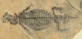
Very occasionally, exceptionally well-preserved fossils reveal new clues about poorly understood creatures. Complete, articulated skeletons are rare and, when found, offer rich insight for palaeontologists. One of our research fellows, Dr James Neenan, has been involved with just such a discovery and description, of an armoured reptile from the Middle Triassic named Eusaurosphargis dalsassoi.

A beautifully-preserved fossil found in the Alps in eastern Switzerland has revealed the best look so far at this animal. The findings about its anatomy and probable lifestyle were somewhat unexpected, according to a paper published in Scientific Reports today, led by Dr Torsten Scheyer at the University of Zurich and co-authored by James.

At just 20 cm long, the specimen represents the remains of a juvenile. Yet large portions of its body were covered in armour plates, with a distinctively spiky row around each flank, protecting the animal from predators. Today’s girdled lizards, found in Africa, have independently evolved a very similar appearance even though they are not closely related to Eusaurosphargis.

Life reconstruction of Eusaurosphargis dalsassoi based on new specimen PIMUZ A/III 4380 (Credits: Beat Scheffold; Palaeontological Institute and Museum, University of Zurich, Switzerland).

The new fossil, found in the Prosanto Formation at Ducanfurgga, south of Davos in Switzerland, is not the first material of Eusaurosphargis to be discovered. The species was originally described in 2003 based on a partially complete and totally disarticulated specimen from Italy. This was found alongside fossils of fishes and marine reptiles, leading scientists to believe that Eusaurosphargis was an aquatic animal.

However, the detail preserved in the new specimen shows a skeleton without a streamlined body outline and no modification of the arms, legs or tail for swimming. This suggests that the reptile was in fact most probably adapted to live, at least mostly, on land, even though all of its closest evolutionary relatives lived in the water.

Until this new discovery we thought that Eusaurosphargis was aquatic, so we were astonished to discover that the skeleton actually shows adaptations to life on the land. We think this particular animal must have washed into the sea from somewhere like a beach, where it sank to the sea floor, was buried and finally fossilised. – Dr James Neenan

The findings from the research team are published in Scientific Reports as ‘A new, exceptionally preserved juvenile specimen of Eusaurosphargis dalsassoi (Diapsida) and implications for Mesozoic marine diapsid phylogeny’.

The Big Brain Competition

What happens in your brain when you receive compliments? And what’s going on in your mind when you watch your football team win a match? Does the brain respond differently when recalling music, compared to listening to it? All these questions, and more, have been posed in our Big Brain Competition

Coinciding with the Museum’s Brain Diaries exhibition, the Wellcome Centre for Integrative Neuroimaging is inviting you to ask your own question about the brain to be in with a chance to have it tested by neuroscientists using Oxford’s state-of-the art Magnetic Resonance Imaging (MRI) scanner.

The advanced MRI scanner at the John Radcliffe Hospital in Oxford is one of the strongest in the world. It allows scientists to carry out functional MRI (fMRI) scans to see the brain in action. This mind-blowing procedure can reveal how the brain changes when learning a new skill or how it compensates when someone recovers from brain damage. It can also reveal which areas are used when people speak, move or laugh, to give just a few examples.

This fMRI scan shows how blood flows to the visual cortex region at the back of the brain when viewing a visually-stimulating checkerboard pattern
layouttile
Dr Stuart Clare of the Nuffield Department of Clinical Neurosciences is asking you for questions about the brain

Functional MRI shows when a brain area is more active by detecting the changes in blood oxygen levels and blood flow that happen in response to neural activity. The technique can be used to produce activation maps showing which parts of the brain are involved in a particular mental process.

The scientist behind the Big Brain Competition is Dr Stuart Clare, whose research involves pushing the technological boundaries of the fMRI technique to reveal new insights about how the brain functions normally and how it is affected by disease. There is still so much that the fMRI scans can bring to light, so Stuart is asking you for ideas!

Over several years of inviting people in to see the beautiful pictures that our MRI scanner can produce, I’ve been fascinated by the questions they have about the brain and whether you can see this thing or that thing in our fMRI scans.  With this competition we want to give people the unique access to our scanner and the chance to try an idea out for themselves.

When coming up with an idea for investigation there are a few practical things to bear in mind. Any activity has to be something people can do when lying down in the scanner and it has to be clear when they start and stop doing the activity. But Stuart is very open to ideas for experiments that they haven’t come across before – something that scientists really don’t already know the answer to.

The animation below explains how fMRI works and what it can do. So take a look, think up an experiment of your own and enter your idea via this form. The best one will be put into action by the research team and you will be able to watch the scans take place at the John Radcliffe Hospital yourself!

Credit: Mike Peckett

Great minds don’t think alike

Credit: Mike Peckett

Museums are a place for many things: inspiration, learning, conservation… the list goes on. But we believe that they should also be a forum for debate and discussion. One of our aims, as part of our Contemporary Science and Society exhibition and event series, is to bring controversial or challenging ideas to our visitors and to encourage a lively, informed and balanced debate.

Thomas Henry Huxley

Controversy is nothing new to this institution; there is a history of debate going right back to 30 June 1860, the year the Museum was founded. The Great Debate is believed to have been the first ever public debate on Charles Darwin’s theory of evolution – certainly the thorny issue of the time.

The debate is now notorious for the clash of ideologies between Samuel Wilberforce, the Bishop of Oxford, and Thomas Henry Huxley, a young biologist known as ‘Darwin’s bulldog’.

Samuel Wilberforce

Reports from the time are a little sketchy, but tempers are believed to have flared and insults were traded, climaxing with the shocking moment where Wilberforce compared Huxley’s grandparents to an ape. This was obviously outrageous to the delicate Victorian temperament, and people were believed to have fainted with shock!

This short video reveals how we think the debate may have gone (with a little artistic license thrown in):

The anniversary of the Great Debate falls next week, and this year it’s an extra special one. We’re reigniting the tradition of a good lively discussion with The Great Debate: Smart Drugs – Is It Cheating? On Thursday 29 June, Claire Fox of the Institute of Ideas and BBC Radio 4 chairs a multidisciplinary panel as in a debate about the ethics, fairness, and effects of so-called smart drugs and their impact on society.

Smart drugs is a name given to prescription drugs, typically used to treat disorders such as narcolepsy and attention deficit hyperactivity disorder (ADHD), which are now also commonly being used to improve cognitive ability and concentration. Some studies suggest that these drugs are now widely being used by university students, in a climate of increased academic and financial pressure.

Many students are said to see these drugs as a normal aid to study, but some experts have serious concerns about increasing levels of self-prescription and the long-term safety of their use, as well as the impact on competitiveness. Increased use in other areas of society may also have implications.

The panel for this debate includes world experts in the fields of neuroethics, evolutionary psychology, and philosophy, each representing different sides of this challenging subject.

Tickets are free, but you need to book your place. We can promise controversial opinions, expert insights and an eye-opening evening, but unlike Wilberforce and Huxley, we can’t necessarily guarantee that the panelists will be flinging insults about each other’s grandparents.

Comedy rears its ugly head

Logo blob fish
Is the Blobfish the ultimate ugly animal?

Animals with big eyes, fluffy tails and cute noses are easy to love. But what about those with tentacles, slimy skin or a large throat pouch?! Luckily the Ugly Animal Preservation Society is here to fight their corner. Education Officer Chris Jarvis shares some tales from a special event…

Comedy met conservation on Saturday night, as the Museum teamed up with Berkshire, Buckinghamshire & Oxfordshire Wildlife Trust, compere Simon Watt and the Ugly Animal Preservation Society. During the evening, six comedians put the case for their own neglected animals in need of conservation: the Scrotum Frog; Christmas Island Frigate Bird; Kaluga Sturgeon; Slimehead; Okapi, and Yak. The audience then voted to choose the official mascot for Oxford, but which of these lovely uglies did they choose?

The Scrotum Frog

Par Samuel Garman (1843-1927) [Public domain], via Wikimedia Commons
Image: Samuel Garman via Wikimedia Commons

Chosen by Iszi Lawrence, this unfortunately-named amphibian’s population in Lake Titicaca is now dwindling. It’s threatened by the introduction of alien salmon for the angling fraternity and is also blended to make a medicinal frog frappe by Andean locals with strong stomachs!

Christmas Island Frigate Bird

By Charlesjsharp (Own work) [CC BY-SA 3.0 (http://creativecommons.org/licenses/by-sa/3.0)], via Wikimedia Commons
Photo: Charlesjsharp via Wikimedia Commons
Some may find its inflatable throat pouch off-putting (in the same way the Scrotum Frog brings certain disturbing images to mind), but not advocate Eleanor Morton. This critically endangered seabird is one of a family of only five species that grace our oceanic skies and deserves our attention.

Kaluga Sturgeon

By User:Cacophony [GFDL (http://www.gnu.org/copyleft/fdl.html), CC-BY-SA-3.0 (http://creativecommons.org/licenses/by-sa/3.0/) or CC BY 2.5 (http://creativecommons.org/licenses/by/2.5)], via Wikimedia Commons
Photo: Cacophony via Wikimedia Commons
Hideous and aggressive, these formidable fish grow to a ton in weight and have suffered a population decline of 80% over the last 90 years: we humans like to eat their eggs and their spawning grounds are now severely polluted. Without action we may lose one of the largest fresh water fish in the world. Kaluga Sturgeon champion Paul Duncan McGarrity made the case for this fugly fish.

Slimehead

By Pengo (Own work) [CC BY-SA 3.0 (http://creativecommons.org/licenses/by-sa/3.0) or CC BY 3.0 (http://creativecommons.org/licenses/by/3.0)], via Wikimedia Commons
Photo: Pengo via Wikimedia Commons
Nigel Lovell chose this sociable, deep sea fish which has been severely overfished due to the remarkable success of a rebranding campaign. You might know it better as the ‘Orange Roughy’ seen in fishmongers since the 1970s. But just remember it’s a ‘Slimehead’ – your dinner guests might not want to see that on their menu!

Okapi

By Raul654 (Own work) [GFDL (http://www.gnu.org/copyleft/fdl.html) or CC-BY-SA-3.0 (http://creativecommons.org/licenses/by-sa/3.0/)], via Wikimedia Commons
Photo: Raul654 via Wikimedia Commons
A rather beautiful (to my mind) relative of the giraffe, which looks like it put on a zebra’s pyjama bottoms by mistake! Rosie Wilby’s case for the Okapi was strengthened by a wonderful demonstration of its courtship behaviour involving a member of the audience and an 18-inch purple tongue*!

*Photographs cannot be shared.

Yak

Photo: travelwayoflife via Wikimedia Commons

Chosen by Teiran Douib, and not to be confused with the domestic yak, the wild yak is listed as vulnerable in its native Himalayas due to poaching, cross-breeding and climate change. These wonderful creatures are blessed with electrically non-conductive fur to survive electrical storms. They are one of the largest of all cow species, reaching up to 2.2 metres at the shoulder. However, their udders and scrotums are particularly small and hairy as protection against the cold.

Once the comedians had made their pitch it was time to decide on an ugly animal mascot for Oxford. Voting was frenetic and passionate as the issues at stake clearly hit home. But finally a champion emerged…

The winner was…the Kaluga Sturgeon! Yes, it’s official, Oxford’s Ugly Animal Mascot is a one-ton, aggressive fish that occasionally upends boats. As Paul Duncan McGarrity pointed out, it’s a shame that Oxford doesn’t have a long-standing boating rivalry with another city where this might come in handy…

Skeletons in the closet

Once found in almost every museum and university in the western world, human anatomy collections have slowly disappeared over time, as medical practices have changed. These collections are historically important and medically relevant, providing direct evidence of patients’ lives and illustrating a pivotal period in the history of medicine.

Over the past seven months, Dr Kathryn Krakowka has been going through the Museum’s human remains specimens to create a new detailed database of the contents. Here, Kathryn talks about the fascinating material she is working on…

The Museum holds many specimens that are not on public display, and perhaps one of the largest collections hidden from view is the human remains. This collection was started in the first half of the 18th century by Dr Matthew Lee, and eventually passed to Dr Henry Acland, founder of the Museum. Acland added a lot to the collection, acquiring his own samples as well as purchasing specimens and collections from other physicians.

When Acland gathered material for the new museum building in 1860, he brought this anatomical and physiological material with him, including the human remains. The collection continued to develop throughout the 19th century and grew to contain over 1,400 specimens. It is now one of the few collections from this period still in existence in Britain.

As a specialist in human osteology, I am trained to assess human skeletonised remains for such characteristics as sex, age-at-death, and any pathologies or trauma that the individual was affected by in life.

The Museum’s collection revealed specimens that tell a tale of health and medicine at Oxford in the 18th and 19th centuries. Many of the specimens were used to teach medicine, so many different skeletal pathologies are represented, including bacterial infections, genetic deformities, nutritional deficiencies, and traumatic injuries.

A skull fragment showing a large growth. The four round marks are thought to have been made by a tool during investigations after death.

One example is the frontal bone of a possible child, pictured above. This presents a large porous growth affecting both the outer and inner table of the bone. The characteristics of the lesion suggest osteosarcoma (bone cancer). There are then what appear to be four tool markings on the outer table of bone that indicate some sort of drill may have been used to take samples of the bone after death, possibly in an attempt to determine what was ailing the individual during life.

As this growth is quite large and just above the forehead, it would have been very noticeable while the individual was alive. This specimen provides an interesting example of how medical practices were often developed through exploratory postmortem analysis.

Now that the collection has been fully catalogued and digitised, it is hoped that further research into specimens like this one can help shed light on historical pathologies and the development of medicine as a discipline.

The database and digital images of the specimens will soon be available on the Museum’s website, thanks to funding by the John Fell Fund.