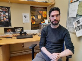
Ricardo Pérez-de la Fuente, Deputy Head of Research here at the Oxford University Museum of Natural History, tells us how he first became interested in natural history. Hear about his work as a palaeoentomologist specialising in the study of insects fossilised in amber, including the discovery of a fascinating lacewing species from around 100 million years ago with an ingenious method of camouflage and defence.
Ricardo’s research into the fossilised lacewing species, Hallucinochrysa diogenesi, has shown that the strategy of covering the body with materials from the environment as a way to camouflage and defend, known as trash-carrying, evolved at least 100 million years ago. Several species of invertebrates use this strategy today, including green lacewing larvae, and some sea urchin and crab species. Can you think of other ways in which insects defend themselves from their prey? Let us know in the comments below or in the Contact Us section of the blog.
You may already know that the museum contains millions of natural history specimens, but did you know that it also houses a world-class collection of books and journals, plus a priceless archive? Held in 38 different locations across the museum, the collection of over 20,000 books covers all areas of natural history: zoology, geology, mineralogy, palaeontology, and is particularly known for its collection of entomology books and journals. In fact, we believe it is the third largest entomology library in the UK after the Natural History Museum in London and the library of the Royal Entomological Society.
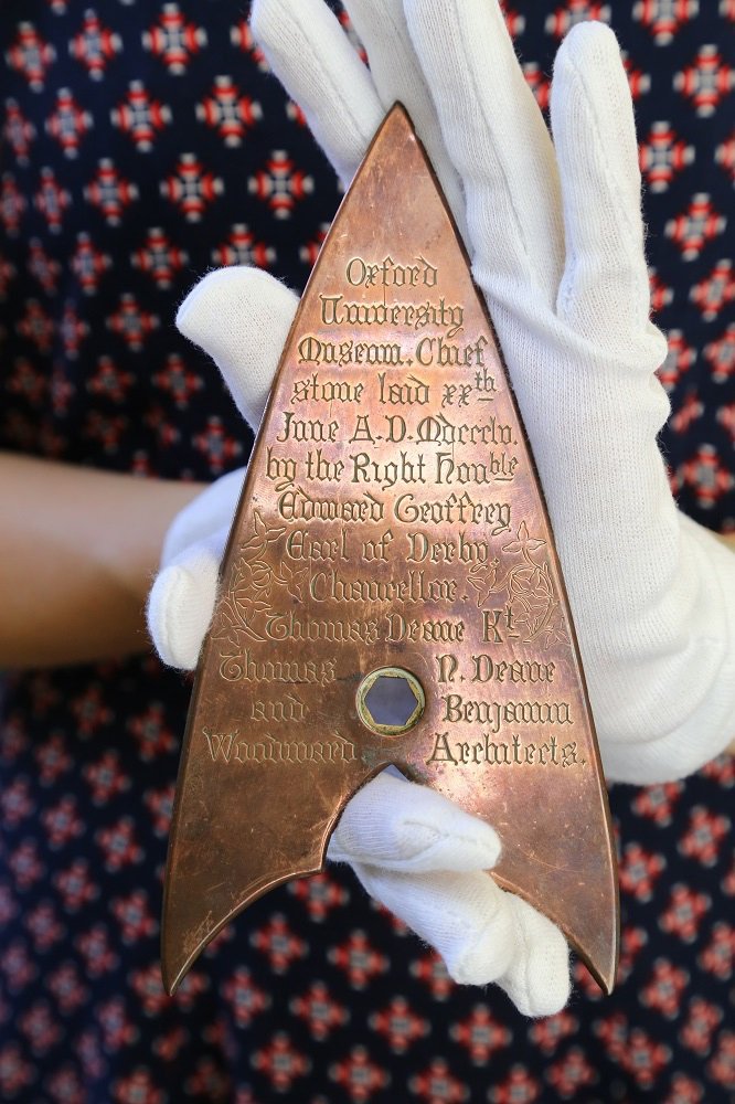
In addition, there is a priceless archive of letters, maps, field notebooks, photographs, artwork and other items of interest. Where else would you find letters from Charles Darwin, the death mask of the Swiss biologist and geologist Louis Agassiz, and the trowel that laid the foundation stone of the museum’s building in 1855.
Trowel used to lay the Museum’s foundation stone
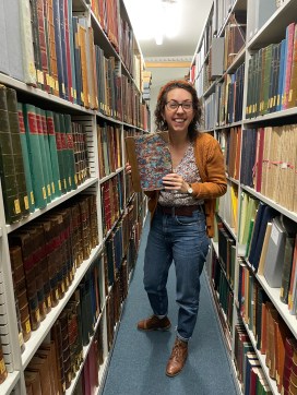
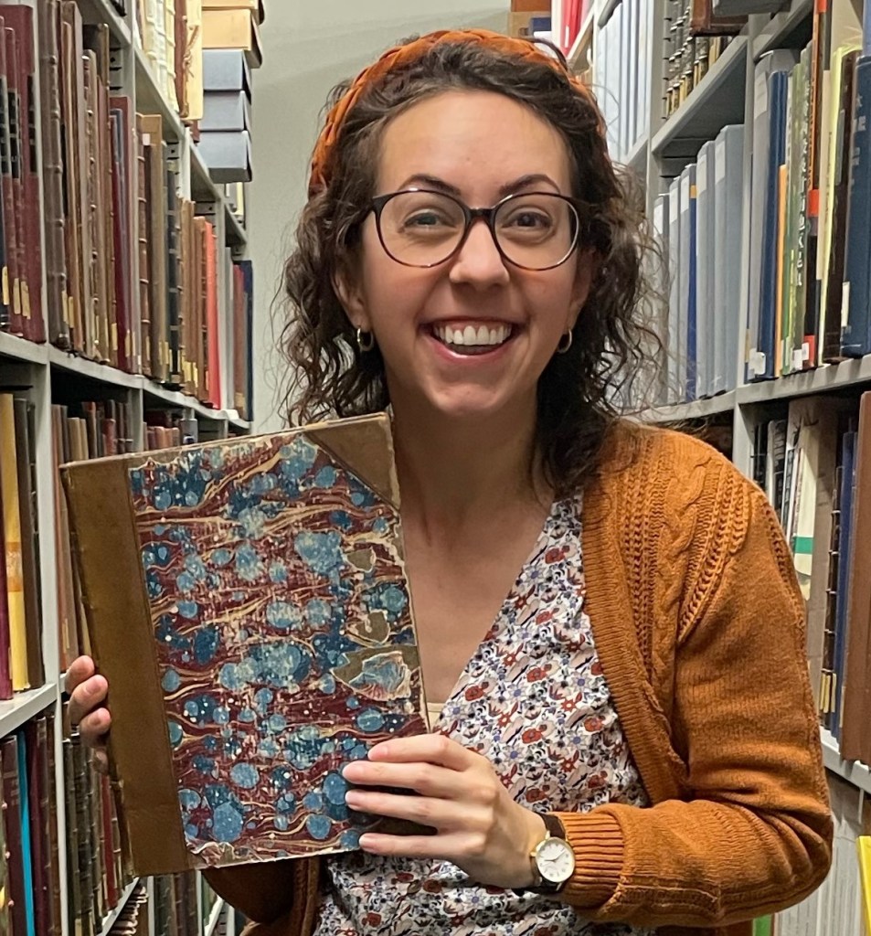
So, who is in charge of this fascinating treasure trove? That job falls to Danielle Czerkaszyn, the museum’s Librarian and Archivist. I went along to meet Danielle to find out how she came to work at the museum, what her job involves and to learn more about the amazing collection in her care.
Danielle Czerkaszyn
From History to Natural History
Having first completed a degree in History in her native Canada, Danielle came to the UK to do a Master’s degree in Museum Studies. Following this, she was selected for a place as a Graduate Library Trainee at the Bodleian History Faculty Library here in Oxford and secured her post at the Oxford University Museum of Natural History in 2017. Danielle revealed:
“When I came for the interview, I was asked what my weakness would be in the role and I replied that I didn’t really know a great deal about natural history! The good thing is that I learn new things every day and my knowledge of natural history has expanded!”
As well as a valuable resource for staff at the Museum and other members of the University, the library and archive is open to anyone with an interest in natural history. On a day to day basis, Danielle deals with many enquiries from the public requesting visits or requests for articles and chapters of books to be provided digitally to help with research. High-use collections in the library and archive are currently in the process of being digitised which will make it even more accessible to natural historians around the world. Danielle is also responsible for keeping the collection up to date with new publications and dealing with donations that come in from members of the public.
Highlights of the Collection
I asked Danielle to choose some of her favourite entomology items from the collection and she suggested two incredible and unique works – one from the archive and one from the library.
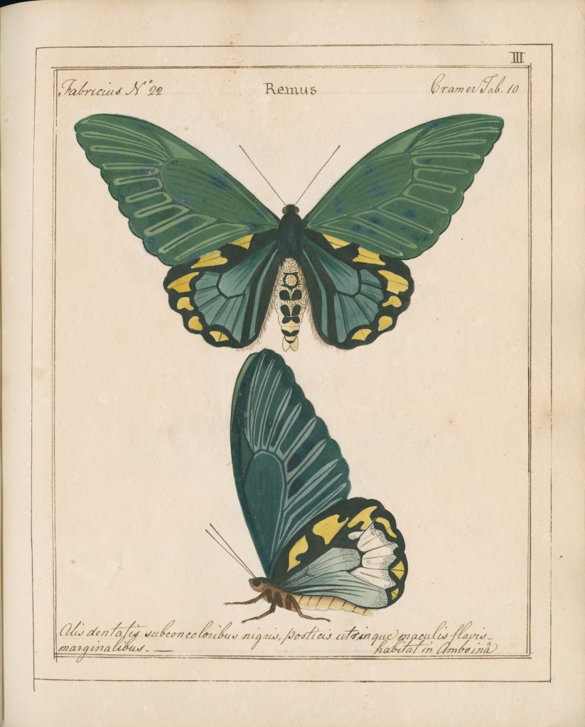
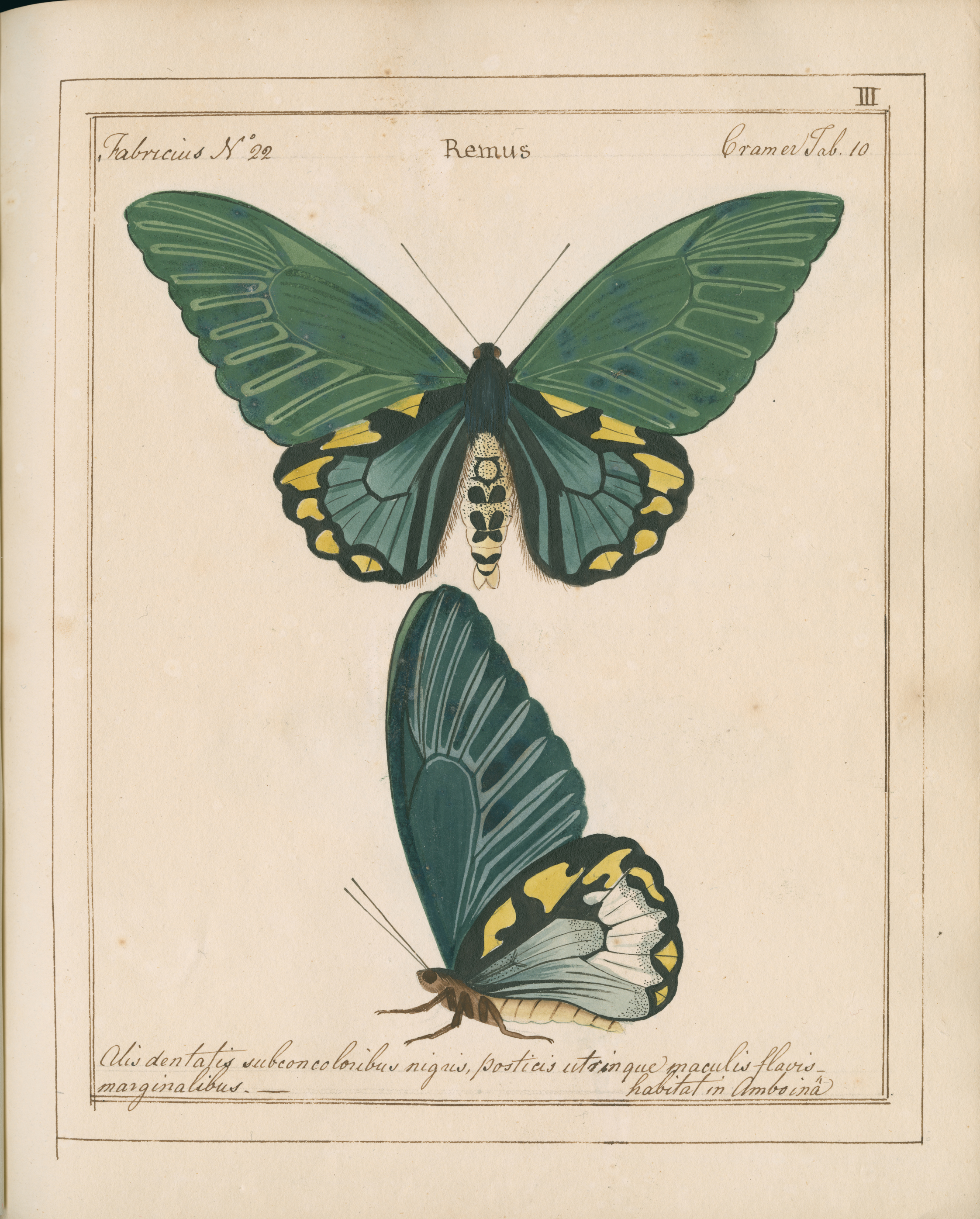
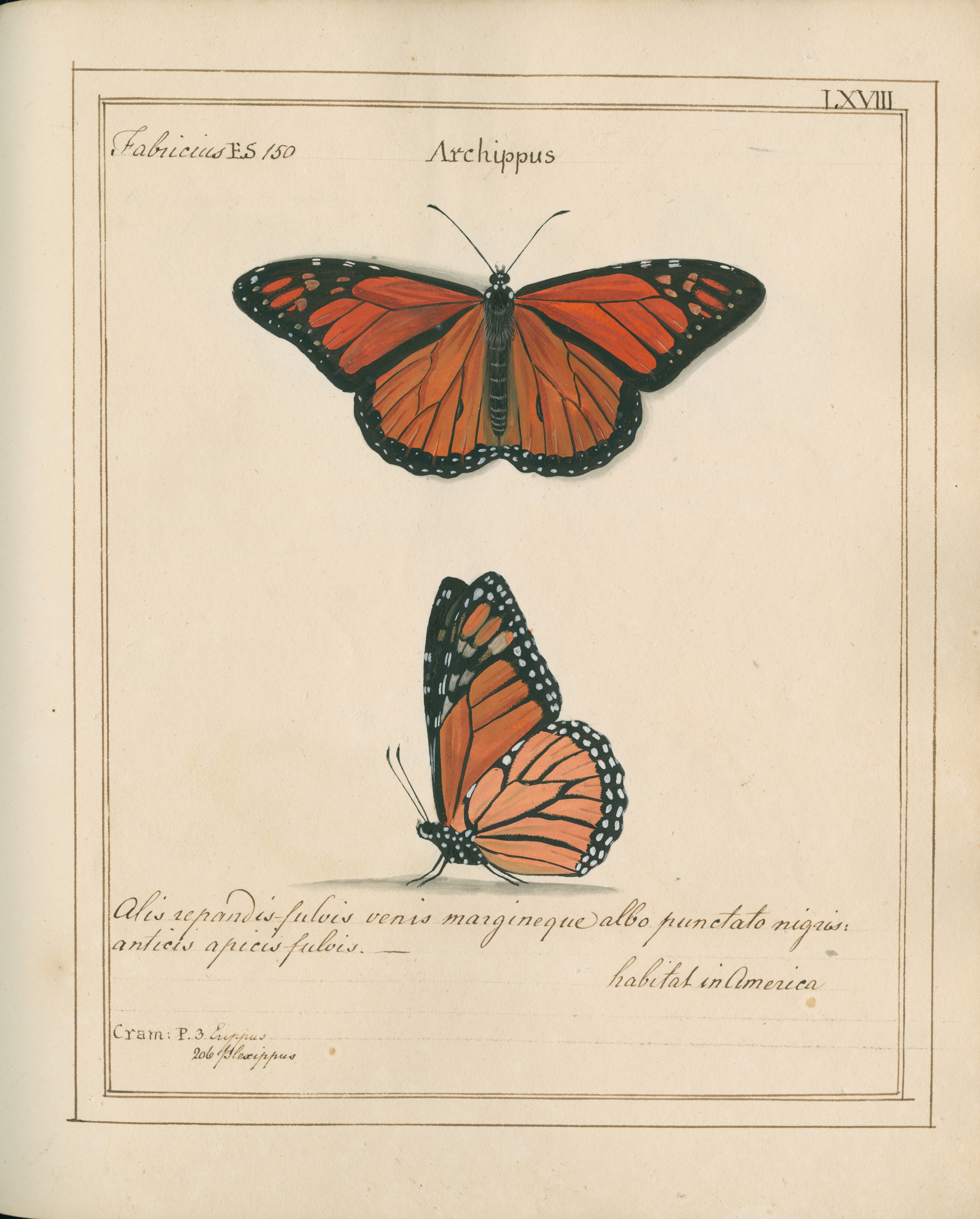
The first is William Jones’ Icones, one of the most scientifically important and stunning works on butterflies and moths ever produced.
William Jones’ Icones
“This work as one of my favourites because the paintings are so
detailed and beautiful. Plus it is completely unique.”
Thank you so much to Danielle for giving us a fascinating insight into the library and archive here at the Museum of Natural History, and for sharing some of the incredible works in her care.
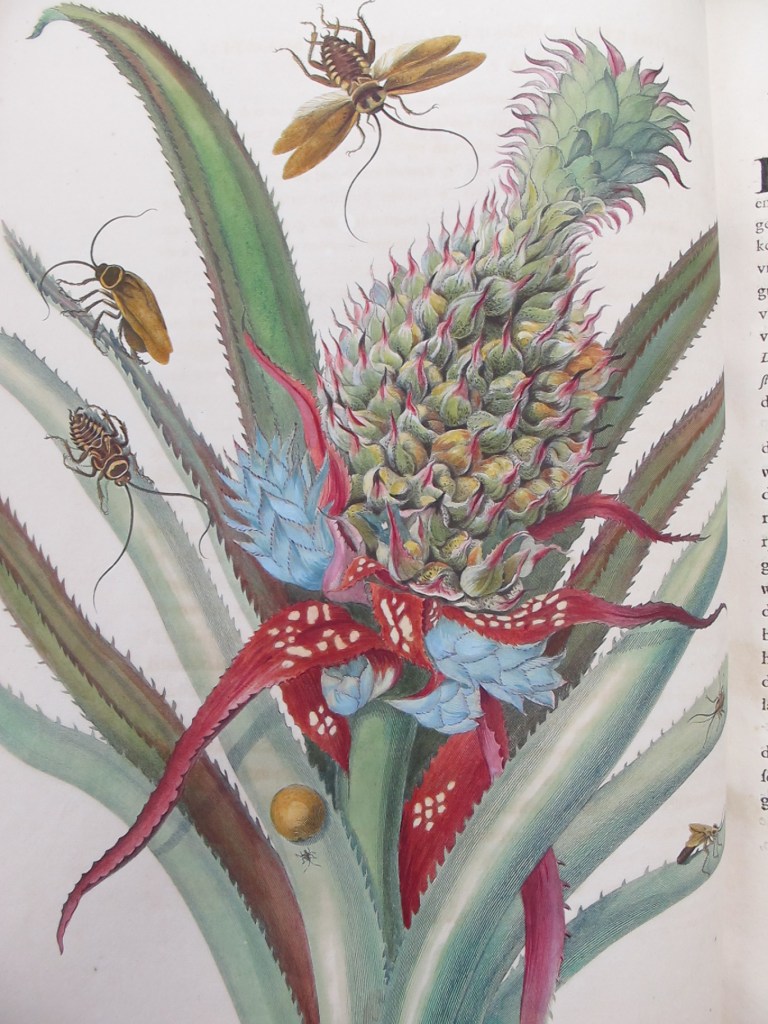
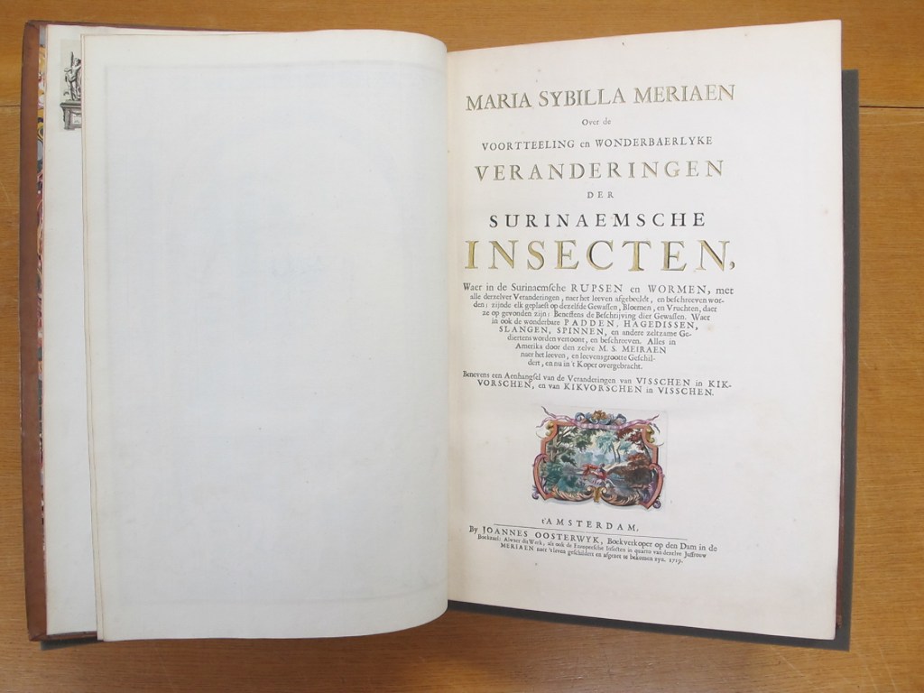
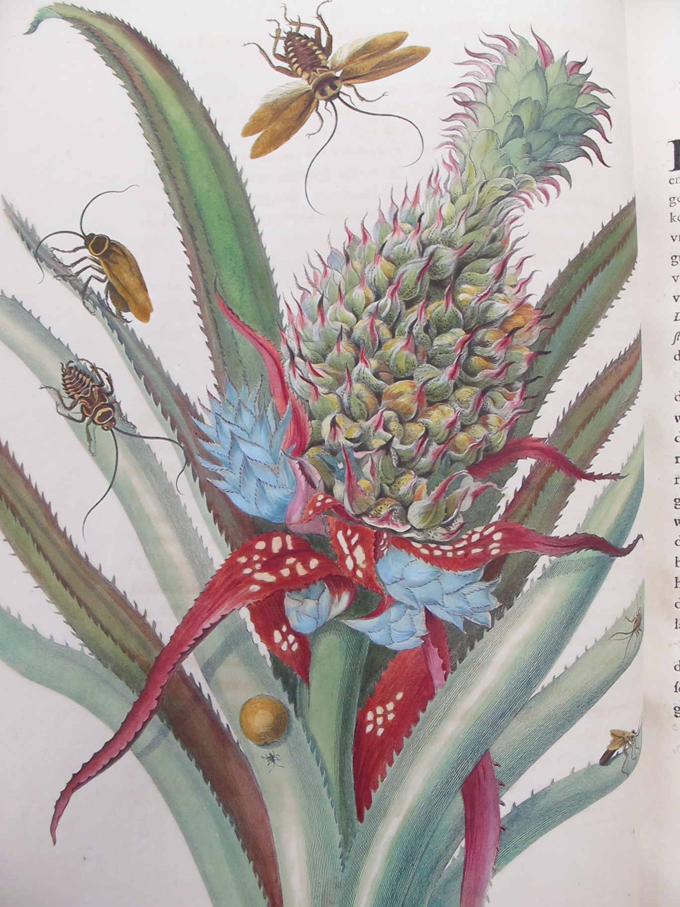
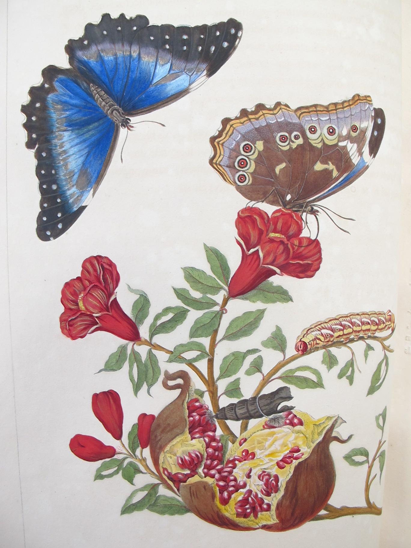
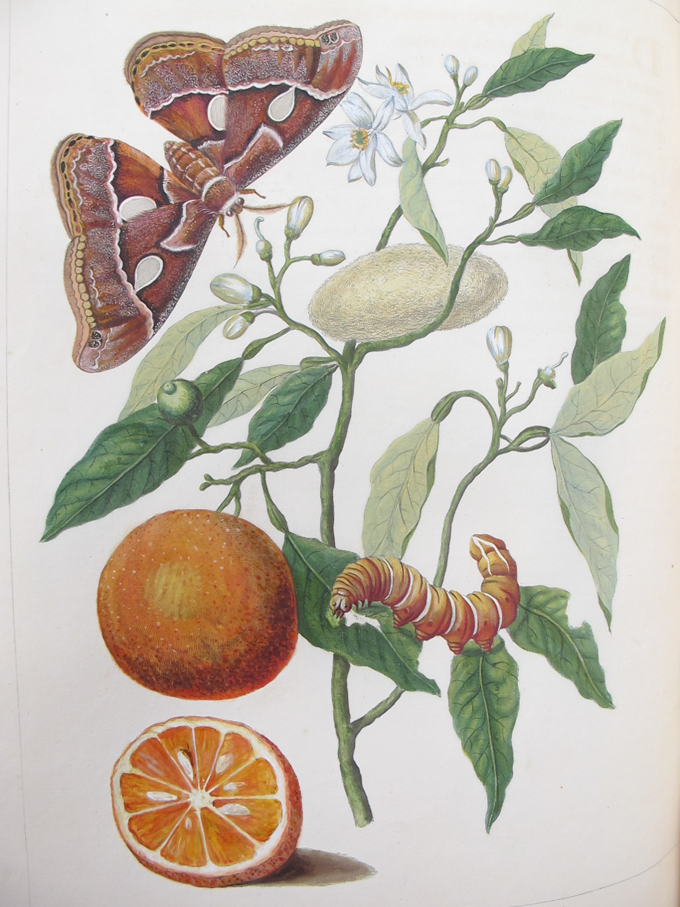
The Library here at the Museum of Natural History is lucky enough to hold a copy of Maria Sibylla Merian’s Metamorphosis Insectorum Surinamensium (Insects of Surinam). This large and stunningly beautiful book by the German artist, scientist and adventurer, Maria Sibylla Merian (1647-1717), was published in 1705. Trained as a professional artist, Maria studied insects and observed their metamorphosis from an early age. She could be described as one of the earliest entomologists.
A woman truly ahead of her time, at the age of 52 she travelled with her daughter to the newly-founded South American colony of Surinam to study insects. Maria carefully recorded what she found in the most fantastically detailed drawings and paintings which were later published in the form of a book.
The illustrations show insects in all stages of their life cycles, increasing knowledge of metamorphosis at the time. By showing insects in their natural settings and on their preferred vegetation, she also highlighted the relationship between insects, plants and other animals. Not only are the illustrations beautiful but they are also very accurate and detailed enabling them to act as the basis for the naming and identification of a number of species.
“Maria Sibylla Merian must have been an incredible woman to undertake such a journey in 1699 without patronage or being accompanied by a man. Once in Surinam, she also had to withstand the conditions of a newly-formed colony, which must have been very basic. She is definitely a figure to be admired as a pioneering role model for women
and I love showcasing her work.”
Danielle Czerkaszyn, Librarian and Archivist, Oxford University’s Museum of Natural History
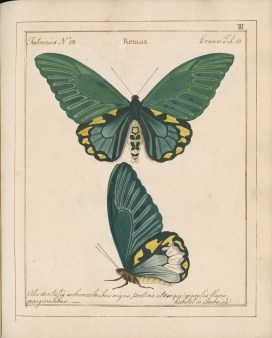
Icones is the work of 18th-century wealthy wine merchant and entomologist William Jones (1745–1818). It is one of the most scientifically important and stunning works on butterflies and moths ever produced. In it, Jones carefully painted over 760 species, describing many for the first time. What makes this book even more special is that it was never published and the copy held in the Oxford University Museum of Natural History archive is the original and only copy.
Two of the hundreds of butterfly and moth species carefully recorded in Icones.
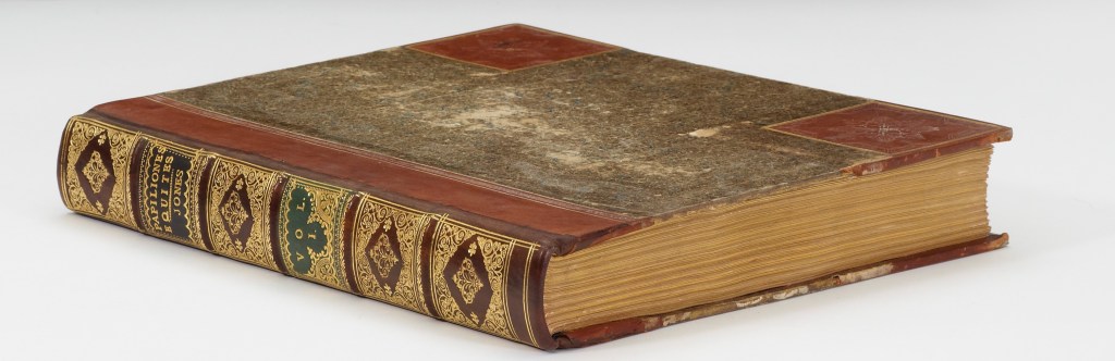
Volume I, William Jones’ Icones
Completed between the 1780 and 1810, Icones is now bound in six leather volumes. Using his own Lepidoptera collection and the collections of other naturalists, Jones create the book at a time when collecting natural history specimens from newly discovered parts of the world was a fashionable pastime among the wealthy. Many of the species are described for the first time in this work and are classed as Iconotypes. This means that the illustrations act as type specimens against which all other specimens of that species are compared for the purposes of identification.
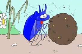
Previously, Chris Jarvis showed us how to draw a mighty Minotaur Beetle (Typhaeus typhoeus) complete with its own ball of dung! In this video Chris returns to add some finishing touches to this picture. He shows us how to convey the size of the beetle, and how to use colour to great effect.
We’d love to see any pictures you draw of insects, whether it is an ant, bee, caterpillar or your very own Minotaur beetle. Share with us at hopelearning@oum.ox.ac.uk.
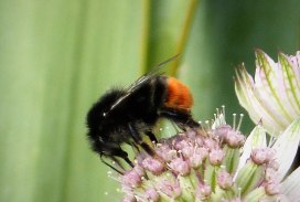
If you see a big jet-black bumblebee with a red tail in Britain, its probably the Red-tailed Bumblebee, Bombuslapidarius. If it has a scruffy white collar, then its a male.
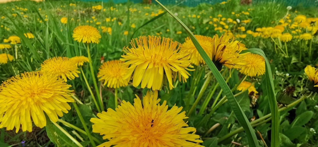
The Red-tailed Bubmblebee has extended its range northwards in recent years and is now common throughout Britain. It can often be seen in parks and gardens. The workers have short tongues and forage on flowers like daisies and thistles which have a big area for them to land on and are made up of small florets, each with a little nectar. Leaving dandelions to grow in the spring helps provide food for these and other insects early on in the growing season.
Let dandelions grow in spring to help insects. Image credit: Pixabay / Claudiu Mladin CC0
The Queens nest under stones and so are sometimes disturbed when these are moved. If you find one like this, have a look without disturbing it then just put the stone back. They may also enter our homes through open windows in search of nesting sites.
The Red-shanked carder bee, Bombus ruderarius, can look similar but is much scarcer in the UK. While the Queens of B. ruderarius are smaller. the workers of the two species can be a similar size. You can tell the difference by looking at the back legs. B ruderarius has red pollen baskets on its otherwise black legs, whereas the leg hairs of B. lapidarius are all black.
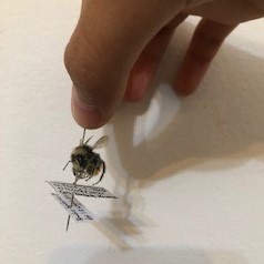
Lincoln Kwong was one of the participants at our Summer school in August. He was so inspired by the HOPE collections team at the museum that he decided to start his own insect collection. One of his first specimens was a dead red-tailed bumblebee that he found in his garden. He preserved and pinned this and recorded the data following advice from James.
Bombus lapidarius. Image credit: Lincoln Kwong
Let us know if you have your own collection. You can get in touch using the CONTACT US page. We’d also love to see any pictures any would be happy to feature them in our PHOTO GALLERY!