There was a time, more than 160 million years ago, when most of Oxfordshire was covered by a warm, clear, shallow sea. Offshore, the waters were agitated by waves and storms, but closer to land these forces were buffered by a submerged sandbank, and calm lagoons developed.
The area that is now Stonesfield was part of this lagoonal environment, and the fossils that have been found there provide a wonderful window into the living world of this Middle Jurassic sea.
So how would these sea creatures compare with British marine life today? Some things would have been very familiar. On the sea bed you would have found a huge variety of bivalves, or clams, along with lobsters, crabs and sea urchins; the waters above would have been full of fish, including several different types of shark.
This fossilised limpet shell has its original colouration preserved
But there would have been other things too: squid-like belemnites, and nautiloids and ammonites with coiled shells, and tentacles like an octopus. Instead of dolphins and porpoises there would have been sea crocodiles and sea turtles, and in the skies above, flying reptiles known as pterosaurs rather than the usual sea birds.
And there’s more. Stonesfield fossils also include plants and animals washed in from nearby land: the leaves and seeds of conifers and cycads, beetle wing cases, reptile eggs, and the remains of both dinosaurs and mammals.
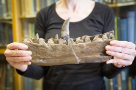
The lower jaw of the first named dinosaur, Megalosaurus bucklandii, found in Stonesfield, Oxfordshire in the early 19th century
One particularly spectacular find was the lower jaw of the carnivorous dinosaur Megalosaurus, nine metres long in life and weighing about a tonne. Megalosaurus became the first creature to be named a ‘dinosaur’, in 1824. Less obvious, but equally significant, are the tiny jaws of some of the shrew-like mammals that would have lived alongside the dinosaurs: Phascolotherium, Amphitherium, Amphilestes and Stereognathus – the first Jurassic mammals known to science.
Along with the preservation of delicate items such as dragonfly wings and the leg of a cricket, and the original colour patterns on some sea snails, limpets and barnacles, the fossil material from Stonesfield is really quite special.
Within the order of insects Coleoptera – the beetles – is a family called Histeridae, also known as the Clown Beetles. This family is found worldwide and so far around 3,900 species have been discovered. Of these, 52 species are found in the UK and it’s these that I’m currently working on.
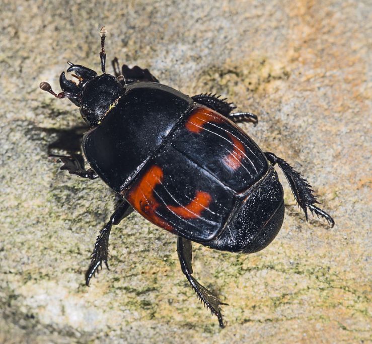
Histeridae aren’t the most appealing beetles to look at. They’re not very big, or round, and they don’t come in pretty colours. Basically they lack the wow factor… until you look at bit closer! Hister quadrimaculatus is probably the biggest and most colourful of the British species; it is black with four red dots (it’s all in the name).
Histeridae live in a variety of habitats, but are most commonly found in dung and carrion. Surprisingly though, they are not eating the dung or the dead animal; they are there for the fly larvae. Flies are attracted to decomposition, and the Clown Beetles take advantage of this. The adult female Histerid lays her eggs within these environments and three days later the larvae emerge to feast on the fly eggs and pupa.
Some of the more obscure species, such as the rare Haeterius ferrugineus live in ants nests, particularly those of the Slave-maker Ant (Formica sanguinea) and the Black Ant (Formica fusca). These types of Histerids vary in colour and modifications compared to the dung- and carrion-dwelling Histerids because they have evolved to live successfully with ants.
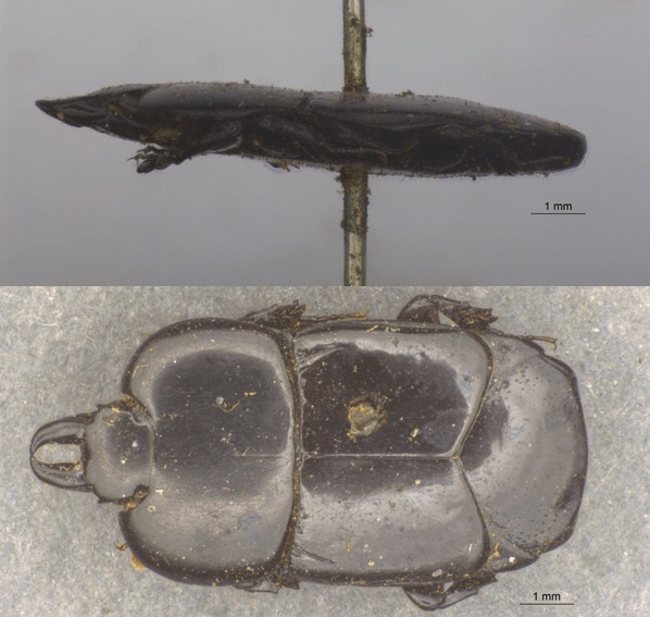
Hololepta plana is little more than 1mm thick, perfectly adapted for living under bark
Another species that differs from the norm is Hololepta plana; as an adult it is completely flat, adapted to live under bark and feed on the larvae of other invertebrates.
The Histeridae are a wonderfully diverse family, and we’ve got some pretty amazing examples of them in the UK. They may not be beautiful to everyone, but they are fascinating creatures that play a vital role within our environment and we’ve got a lot more to learn about them.
by Juliet Hay, Earth Collections preparator and conservator
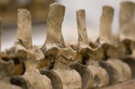
I feel myself very lucky to have a job that involves working with the fossil remains of long-extinct animals. One of the things my colleagues and I are currently working on is a plesiosaur – a marine reptile that lived in the sea millions of years ago.
This particular specimen was found in a clay pit near Peterborough by members of the Oxford Clay Working Group in 2014, and is a near-complete example of its kind. The palaeontologists who found the specimen named it Eve, although we don’t know if it was male or female, and perhaps never will.
The discovery of large fossil vertebrates like this is rare, so we are fortunate to have had the specimen donated to the Museum by the quarry owners Forterra.
Juliet at work on the plesiosaur skull
The plesiosaur is 165 million years old and, when alive, was around 5.5 metres long. It had a long neck, a barrel-shaped body, four flippers and a short tail. The find is particularly exciting as the skull was also discovered. It is encased in a clay matrix, which is relatively easy to remove, but the work has to be carried out under magnifying lenses and microscopes.
As the skull is quite small relative to the size of the body, the features are very delicate and it is a painstaking process to remove the sediment without damaging the fossil bone or losing any tiny fragments. Fortunately, pictures of the skull have been produced using CT scanning technology, and the images are proving invaluable as an aid to assist in its preparation. It’s a bit like having a jigsaw puzzle with the picture on the lid to refer to!
A belemnite hooklet at 12x magnification, found with the plesiosaur remains and possibly part of Eve’s last meal
The clay covering the skull is being sieved and examined and tiny hook-shaped fossils have been found. These came from the arms of squid-like creatures called belemnites, which may have formed a large part of the plesiosaur’s diet.
It is too early to say for sure, but Eve could represent a species new to science, as some features, such as the shape of the flipper bones and some of the surfaces of the bone in the skull, are quite unusual. Further research needs to be done before the findings can be published in scientific journals – watch this space.
And if you’re visiting the Museum before 25 July, you can see some of the fossilised remains of Eve for yourself, in our Presenting… display case.
Starfish are among the most distinctive animals found along the seashore today. Together with other well-known forms such as sea urchins, sea cucumbers and brittle stars, they belong to a major group called the echinoderms, which is characterized by a unique type of symmetry — called fivefold symmetry. This means they can be divided into five roughly equal parts.
In contrast, the closest living relatives of echinoderms are worm-like animals that have bilateral or mirror-plane symmetry, where they are divisible into mirror-image halves. It’s widely-thought that the common ancestor shared by echinoderms and other animals also had bilateral symmetry. Because they are so different to all other living animals, deciphering the evolutionary history of echinoderms, and their path from worms to stars, has proven a major challenge for scientists.
The closest living relatives of echinoderms are worm-like animals like these acorn worms Balanoglossus sp.) from Naples
Fortunately, fossils can shed light on echinoderm evolution. Echinoderms have an excellent fossil record because they possess a hard, mineralized skeleton, which greatly enhances their chances of being preserved as fossils compared to soft-bodied organisms. The first fossil echinoderms are over half a billion years old, and include extinct groups that show both bilateral and five-fold symmetry.
In addition, fossils are known that exhibit three-fold symmetry, as well as others that lack a clear plane of symmetry – they are asymmetrical. These fossils document the earliest history of echinoderms, and so could help us to better understand their evolution.
The fivefold symmetry of the starfish (Randasia granulata from Madagascar)
Based on our understanding of living animals, and using modern methods for reconstructing the relationships of different species, it’s possible to infer that the early fossil echinoderms with bilateral symmetry belong at the base of the echinoderm evolutionary tree.
The next branches in the tree lead to the asymmetrical fossil groups, and these are followed by those forms that show three-fold symmetry. Lastly, we see the diversification of forms with fivefold symmetry, including species belonging to the groups that still exist today, such as the starfish.
Using the fossil record, we can therefore see a clear picture of how echinoderms evolved from worm-like organisms into star-shaped creatures.
One cow can produce over nine tonnes of dung per year. With a population of about 3.4 million cows in the UK alone, that’s a heck of a lot of dung deposited on our grasslands. Just imagine how much dung is produced every year if we include the output of horses, sheep, pigs, and all the wild animals out there.
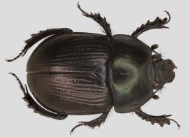
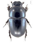
Dor Beetle – Geotrupes mutator
All of this dung is broken down by a multitude of invertebrates, including flies, worms, and beetles, as well as bacteria, fungi, and weathering. One of the key groups involved in the removal and degradation process is the aptly named ‘dung beetles’.
In the UK there are 61 species of dung beetle, though sadly just over half of these are now in decline and some have already become regionally extinct. UK dung beetles vary in size from just 3 mm to over 25 mm and occur wherever dung is found, though some prefer sandy soils and others like to live in woodlands.
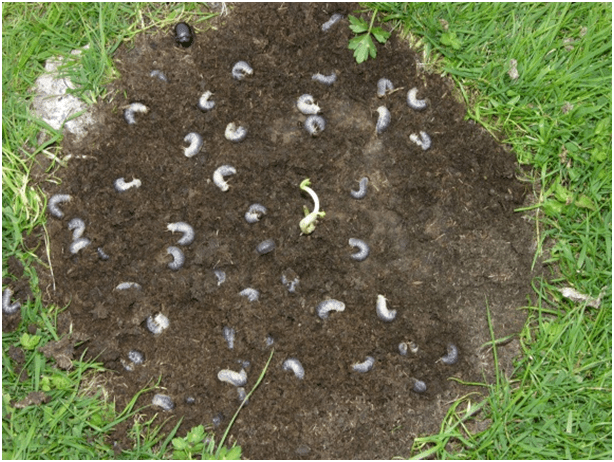
Dung beetle larvae (Aphodius fossor)
As adults, dung beetles feed on the liquid part of dung. The larvae of most of our species live inside the dung pile and are called the dwellers. These munch their way through the solid matter of the dung pile, gradually breaking it down over a few months. Other species such as Geotrupes mutator, pictured above, excavate a tunnel and bury the dung below ground. These tunnellers construct a brood chamber in which their young develop.
Aphodius fossor
Through their actions, dung beetles perform a number of valuable ecosystem services. The most obvious is dung removal and degradation which leads to improved soil health by nutrient cycling and soil movement. By burying the dung they reduce the amount of available breeding habitat for pest flies and livestock parasites too.
All of these important services have been estimated to save the UK cattle industry £367 million per year. The value of dung beetles doesn’t end there as they also provide an important source of food for farmland mammals and birds. So next time you see a pile of dung in a field, just think of all the hard working beetles within…
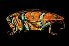
Today we are excited to be opening our new special exhibition, Microsculpture: The Insect Photography of Levon Biss. You may well have already heard about Microsculpture, and have perhaps watched the video showing how Levon Biss made these incredible portraits of insects from the Museum’s collection.
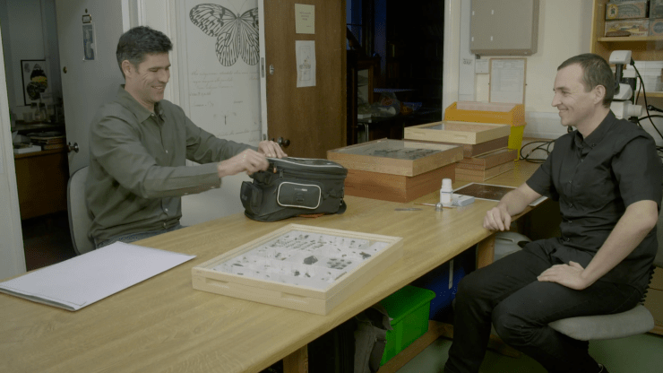
As we open the show in the Museum’s main court we wanted to give a little more insight into the process that Dr James Hogan, an entomologist in our Life Collections, went through to select and describe the specimens for the project. So I put a few questions to James to explain the making of Microsculpture from his point of view.
James Hogan (right) selecting specimens with Levon Biss
There are 23 specimens on show in the exhibition: how did you choose these particular ones? We wanted a wide variety of specimens that would all show some interesting surface detail. Some were also chosen for their spectacular colours, interesting shapes or strange appearance.
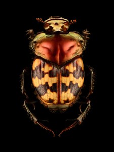
Splendid-necked Dung Beetle (Helictopleurus splendidicollis) from Madagascar. Length: 10 mm
But perhaps 95 per cent of the specimens we initially considered were rejected because we had some pretty strict criteria; very hairy specimens, which includes most bees, were out as the image processing was too difficult; the specimens had to be intact, so no broken antennae or legs.
Probably the biggest problem was finding specimens which were clean enough. Because Levon’s technique reveals so much detail, any dirt on the specimen is glaringly obvious. And the whole idea was to look at the surface structures, which are obviously very small and easily obscured by any dirt.
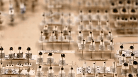
How did you prepare them for their close-ups? All the specimens needed a bit of preparation work before being ready for Levon’s photography. First they needed to be remounted on the end of long pins to allow more clearance space for the lighting setup. Some of the specimens are very small and fragile and this is the stage where damage is most likely to occur.
After remounting, all the insects needed at least some degree of cleaning under the microscope. For this I used a range of fine paintbrushes; we also make our own tools from micro-pins bent into different points and hooks. Again, I had to be careful to not damage the specimens – easily done by over-enthusiastic cleaning!
Where do the specimens come from? The specimens in the exhibition are from the Museum’s very large insect collection, stored behind the scenes in many different rooms. The insects in the show are from all over the world, from a back garden in England to a remote island near Antarctica. Some were collected over 150 years ago while others were collected very recently by Museum staff.
Of particular historical significance is the Tricolored Jewel Beetle (Belionota sumptuosa) which was collected by the Victorian naturalist and explorer Alfred Russel Wallace, co-publisher of the theory of evolution with Charles Darwin in 1858.
Tricolored Jewel Beetle (Belionota sumptuosa), collected by Alfred Russel Wallace in Seram Island, Indonesia. Length: 25 mm
How big are the actual specimens in comparison with the photos? The actual specimens are mostly pretty small, ranging from 6 mm to about 30 mm. Some of the larger printed photographs will be about a thousand times larger than the specimen!
Orchid Cuckoo Bee (Exaerete frontalis) from Brazil. Length: 26 mm
What do you hope people will get out of the pictures in the exhibition? To me insects are beautiful creatures and Levon has done an outstanding job of revealing this. I hope the photographs will spark people’s interest in insects and inspire more people to study them.
The photographs certainly pose some interesting questions, and reveal that there is so much we don’t know, particularly about the functions of all their strange and varied adaptations.
Is it valuable to showcase specimens from the collections in this way? One of the most exciting parts of this exhibition for me was the opportunity to show part of the Museum’s collection which visitors don’t normally see.
The collection is used all the time by scientists, artists and educators, but unfortunately most of it is unsuitable for display because the specimens are so small. Levon’s ultra-high resolution photography has in a way solved this problem, allowing us to showcase some of these spectacular, but tiny and fragile creatures in all their beauty.