Marble-effect frame inlaid with a marble-effect stone showing the outline of numerous cross-section gastropod shells

The Continuing Importance of Corsi’s Legacy

Four Crowns is a studio based in Oxford which is dedicated to keeping the craft of scagliola alive. But what exactly is scagliola, and how does it relate to the Museum’s collections? Freddie Seddon, a University of Oxford Micro-Internship Programme participant at Four Crowns, tells more about this fascinating process…

Sculpture of the front half of a foot in brown/yellow marble effect, showing cracks and damage to some of the toes
Foot, Four Crowns, 2020
145mm

Scagliola is the technique of imitating the beautiful patterning and colours of marble. With roots in the ancient world, scagliola saw a revival from the 17th century, when European artists and architects returned from their Grand Tours of the continent wishing to replicate the marbles of Classical and Renaissance Europe.

Several techniques can be used to reproduce the appearance of marble in plaster, with the addition of other natural pigments and larger chips of coloured plaster. The artist must try to replicate the conditions under which particular marbles form: compressions, twists and layers applied to the plaster to give the image of breccia, veins, and even fossils.

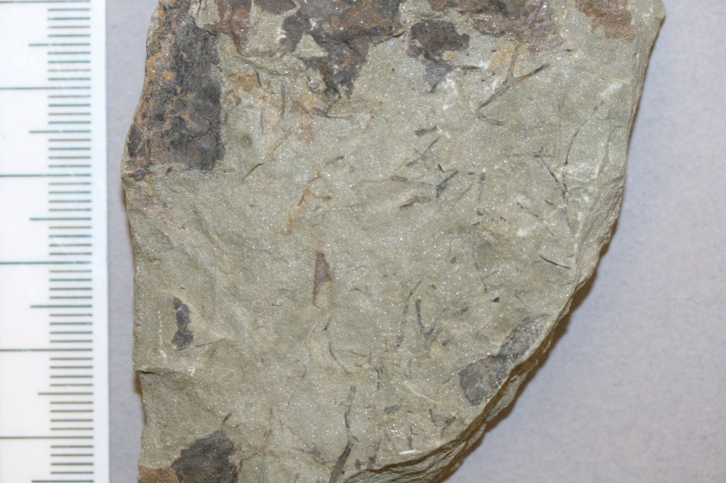
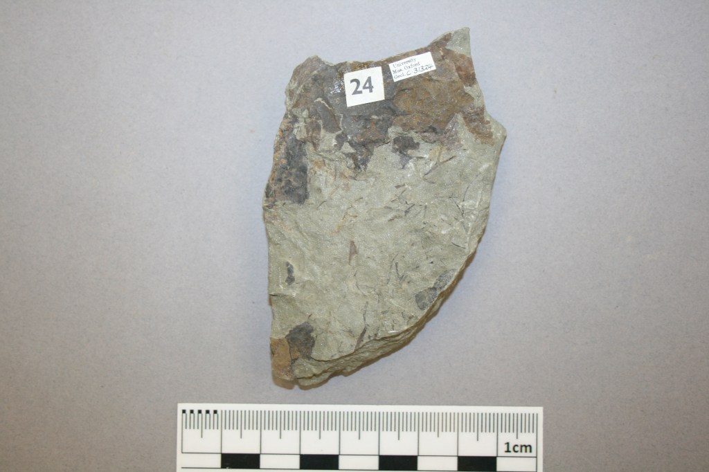
The Museum has a large collection of decorative stones, including the Faustino Corsi collection, acquired in 1827. The Corsi collection holds 1,000 samples of ancient and modern decorative stones, including polished marbles, granites, serpentines, and jaspers. Faustino Corsi (1771–1846) built the collection in the early 19th century, first by gathering material used in ancient times across the Roman Empire, and later adding decorative stone from contemporary quarries, mainly in Italy, but also Russia, Afghanistan, Madagascar and Canada.

Marble-effect frame inlaid with a marble-effect stone showing the outline of numerous cross-section gastropod shells
Lumachellone, Four Crowns, 2018 990x485x60mm

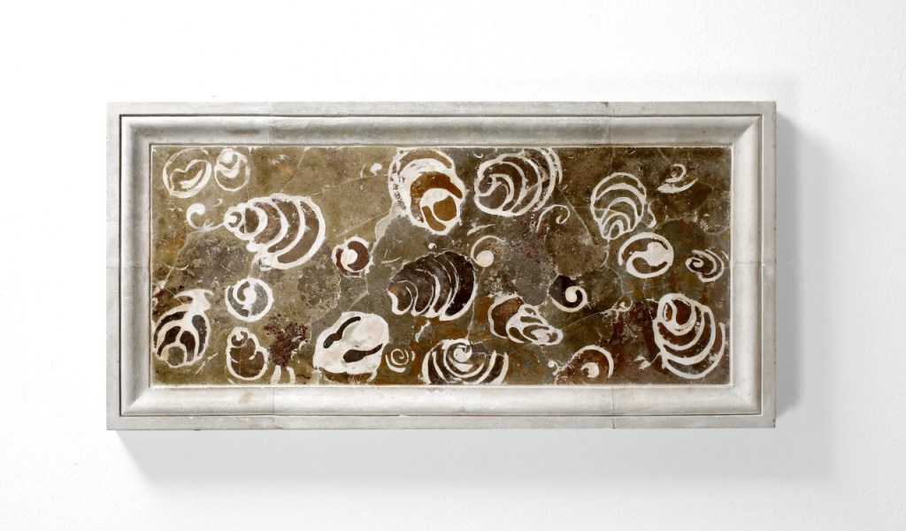
The Corsi collection is valuable tool when it comes to scagliola. Images and marble descriptions from the Corsi database help determine the processes a certain scagliola sample should undergo and the natural colours that these would produce. To accurately depict marble, an artist might need to create upwards of twenty colours and clarity levels – even then, only high-quality, natural pigments will produce natural results. The piece is polished to obtain a shine like that possible on natural marbles, and cross-checked against Corsi’s samples one final time to guarantee a faithful replication of the stone.

Statue of a robed figure standing on a plinth and holding a golden lizard-like reptile in one hand
Codazzi, Four Crowns, 2017
270x200x760mm

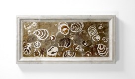
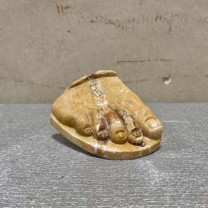
In this way, the selection of which stone to imitate is a creative challenge in itself for the artist. Each item in the Corsi collection offers different aesthetic and cultural experiences. Lumachellone antico, for example, is limestone with large fossilized gastropods, admired in classical Rome for its richness and complexity. The collection contains only one example of this stone, composed of samples from two different locations, which the Four Crowns artist has been able to faithfully replicate. As this marble type has never been available on any commercial scale or markets, it is up to the emerging generation of scagliola craftsmen to painstakingly reproduce this ancient stone.

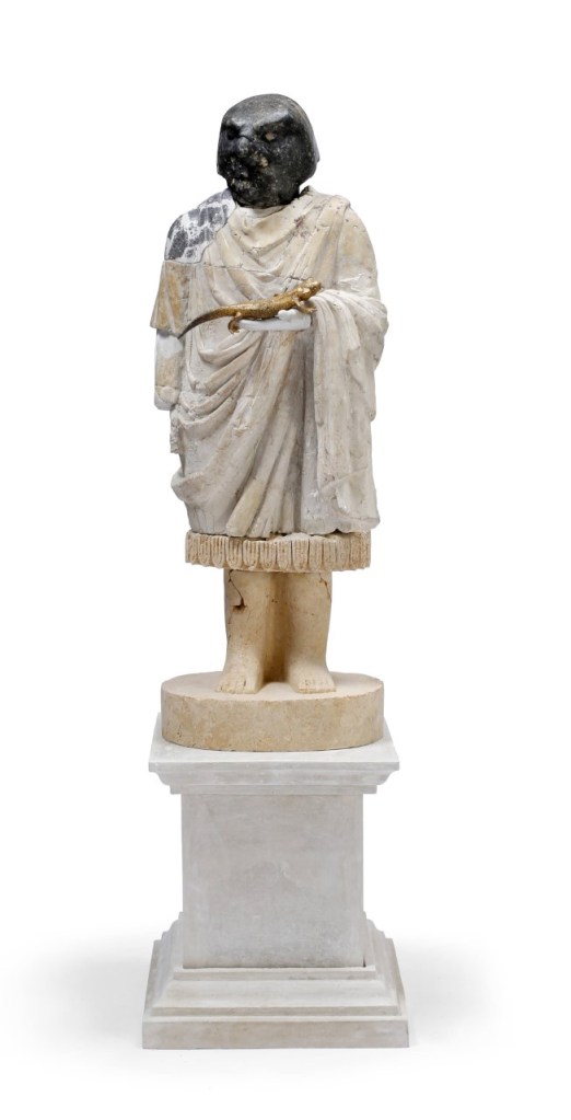
The most ambitious and impactful presentations of scagliola can even mirror a combination of marbles. The Four Crowns’ Codazzi emulates four different stone types: the head is bigio antico, the drapery is giallo antico, and the legs and feet replicate a limestone common in Sumerian sculpture, with a shoulder inlay of bianco e nero.

Through the art of scagliola, and the unique reference resource of the Corsi Collection, rare, beautiful or lost marbles are able to be recreated time and again.

Freddie Seddon is a second year student, reading Ancient and Modern History (BA) at Wadham College, Oxford.

Ink drawing showing the skeleton of dinosaur

Tales of Iguanodon Tails

By Leonie Biggenden, Volunteer

As one of our many invaluable volunteers, Leonie Biggenden has regularly helped to run our Science Saturdays and Family Friendly Sunday activities, both of which take place under the watchful eyes of the large T. rex and Iguanodon skeletons in the Museum’s main court. Having spent so much time beside the Iguanodon, and with a lack of in-person volunteering opportunities in recent months, Leonie decided to find out some of the history of this striking cast. For Volunteers Week this week, she shares what she discovered…

Next year will be the 200th anniversary of the discovery, by a roadside in Sussex, of the first Iguanodon teeth. Found by Mary Mantell in 1822, her husband Gideon saw their similarity with the teeth of modern iguanas and suggested they were from a huge, ancient, herbivorous lizard. He called the animal Iguanodon, and you can see his sketch reconstruction at the top of this post.

However, as an amateur palaeontologist, Gideon Mantell was not initially taken seriously by the scientific establishment. Some claimed the teeth were actually from a rhinoceros, or even a pufferfish! But in 1834, more complete remains were found by workmen who had accidentally blown up a slab of rock in a quarry near Maidstone, Kent. Iguanodon became a rock star of the dinosaur world, being only the second dinosaur – and the first herbivorous one – to be named (the first was the carnivorous Megalosaurus – another famous Museum specimen).

The Iguanodon bernissartensis cast in the centre court of the Museum.

Twenty years later, a model of an Iguanodon was constructed by sculptor Benjamin Waterhouse Hawkins as one of a set of 30 life-sized models of extinct animals for the relocated Crystal Palace Gardens in South London. It was mounted in a rhinoceros-like pose, with what we now know as a thumb spike placed as a nose horn. Scientists always look to the information they have available to them, including observation of living animals, and there is an iguana called Cyclura cornuta – the Rhinoceros Iguana – which does indeed have nose horns, so at the time the nose horn made sense.

Close up photo of iguana head
Rhinoceros Iguana, showing a nose horn. Image: H. Zell, CC BY-SA 3.0 , via Wikimedia Commons

Another 20 years on and a most significant find was made in southern Belgium. In February 1878, more than 30 fully articulated, adult Iguanodon fossil skeletons were found by miners Jules Créteur and Alphonse Blanchard, 322 m deep in the Sainte Barbe coal mine. Louis de Pauw from the Belgian Royal Museum of Natural History started to excavate the skeletons. It was a risky undertaking. In August an earthquake cut them off for two hours, and in October they were forced to return to the surface as the mine flooded.

The fossils were wrapped in damp paper, covered in protective plaster, and divided into 600 blocks. Each specimen was given a number and each block a letter, to record their exact positions in the mine. The 130 tonnes of specimens, rock, iron reinforcing rods, and plaster were then brought to the surface of the mine by horse drawn trucks and transported to Brussels.

For the first time, scientists, and later the public, could see complete dinosaur skeletons. This was important because scientists learned that the unusual spike found in the scattered fossils in the UK was a thumb spike rather than a nose horn, and they ditched rhino resemblance too, though not in time for the Crystal Palace reconstruction!

In 1882, de Pauw began assembling at least 38 Iguanodon skeletons under instruction from Louis Dollo, another famous Belgian palaeontologist. The aim was to put them in their most probable living position. A room with a high ceiling was needed because of their size, and a chapel was chosen. Scaffolding was built with hanging ropes being adjusted so the fossilized bones could be moved into their most likely position and then fixed and reinforced with iron rods.

Iguanodon bernissartensis, like the one on display here in the Museum, was a new species, named in 1881. It lived about 125 million years ago. The first assembly was revealed in 1882 and went on public display in Brussels in 1883. Points of reference used for the pose were the skeleton of a cassowary and a kangaroo.

On the Museum’s cast skeleton you can see rod-like structures going across the blade-like, bony processes on the back. These are ossified, or hardened, tendons and would help to stiffen the tail and therefore restrict its movement. They have been broken where the bend in the tail was made to resemble a kangaroo-like stance. The displacement shows that the true position of the tail should be straight.

But having such a straight tail would mean that the Iguanodon would need its head and arms nearer the ground for better balance. The strong hind limbs suggest it would usually walk on two legs with its tail held aloft, as does the fact that fossil Iguanodon footprints are three-toed, and the three-toed limbs are the back ones.

By the end of 1883, six Iguanodons had been mounted this way and positioned in their own glass cage in the courtyard of the Brussels museum. So Iguanodon was one of the very first dinosaurs to be recovered in its entirety and mounted in three dimensions as though a living animal!

Leonie is a longstanding Public Engagement volunteer at the Museum. Unable to volunteer in the normal way during the lockdown, she researched the history of this favourite specimen and shared what she learned in a talk for other volunteers as part of an online ‘social’. This article has been adapted from that presentation.

Coloured digital models of animals in strange shapes

Revealing Exceptional fossils, one layer at a time

Around 120 years ago, William Sollas, Professor of Geology at the University of Oxford, developed a special technique for grinding down and imaging certain kinds of fossils. Sollas was based at the Museum at the time, and the process he pioneered is still used here today, as our Palaeobiology Technician Carolyn Lewis explains to mark the anniversary of Sollas’ birthday on 30 May.

Rock face with geologists hammer
Site of the Herefordshire Lagerstätte, showing the nodules embedded in soft volcanic ash.

Here at the Museum, I work on a collection of exceptionally well-preserved fossils from the Silurian Herefordshire Lagerstätte. They were deposited on the seabed 430 million years ago when the animals were buried by a volcanic ash flow. The fossils range in size from less than a millimetre up to a few centimetres, and represent a diverse collection of marine invertebrates that includes sponges, echinoderms, brachiopods, worms, molluscs and a wide variety of arthropods.

These Herefordshire Lagerstätte fossils are unusual in that many of them have preserved soft tissues in remarkable detail, including eyes, legs, gill filaments, and even spines and antennae only a few microns in diameter. The key to this extraordinary preservation is that as the fossils developed, calcium carbonate nodules formed around them, protecting and preserving the fossils since the Silurian Period.

Usually, only the hard parts of fossil invertebrates are preserved – the carapace of trilobites or the shells of brachiopods, for example – so the Herefordshire material provides us with a great opportunity to work out the detailed anatomy of these early sea creatures.

Split rock nodule showing fossil of Offacolus kingi inside.
Close-up of the fossil of Offacolus kingi

But the problem we face is how to extract the specimen from the rock nodule without losing the information it contains. The fossils cannot be separated from the surrounding rock by dissolution, because both fossil and nodule are made mainly of calcium carbonate, so would dissolve together. And they are too delicate to be extracted mechanically by cutting and scraping away the surrounding nodule. Even high resolution CT scans cannot, at present, adequately distinguish between the fossils and the surrounding rock material.

To get round this problem we use a method of serial grinding and photography based on the technique developed by William Sollas in the late 19th century. We grind the fossils in increments of 20 microns then photograph each newly ground surface using a camera mounted on top of a light microscope. This generates hundreds of digital images of cross sections through the specimen.

Then, using specially developed software we convert the stack of two-dimensional images into a 3D digital model that can be viewed and manipulated on screen to reveal the detailed form of the animal. These 3D models are artificially coloured to highlight different anatomical structures and can be rotated through 360o, virtually dissected on screen, and viewed stereoscopically or in anaglyph 3D.

Although our method of serial grinding is still fairly labour intensive, it is far less laborious and time-consuming than the process used by William and his daughter Igerna Sollas. Compared to the photographic methods of the early 20th century, where each photographic plate required long exposure and development times, digital photography is almost instant, enabling us to grind several specimens simultaneously.

Grid of images show a fossil at different stages of grinding down
Sequential serial grinding images of an ostracod

Computer software also allows us to create 3D virtual models rather than building up physical models from layers of wax. Yet despite our modern adaptations, we are using essentially the same technique that William Sollas developed here at the Museum 120 years ago. And using this technique to study the fossils of the Silurian Herefordshire Lagerstätte has yielded a wealth of new information that opens up a unique window into the evolution and diversification of early life in our oceans.

two swifts looking out from their nesting area

A Swift Return to Summer

By Chris Jarvis, Education Officer

Amidst reports during the last week of Swifts being sighted feeding over the nearby Farmoor reservoir, Museum staff have kept their eyes to the skies eagerly waiting to be the first to spot our resident birds returning to their breeding site in the nest boxes of our tower. A wet and windy weekend caused by a deep depression over Britain meant little opportunity to feed on their diet of small flies and other invertebrates that make up the aerial plankton they relish, and which normally drifts unseen above our heads in large numbers on still summer days.  The high winds would certainly also have made any attempt to land, for the first time in a whole year since they left their nest boxes and for the first time ever for those just reaching maturity, extremely precarious, and so it seems our Swifts headed farther afield, possibly back to continental Europe, for a few days to await better conditions.

swifts flying around the museum tower against a cloudy sky
Swifts flying around the Museum tower by Mark Garrett

However, this morning, the 5th of May and right on cue, we were treated to the first two Swifts performing a low, high speed fly-by of the tower. Having flown around 14,000 miles in the last year from the Museum’s tower to their winter feeding grounds in southern Africa and back again, the Swifts have arrived on exactly the day of their average time of arrival over the last couple of decades.  We know this because the Swifts in the tower are part of an ongoing study which is the longest running study of any bird colony in the world, started by David Lack in 1947, our Keeper of the Swifts, George Candelin still climbs the spiral stone stairs and ladders each week under red lights to carefully and quietly monitor each nest box throughout the breeding season an count and ring each chick noting down all sorts of other data as he does so from wind speeds to egg rejections, weights and even altercations between birds in boxes over rights to nest sites.  Whilst you can’t be involved in the weighing and ringing of the birds, we do offer the next best way of getting involved; our nest box webcams, which you can find on our ‘Swifts in the Tower’ page, allow you to watch all the action live as it happens from the arrival of the adults to the final fledging as the next generation takes wing for the first time. Hidden microphones will also allow you to hear as screaming parties bang their wings against the nest box covers in order to ascertain if they are occupied and the keening noises of begging chicks!  George’s stats and comments will also be downloaded to the Swift’s Diary each week enabling you to get a full picture of what’s happening across the colony’s 147 nest boxes as the season progresses.

Swifts have markedly declined in numbers over the last few decades, and their breeding season is one of the few times anyone has to measure population changes and you can get involved, too.  Check out if there is a Swift City project near you like Oxford Swift City @oxford_swift or @EdinburghSwifts to get directly involved in monitoring projects or just record your Swift sightings to the RSPB at their Swift Mapper site. All your observations give us a really good idea of how these enigmatic summer visitors are doing!

Update-in the half hour it has taken to write this blog post: the number of Swift’s flying around the tower is up to 5-and they’re screaming!

Summer is here!

Hedgehog Awareness Week

For Hedgehog Awareness Week, Zoology Collections Manager Mark Carnall and Museum Librarian and Archivist Danielle Czerkaszyn discuss these prickly and charming creatures.

The 2-8 May is Hedgehog Awareness Week, which give us an excuse, not that one were needed, to talk about these charismatic mammals. Although the West European hedgehog (or common hedgehog if you’re in Europe, these vernacular names get very confusing when geography and language is taken into account), Erinaceus europaeus, is probably the hedgehog that springs to mind to many of our readers, there are nearly twenty living species of hedgehog and many fossil species are known.

Hedgehog specimen at OUMNH

In terms of evolutionary relationships they share a family with the moonrat and the rather wonderful gynmures, distinctly un-hedgehog-like relatives.

Their characteristic spikes that run across the back of hedgehogs are modified hairs which are periodically replaced and each individual hedgehog has around 7000 spines at any one time, varying slightly with age and size. Behaviourally, they are competent climbers (and have a built in shock-absorbing coat should they fall) and surprisingly perhaps, all species are thought to be competent swimmers.

Although much loved across their native range, Erinaceus europaeus, is considered a pest species in New Zealand where it was deliberately introduced as a form of biological control, by acclimatisation societies and possible as pet animals. They have now spread to all but the highest parts of New Zealand threatening native species of birds, amphibians, reptiles and directly competing with native mammal species.

In 2020, Erinaceus europaeus was added to the Red List for British Mammals as vulnerable across the lists for Great Britain, England, Scotland and Wales informed by analysis of citizen science data although there remains some uncertainty about true population levels.

Unsurprisingly perhaps they are comparatively well represented in the collections at the Museum including specimens donated and prepared for the Museum from the 19th Century through to much more recent specimens acquired from road death animals for display. The specimen pictured above being one such relatively recent acquisition for display in the Museum’s display case on the animals featured in Alice in Wonderland.

We’ll leave you with one more hedgehog from the Museum’s library and archives. Hedgehogs unusual appearance initially led to some odd beliefs about why their quills existed. For example, in his book ‘The History of Four-Footed Beasts and Serpents’ (1658) Edward Topsell wrote:

“The hedgehog’s meat is apple, worms and grapes: when he findeth them upon the earth, he rolleth on them until he hath fylled up all his prickles, and then carrieth them home to his den.”

– Edward Topsell

One of the most common questions about hedgehogs is how do they mate? The answer is of course, very carefully.

The Evolution of Plants

To mark Plant Appreciation Day today, Lauren Baker and Chris Thorogood of the University of Oxford Botanic Garden and Arboretum take us on a quick tour of the evolution of plants: from primitive water-dwelling algae to the colonisation of land, and the eventual success of angiosperms – the flowering plants.

The Earth formed around 4.6 billion years ago, and around 2.7 billion years ago the very first plants evolved. These were the algae, a diverse group that live mainly in water. The ancestor of all modern algae – and the first organisms to photosynthesise – were cyanobacteria. Green algae evolved from these cyanobacteria and are the ancestors to all modern plants.

We owe the air we breathe to plants. With the production of oxygen through photosynthesis came a drastic climatic shift around 2.4-2.0 billion years ago. Known as the Great Oxygenation Event, it dramatically increased oxygen and decreased carbon dioxide in the atmosphere.

Non-flowering plants

Jump ahead 1.5 billion years and the evolution of plants really takes off. To leave the water, plants needed to develop protection from drying out. The group that colonised the land is called the bryophytes, and includes the liverworts, hornworts and mosses.

Bryophytes are simple plants that lack true roots or ‘plumbing’ vascular tissue such as xylem or phloem. Bryophytes may have evolved from green algae in shallow, fresh water and developed the ability to survive on land when these pools dried out: 470 million years on, you can still see many bryophytes growing in damp habitats today.

A living bryophyte: Marchantia species growing in the Carnivorous House at Oxford Botanic Garden

The first vascular plants appear around 430 million years ago. One of the earliest examples was Cooksonia, consisting of a simple branching stalk without leaves.

Lycophytes, which evolved around 350 million years ago, also have vascular systems that enable water and nutrients to be moved around the plant. This drove the evolution of more complex, multicellular plants.

The ability to pump water allowed lycophytes to grow to heights of 45 m and they formed vast forests. Their remains also make up the coal, oil, and natural gas we use for energy today. More than 1,200 species of lycophytes exist now, grouped into three orders: the club mosses, quillworts and spike mosses.

A ‘living fossil’ that can be seen growing at the Botanic Garden is Equisetum, commonly called the horsetail. Horsetails evolved around 350 million and although the species alive today are herbaceous, extinct horsetails such as Calamites once formed large trees. The fossilised remains of Calamites in the collections of the Museum show the vascular tissues that would have carried water and nutrients up the vast trunk of the tree.

​Cycads also evolved around the same time as the lycophytes and horsetails. They could easily be confused with palms, but unlike palms they are not flowering plants. Cycads belong to a group of plants called the gymnosperms, a name that literally means ‘naked seed’, and refers to the plants’ reproduction with seeds that are not encased in an ovary. Cycads can survive for over 1,000 years and are very slow growing. Today, the majority of the 200 surviving species are threatened with extinction.

​Another ancient and unusual group of gymnosperms that evolved alongside cycads and lycophytes are Gnetophytes, which include plants such as Ephedra, Welwitschia, and Gnetum. There are about 40 living species of Gnetum, and they are tropical evergreen trees, shrubs and lianas. Before DNA sequencing technology, they were believed to be the closest living relatives of flowering plants due to the sugary sap they produce to attract pollinating insects, like the nectar produced by flowers.

Fossils of Ephedra date back as long as 120 million years ago. They are pollinated by both wind and insects, and are found across all continents except for Australia. With small, scale-like leaves they are highly adapted to arid environments, growing in sandy soils with direct sun exposure.

But perhaps the most familiar gymnosperms are the conifers. Conifers include the world’s oldest tree, the bristlecone pine, and the world’s largest tree, the giant Sequoia. There are over 615 species of conifers, most belonging to the pine family, Pinaceae.

Flowering plants​

The evolution of flowering plants – the angiosperms – 125 million years ago, was the start of a global botanical competition with gymnosperms, and it changed the appearance of our planet forever. The fossil record shows the earliest flowering plants bloomed alongside the dinosaurs, and probably looked something like a magnolia.

Magnolia stellata blooming at Oxford Botanic Garden

Unlike the gymnosperms, the angiosperms reproduce with flowers and their seeds are contained within protective ovaries. Despite their relatively late emergence, the diversity of flowering plant species was accelerated by their evolution alongside insect pollinators. Today, of the roughly 350,000 known plant species, 325,000 are flowering plants.