Many years ago, when re-identifying dung beetles in the collections of the British Entomological and Natural History Society, I found a specimen that I didn’t immediately recognise. So I borrowed it, and after a few hours of checking the European literature back in Oxford, I realised that I’d found a beetle that had not been recorded anywhere in Britain before.
The small black circles show the locations of known records for Melinopterus punctatosulcatus.
The dung beetle in question was Melinopterus punctatosulcatus, a species widely distributed across Europe but until this discovery unknown in Britain, despite its presence in the BENHS collection. This is because it had been misidentified as a different species: the beetle superficially looks like two closely-related species, and so had been overlooked by beetle collectors for over a hundred years.
Since that initial specimen, I have scoured numerous UK museum collections and to date have found a total of just 20 specimens, distributed across the World Museum in Liverpool, the National Museum Wales in Cardiff, and here in the Museum of Natural History in Oxford. All these specimens are from Deal, Kent and were caught between 1891 and 1910.
The last known record is of a single specimen from Ryarsh, Kent collected in 1938, which just happens to be the first specimen I found some 20 years ago in the BENHS collection.
The male genitalia of Melinopterus punctatosulcatus. The appearance of the genitalia is one of the best ways of identifying one species of beetle from another.
But this week, the 21st known specimen was discovered in our collections by Mary-Emma, a placement student who is with us from the University of Reading. She uncovered the beetle during the re-curation and identification of a collection made by A. J. Chitty. Thankfully the specimen was a male, so we were able to confirm the identification using the genitalia – one of the best ways of determining a species.
It seems that Mr Chitty had a knack for finding this particular species of dung beetle, since 14 of all the known specimens were caught by him at Deal. It’s just a shame that he didn’t realise his amazing discovery at the time.
Mary-Emma identifies Melinopterus punctatosulcatus by examining the dissected genitalia, visible on the right hand side of the monitor screen.
In the recent Conservation Status Review of dung beetles, Melinopterus punctatosulcatus was designated as Regionally Extinct in the UK because there have been no known sightings since that one in 1938. So this species possibly went extinct in Britain before we even realised that it was here. And were it not for museum collections we may never have known it once lived in Britain at all.
Not many summer placements involve being face to face with a grey wolf. The latest intern getting her hands dirty in the Life Collections Conservation Lab is Kathryn Schronk, from the BSc Conservation of Objects in Museums at Cardiff University. Here she tells us a little bit about herself and what she’s been working on during her time at the Museum…
Desiring a bit of a respite from broken pottery and rusty metal, I came to the Museum of Natural History to gain some experience with different objects and materials: namely taxidermy. I mean, why not? The possibility of getting up close and personal with wild animals was tempting, and I wouldn’t get a limb gnawed off or an eye poked out either, as might be the case with live creatures. A win-win situation!
Kathryn airbrushing synthetic hair in the Conservation Lab
Natural history specimens were always off in some strange yet fascinating realm I knew nothing about until a few weeks ago. Curiosity got the better of me, and here I am, surrounded by dead things and not the least bit freaked out. Except for the spiders; they’re still creepy, dead or alive.
The wolf who cried for help, before treatment.
My first project was a taxidermy grey wolf (Canis lupis). After many years on display, the skin has dried out and become brittle, causing it to crack and tear. These tears were visible around each hind legs, the neck, and at the tail, actually separating it from the body.
Some of the filthy cotton pads
I first cleaned it to remove dust and dirt; using a museum vacuum followed by 50:50 alcohol and water on large cotton pads for the more stubborn, ingrained dirt.
My attention was then turned to the tears at the legs. While quite long in length, there was not much of a gap between the two pieces of skin, which would make a repair easier to undertake. These were repaired with adhesive film and polyester cloth as a support material, which I slid underneath the skin and behind both sides. This was done to reduce the stress upon the brittle, dry skin and prevent the tears from increasing.
The tear around the left leg, before (left) and after (right) repair
There were massive cracks inside the mouth, where the old fill material had failed. After some testing, I chose a fill material that was flexible and able to withstand a fluctuating museum environment. This was an EVA adhesive, coloured with pigments to match the surrounding gum.
The muzzle needed substantial retouching, due to fur loss around the nose and eyes. Using conservation grade acrylic paints, I layered the colours, matching the various shades of the wolf’s coat. A very fine bristled brush was used to create the natural texture, painting on each hair practically one by one.
Lastly, I created a synthetic patch of fur made out of polyester teddy bear stuffing to combat the bald patches in front of the legs. The fibres were airbrushed with acrylic paints to match the coat and then felted onto a backing material which was adhered to the wolf using EVA adhesive. These repairs made the tears less noticeable and the wolf more aesthetically pleasing and realistic.
The wolf after conservation treatment
The wolf has now returned to the museum display, looking much livelier. Let’s hope he attracts a wolf whistle or two.
Extinct or not extinct; that is a question raised by a report into the status of the beetles of Great Britain, published last year by Natural England. It may sound easy to determine whether a species is extinct or not, but tiny insects can be very hard to spot, despite the best efforts of many people.
The results of the report were alarming: using the International Union for Conservation of Nature criteria, just over half of our dung beetles are in decline, five have gone regionally extinct, and a further four were classified as Critically Endangered (Possibly Extinct) in Great Britain.
Prompted by this assessment, targeted surveys were made at known historic sites for some of our rarest and possibly extinct species. Over the past two years we have already made some exceptional discoveries, including new sites and new county records for several rare dung beetles.
My favourite finds from recent field exploits are the discovery of two new populations in Gloucestershire for the Critically Endangered Aphodius quadrimaculatus, and the rediscovery of Heptaulacus testudinarius in the New Forest, Hampshire after 35 years with no records. But sadly we have failed to find four of our target species at their last known sites.
Finally, after ten years of repeated site visits, we did finally find one of our rarest species, the Ainsdale dung beetle Amoecius brevis. This small beetle, just 3.5-4.5 mm long, was first found in Britain in 1859. It’s restricted to the Ainsdale and Birkdale sand dunes of Lancashire, where there were several records from the early 20th century, one record in 1962, and four records from the 1990s.
A specimen of Amoecius brevis from the Museum, collected in 1903
The last known record was of a single specimen caught in 1996. The lack of recordings for the past 20 years, despite a large number of surveys, led us to proclaim it Critically Endangered and ‘Possibly Extinct’ in the Natural England report.
Unlike many of our other dung beetles, which prefer fresh dung, Amoecius brevis breeds in older dung of large herbivores, such as cattle and horses, and rather unusually, in the UK it is also found breeding in rabbit latrines.
So it was in pursuit of rabbit latrines that we spent five days walking up and down sand dunes, covering an area of about 5km2. We then used a fine mesh sieve and tray to search through the dung and sand beneath. When our first beetle appeared it took a few minutes for the euphoria to fade, and then to our delight a further three were found in the next handful of sand and rabbit dung, along with a few more a little way down the coast.
In one sense, proclaiming a small, inconspicuous and evidently hard to find beetle as ‘Possibly Extinct’ is premature, but without that designation who would bother to go and look? Would wildlife conservationists give it any attention?
Since the Natural England Status Review was published, surveys have been commissioned for four rare dung beetles; in the case of the Ainsdale dung beetle at least, this has proven very successful.
I hope that the rediscovery of this very rare beetle will highlight the importance of invertebrate conservation as a whole. In the meantime, our data will feed in to conservation management plans for the Ainsdale site, safegaurding this little beetle’s future.
Our next exhibition – Brain Diaries: Modern Neuroscience in Action – opens on 10 March and in preparation we have indulged in a little bit of brain-washing… This article contains an image of a preserved human brain.
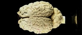
One of the first displays visitors will encounter is a ‘wall’ of 23 fluid-preserved mammal brains – from a Short-nosed Bandicoot to cow. The style of jar, with its black bitumen and paint backing, tells us that these were once used for display so it is exciting to put them in the public galleries again. Museum conservator, Jacqueline Chapman-Gray, runs us through the meticulous process she undertook to ensure these brains will look their best for their return to the limelight.
Cow brain before conservation treatmentA number of the brains had become dehydrated over time as the level of fluid – alcohol – had dropped. These needed to go through a rehydration programme to ensure their long-term preservation. This is more complex than simply adding more fluid to the jar. Instead the alcohol level needs to be increased gradually to avoid damaging the tissues.
Brains soaking in alcoholOthers had started to detach from their glass mounts, or anatomy labels that marked each of the different areas or sections of the brain had come loose. These were carefully remounted using specialist conservation-grade materials and a steady hand! Three brains had become completely detached and were repaired using a polyester monofilament thread, otherwise known as fishing line.
Repairing a human brain with a beading needle Labels found detached at the bottom of the jarFor the smallest of the brains a normal sewing needle was enough to pass through the tissues but for the larger two either a flexible 10cm beading needle or large 25cm mattress needle was needed. The original threading points were reused wherever possible though in one case this proved to be too difficult, as the tissue was soft and susceptible to breaking. With precision and patience I was able to gently stitch them back into place on the backing plate so they look as good as new.
All of the jars were given a thorough clean to ensure that seals were tight fitting and that the contents were shown off to their best. They were then filled with fluid to 4/5ths from the rim and the brains gently placed back inside.
Lids were sealed with clear silicone and each jar was topped up with a syringe through a small hole in the lid that is there for this very purpose – once full, this hole is also sealed.
Lastly, after the seals had dried, for the final finishing flourish black paint was reapplied to the backs and tops of the jars to provide a contrasting backdrop.
Ta-dah… the cow brain after conservation treatmentBrain Diaries opens on Friday 10 March and runs until Monday 1 January 2018. Take a look at the website to find out more about the exhibition and accompanying programme of events at braindiaries.org
For the past nine months there has been a lot of moving going on around here. Imagine moving house endlessly for weeks on end, but where your house is full of bones, insects, fossils, rocks, and weird and wonderful taxidermy. And the location of everything has to be precisely recorded. The museum move project was a bit like that.
Project assistant Hannah Allum explains…
The museums are migrating, we declared in May 2016. And so they have. The first major stage of the stores project has been completed. After we had created inventories for the largely unknown collections held in two offsite stores, the next stage was to pack them safely and transport them to a new home nearer the museum, a job which demanded almost 70 individual van trips! We now have over 15,000 specimens sitting in vastly improved storage conditions in a new facility.
A miscellany of boxes for a collection of shells
Let’s revel in some numbers. All in all there were over 1,000 boxes of archive material, mostly reprints of earth sciences and entomological research papers; over 1,300 specimens of mammal osteology (bones); and more than 1,000 boxes and 650 drawers of petrological and palaeontological material (rocks and fossils).
Some of the more memorable specimens include old tobacco tins and chocolate boxes filled with fossils and shells; a beautifully illustrated copy of the ‘Report on the Deep-Sea Keratosa’ from the HMS Challenger by German naturalist Ernst Haeckel; and the skull of a Brazilian Three-banded Armadillo (Tolypeutes tricinctus), complete with armour-plated scute carapace.
The skull and carapace of a Brazilian Three-banded Armadillo (Tolypeutes tricinctus)
There were also a few objects that have moved on to more unusual homes. A 4.5 m long cast of Attenborosaurus conybeari (yep, named after Sir David) was too large to fit in our new store and so made its way to another facility along with a cornucopia of old museum furniture. A set of dinosaur footprint casts, identical to those on the Museum’s lawn, have been gifted to the Botanical Gardens for use at the Harcourt Arboretum in Oxford.
And last but not least, a model of a Utahraptor received a whopping 200 applications from prospective owners in our bid to find it a suitable home. After a difficult shortlisting process it was offered to the John Radcliffe Children’s Hospital and following a quarantine period should soon be on display in their West Wing.
Casts of footprints by made Megalosaurus, queuing for a lift to Harcourt Arboretum. Image: Hannah Allum
Fittingly, the final specimen I placed on the shelf in the new store was the very same one that had been part of my interview for this job: The skeleton of a female leopard with a sad story. It apparently belonged to William Batty’s circus and died of birthing complications whilst in labour to a litter of lion-leopard hybrids before ending up in the Museum’s collections in 1860.
The sad story of a performing leopard
Though the moving part of this project is now complete there is still plenty of work to do. We are now updating and improving a lot of the documentation held in our databases, and conservation work is ongoing. The new store will also become a shared space – the first joint collections store for the University Museums, complete by April 2018.
The Utahraptor model in the old store awaiting collection by its new owners.
Illustrated plate from the Report on the Deep-Sea Keratosa from the HMS Challenger by Ernst Haeckel
The title page of the Report on the Deep-Sea Keratosa from the HMS Challenger by Ernst Haeckel
On the van, off the van; on the van…
Boxes of Earth collections material stored safely and in sequence in the new store
Boxes, glorious boxes…
Rack ‘n’ Roll: Hannah deftly working the racking
Lily Wilks, an intern assisting with the inventory of bound reprints. Image: Hannah Allum
Hannah Allum working in the old store. Image: Edward Adcock
Life Collections Manager Mark Carnall and Project Assistant Hannah Allum carrying one of the final specimens, a Mirounga leonina (Southern elephant seal) skeleton, into the new store. Image: Edward Adcock
The Attenborosaurus fossil cast, in its unusually shaped case, en route to a new custom built support frame. Image: Hannah Allum
The museum holds the only remaining soft tissue of the extinct dodo known anywhere in the world. The partially dissected skin of the head and scales from the feet of a single dodo represent one of natural history’s most iconic specimens. In fact, it is so tied to the museum’s identity and history that we use the dodo as our logo and it is even incorporated in the name of this blog.
Although the dodo head had been at Oxford University since the formation of the original Ashmolean Museum in the 17th century, it wasn’t really until the 19th century that the specimen really became celebrated.
Around this time, publications confirmed the extinction of the dodo from the island of Mauritius, where it was endemic. To capitalise on the rising interest in the animal, Ashmolean Museum Keeper John Duncan commissioned a number of casts of the Oxford Dodo head to give to, and exchange with, other museums.
One of the earliest of these casts was presented to the British Museum in 1828; later casts are recorded as being sent or exchanged with leading scientists of the time, as well as with Leiden Museum and the Royal College of Surgeons.
From these original and later casts further casts and models were presumably made, and eventually, dodo specimens spread to virtually every major natural history museum in the world. Today, many museums display casts of this head, all stemming from the single specimen held here in Oxford.
One of the many casts in the Oxford University Museum of Natural History, this one has been painted to match the original specimen
The Museum contains a number of models and casts of the head too; some are made from plaster and resin, some are painted to resemble the original specimen. The head of the dodo was actually dissected in 1847, by Henry Acland. He removed the skin from one side of the face so the early casts are a record of how the specimen would have looked originally.
In preparation for the Presenting display in the Museum I contacted natural history museums through the Natural Sciences Collections Association asking people to share information and photos about their casts and models of the dodo head. I wanted to try and construct a picture of how the dodo head was disseminated, as well as capture the diversity of quality and colours of representations of the original specimen. Here’s how far some of the dodos have flown:
Cast of the head labelled as coming from Cartwright Hall. Curator Gerry McGowan suspects this may have come via the Bradford Philosophical Society collections. The first curator of the society, Louis Compton Miall was friends with Thomas Henry Huxley and through him had contacts with many other geologists who may have gifted or exchanged this cast.
Bristol Museum & Art Gallery Bristol received a cast of the head directly from Oxford from Philip Duncan in 1834, keeper of the Ashmolean Museum between 1826 and 1855. Unfortunately, the head was likely destroyed in bombings of Bristol in 1940.
Canterbury Museum, New Zealand
Received a cast of a head from Professor Rolleston on 21 July 1871 in exchange for two kiwi skeletons which are still in the museum collections today.
Cast of head and foot presented to the museum by E.Ray Lankester in 1891/1892 just after leaving UCL and being appointed the Linacre Professor of Comparative Anatomy at Oxford.
The Great North Museum Hancock’s cast was presented by George Townsend Fox. This specimen had been presented to the Natural History Society of Newcastle in 1841 by Fox and had originally been presented by Philip Duncan.
Cast of the head that had quite a circuitous route to the Horniman Museum. The Horniman received the cast from the geology department of Queen Mary’s University of London in 1964 which received the cast from the Saffron Walden Museum in 1962.
Manchester Museum Cast of a head, presumed to have been presented by William Boyd Dawkins. The cast is currently on display in the Living Worlds gallery in Manchester Museum.
The National Geological Repository British Geological Survey
The Natural History Museum’s collections contain three casts of the Oxford Dodo head – two in the ornithology collections (pictured) and one in the palaeontology collections. The unpainted cast in the ornithology collections has the name ‘Johnson’ inscribed into the base.
Colleague Adam Smith got in touch with some interesting specimens from Wollaton Hall. The first one looks like another cast in this series but the second cast is unlike any of the others gathered here. The cast shows an open eye, detail on the beak as well as a more defined hook to the end of the bill.
Unfortunately, there’s not much information about the origins of these two casts so it’s probable that the ‘open eye’ cast may be a cast of a model reconstruction or an in progress sculpt. There’s an extremely slight chance it’s a cast of an otherwise unknown dodo head… If you recognise this dodo head do get in touch so we can solve this mystery for colleagues in Nottingham (it’s not the model dodo that we have on display here!).
Cast of a head at Warwickshire Museum with damage to the beak. Donated to the museum by clergyman and naturalist Reverand Andrew Bloxham in the 19th century. As the museum is currently moving stores, further information about when this cast was acquired is inaccessible.
**
If you work at a museum and have a dodo head cast to share, please do get in touch and we’ll update this blog accordingly.
Last updated: 10/11/17
‘Presenting… The Flight of the Dodo’ was on display at the Museum of Natural History from the 25 January to 22 March 2017.
Acknowledgements
With many thanks to colleagues across the sector who helped with information and images about dodo specimens: Adam Smith, Alice Adams, Jack Ashby, Carol Davies, Bonnie Griffin, Dan Gordon, Yvette Harvey, Mike Howe, Emma-Lousie Nicholls, Laura McCoy, Gerry McGowan, Nigel Monaghan, Henry McGhie, Pat Morris, Paul Scofield, Paul Shepherd, and Paul Sweet.


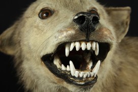


 There were massive cracks inside the mouth, where the old fill material had failed. After some testing, I chose a fill material that was flexible and able to withstand a fluctuating museum environment. This was an EVA adhesive, coloured with pigments to match the surrounding gum.