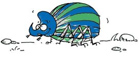
Why the world needs Dung Beetles

To celebrate National Insect Week 2016 we thought we would introduce you to the custodians of the Hope Entomology Collection here at the Museum. Our insect collection is made up of a whopping 6 million specimens, so our resident entomologists definitely have their work cut out. However, they have taken a little time out to tell us all about their specialisms and why their favourite insects are the best.

Darren Mann – Head of Life Collections

Darren out in the field collecting Dung Beetles

Dung beetles have been my passion since my late teens. I started with British species and then gradually broadened my interests to encompass the world fauna. But why dung beetles?

Well, they are beautiful insects, exhibiting an array of shapes and colours; they have been around since the dinosaurs, and have interesting biologies and behaviours, from nest-building and parental care, to stargazing. As a group, dung beetles are also very important in the ecosystem, removing dung and recycling nutrients.

Not only that, but dung removal and relocation offers additional ‘ecosystem services’ of fly control, livestock parasite suppression, plant growth enhancement, improved soil structure, reduction of greenhouse gas emissions, seed dispersal, and pollination. Inevitably, they are a source of food for other animals too.

Darren takes a closer look at a collected specimen

Dung beetles are found in all regions of the world, and consist of three main groups: the dor or earth-boring beetles (Family Geotrupidae) of around 600 species; the ‘lesser’ dung beetles (Family Scarabaeidae, subfamily Aphodiinae) of around 3,500 species; and the ‘true’ dung beetles (Family Scarabaeidae, Subfamily Scarabaeinae) of around 6,000 species.

With just over 10,000 species in total you’d think we have found all the dung beetles out there, but not so: it’s estimated that 40 per cent of species new to science are still to be discovered. In the UK we have just 60 species and over half of these are in decline due to agricultural intensification, pollution, use of veterinary drugs, and changes in livestock farming practises. The Dung Beetle Mapping UK Project (DUMP) aims to highlight the importance of this group and promote research and conservation in this area.

Despite their name, not all dung beetles eat dung, with some species preferring fallen fruit, fungi, or even dead animals. The South American roller (Deltochilum valgum) is an avid predator of millipedes and another South American species (Zonocopris gibbicollis) feeds on snail mucus!

So with their high diversity, fascinating ecology, and great economic benefit, perhaps the question really should be ‘why not study dung beetles?’.

A plesiosaur named Eve

A Spotlight Specimens special for Oxford Festival of Nature

by Juliet Hay, Earth Collections preparator and conservator

I feel myself very lucky to have a job that involves working with the fossil remains of long-extinct animals. One of the things my colleagues and I are currently working on is a plesiosaur – a marine reptile that lived in the sea millions of years ago.

This particular specimen was found in a clay pit near Peterborough by members of the Oxford Clay Working Group in 2014, and is a near-complete example of its kind. The palaeontologists who found the specimen named it Eve, although we don’t know if it was male or female, and perhaps never will.

The discovery of large fossil vertebrates like this is rare, so we are fortunate to have had the specimen donated to the Museum by the quarry owners Forterra.

Juliet at work on the plesiosaur skull

The plesiosaur is 165 million years old and, when alive, was around 5.5 metres long. It had a long neck, a barrel-shaped body, four flippers and a short tail. The find is particularly exciting as the skull was also discovered. It is encased in a clay matrix, which is relatively easy to remove, but the work has to be carried out under magnifying lenses and microscopes.

As the skull is quite small relative to the size of the body, the features are very delicate and it is a painstaking process to remove the sediment without damaging the fossil bone or losing any tiny fragments. Fortunately, pictures of the skull have been produced using CT scanning technology, and the images are proving invaluable as an aid to assist in its preparation. It’s a bit like having a jigsaw puzzle with the picture on the lid to refer to!

sdfsd
A belemnite hooklet at 12x magnification, found with the plesiosaur remains and possibly part of Eve’s last meal

The clay covering the skull is being sieved and examined and tiny hook-shaped fossils have been found. These came from the arms of squid-like creatures called belemnites, which may have formed a large part of the plesiosaur’s diet.

It is too early to say for sure, but Eve could represent a species new to science, as some features, such as the shape of the flipper bones and some of the surfaces of the bone in the skull, are quite unusual. Further research needs to be done before the findings can be published in scientific journals – watch this space.

And if you’re visiting the Museum before 25 July, you can see some of the fossilised remains of Eve for yourself, in our Presenting… display case.

OFoN_logo_green block_small

The museums are migrating!

Night falls in the Museum

by Hannah Allum, Project Assistant

The film ‘Night at the Museum‘ is not as far from the truth as you might think. Museum specimens may not come back to life at night, but they are moving all the time. Whether we’re putting on a new display, loaning a specimen to another museum or using them for teaching sessions, our collections are very active, despite being long dead.

Amphistium fossil fish; an early relative of the flatfish and part of the Earth collection

The Oxford University museum collections are about to begin a huge migration as they move from various off-site stores to a new facility. Generally, museum stores are not open to the public and often house important reference and research specimens, which make up a huge proportion of natural history collections. These valuable stored collections require specific environmental conditions to make sure that they last for generations to come. Taxidermy and articulated skeletons make for eye-catching displays, but tend to be a relatively small part of the collection. In fact, having just 1% of your natural history specimens on public display is the norm.

Lured in by Life Collection labels; this headless brown bear will soon have a new home.

As well as improving the storage conditions and ease of access for staff and researchers, this will also be the first time that the Oxford University museums have shared a storage space. It will be a challenging but rewarding project to re-home such a huge variety of artefacts and specimens that have come from all over the world; each with their own unique story.

Hannah profile
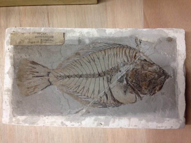
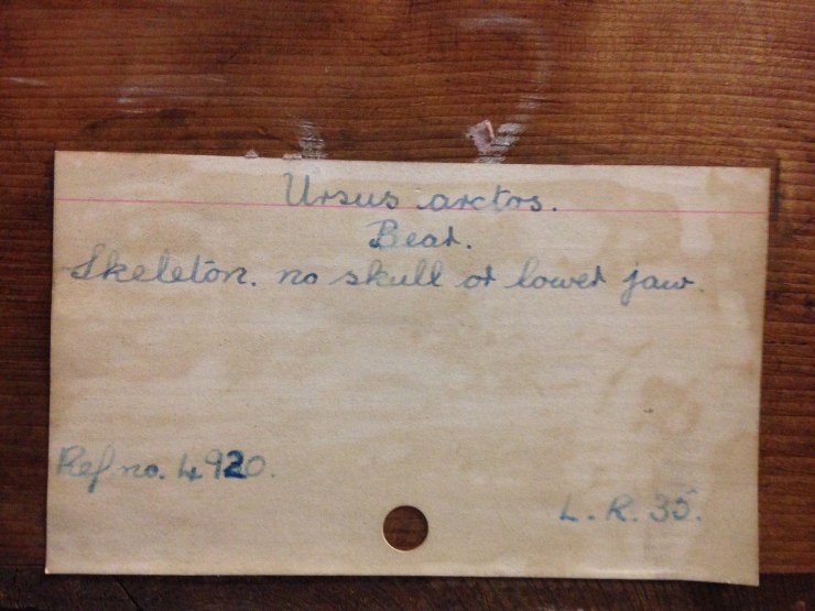
As the new Project Assistant working for the Museum of Natural History, I am the lucky person who gets to discover some of these stories. I will be working with specimens from both Earth and Life collections, as well as some material from the Library and Archives. The first stage will be making a detailed list of everything that needs to be moved, then I can go on to prepare the new store and get the supplies I’ll need to document, pack and transport everything safely.

Some enticing titles from the Library

As you can see, I have already come across some fascinating specimens and look forward to getting stuck in to this project.

There will be blog posts throughout the year to update you on our progress and to reveal some exciting stories from the stores.

You can also follow the hashtag #storiesfromthestores on the Museum’s Twitter feed: @morethanadodo

Secretarial work

After Treatment 2

Our striking Secretary Bird stands at over 1 metre tall and should be one of the most impressive specimens in the Museum, but it hasn’t been looking its best for a while. This African bird of prey was looking rather sorry for itself, with scruffy feathers and moth damage. Conservation intern Ruth Murgatroyd stepped in to bring it back to its former glory.

**

Wrapped and ready for the freezer

The first step for any specimen undergoing conservation treatment is a 72 hour spell in the freezer at -30⁰C. This kills off any inhabiting webbing clothes moths, which can be very destructive to taxidermy specimens. Getting ready for the freezer required some creative packaging to protect the characteristic quill shaped plumage and tail feathers, before wrapping in plastic.

The feathers would need a good groom, which is a lot easier once they’re clean. I used dry methods first, including a brush dust with a vacuum cleaner and the very effective use of cosmetic sponges. The feathers were further cleaned with a gentle non ionic detergent in water and rinsed with a water/ethanol mix. I used a paint brush to dab the solution onto each feather individually.

Ruth cleaning tail feathers

When the feathers were clean and dry they were groomed to realign the filaments of the feathers, known as barbules. Parts of the bird’s tail and right wing were missing, so as this is a display specimen, we decided it was appropriate to recreate these areas to more accurately represent what the bird looks like in the wild. Any additions had to be easily identifiable and reversible. Goose feathers were sourced and colour-matched with Orasol dyes. They are now held in place by adjacent feathers and give a much more natural appearance.

Before treatment, with missing wing and tail feathers

The face of the Secretary Bird had been previously painted but this was quite faded in colour compared to the buoyant oranges and yellows of the animal in the wild. We decided to reflect this with a touch up. The new layer was painted in with acrylics, but a base layer of water soluble adhesive now protects the original paint, so layers of paint could be taken back at any time.

The finishing touch to the conservation of a taxidermy specimen is often to make sure the eyes are clean and gleaming. Saliva on a swab is really effective for this.

The Secretary Bird was then ready to go back in its newly-polished case. This had also been lined with UV film to protect the specimen from light damage. Just before it went back on display, the bird made an appearance at the Museum’s daily ‘Spotlight Specimens’ session where it met visitors keen to hear about its recent conservation.

Secretary Bird  back on display
Secretary Bird back on display

Pop in to the Museum to see the finished Secretary Bird on display and standing tall .

Ruth Murgatroyd, Conservation Intern

TLC (turtle loving care)

Turtle post-treatment

The Green Turtle (Chelonia mydas) above has been receiving some much-needed TLC from Abby Duckor, our first conservation intern from UCL’s MSc in Conservation for Archaeology and Museums. Here Abby explains what she’s been up to…

During my time at the Museum I have been lucky enough to spend a fair amount of time working on this taxidermy turtle. There was plenty to do: the specimen was covered in a dark layer of dust; there was a large tear in the neck, perhaps from a knock; and the taxidermy was generally overstuffed, noticeably on the stomach plate which had become completely detached, revealing the inner filling material.

The turtle shell (carapace) in the middle of treatment. The carapace was cleaned with detergent and de-ionized water. Toothbrushes were used to scrub the hard shell and to help remove any ingrained dirt.
Most of the filling was wheat with a small grain size, dating it to pre-1950, according to Dr Stephen Harris: after 1950 wheat grains were cultivated to be fatter and the stalks shorter. The wheat and other plant materials in the filling suggest an English location for the taxidermy.

The specimen itself is labelled as part of Rev. Buckland’s collection, dating it to the early 1800s, if not earlier. William Buckland, an important early geologist and palaeontologist, was quite a character. During his life he amassed a large collection of living and mounted animals. He claimed to have eaten his way through the animal kingdom, and you have to wonder if he ever tasted this fellow… Green Turtles were a popular food for sailors and locals, reducing their population size. Today they are listed as endangered.

Abby working on the turtle in the conservation lab.
Conservation treatment of this specimen involved cleaning the shell with detergent and deionized water, revealing a colourful shell underneath. You may have noticed that this Green Turtle is not actually green. In fact, the Green Turtle is named after the colour of its fat, not the colour of the skin. Ours had been painted a dark greenish-brown colour, because they lose their skin colour after they die, but I removed the top layer of paint to better reveal the yellow scales on the turtle’s head, tail and limbs.

The final touch was the replacement of the stomach plate, or plastron, which is now held in position with epoxy putty wedges attached to the metal stakes that hold up the specimen. Cleaned, and with everything in its right place, our Green Turtle is now in much better shape, as you can see in the photo at the start of the article.


Twice in a whale

natural-history-museum-oxfordsmall-41

By Mark Carnall, Life Collections

Followers of our Once in a Whale blog a while back may be aware of the huge task that faced our Life Collections conservator Bethany Palumbo and her team as they set out to clean, restore and repair the whale skeletons that hang from the Museum roof.

In my first week here, I received an enquiry about the history of these specimens, and digging through the archives I was pleased to find that they are not just ‘prop’ skeletons acquired for the purpose of display – they are important in the history of whale biology too. So this article is something of a postscript to the Once in a Whale project.

Surprisingly, it wasn’t until the mid-19th century that much was understood about the science of the largest animals to have lived on Earth. Some species were known from strandings; others from accounts – varying in reliability – from fishermen.

img_5415small-copy
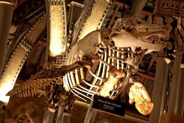
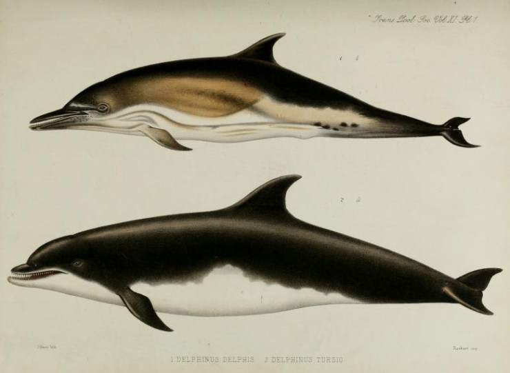
Yet difficulties in preserving and transporting such large creatures (as well as the penchant for eating stranded whales at community festivals) meant that the biology and behaviour of whales was poorly-described and documented until fairly recently. So much so, that in early scientific literature just a few scientists are singled out as having actually seen the animals they were studying.

In the Museum there are five whale skeletons suspended from the roof, along with the skull of a Humpback Whale and the mandible of a Sperm Whale. Some of the earliest ‘whaleologists’ made the trip to Oxford to see these specimens in a race to formally describe new species or new aspects of whale biology.

In particular, two 19th-century anatomists competed to make new discoveries about whales. Dr John Edward Gray, keeper of zoology at the British Museum (Natural History), and Professor Daniel Frederick Eschricht, a Danish comparative anatomist, were so competitive that Gray made sniping comments in formal papers, questioning Eschricht’s observations.

John Edward Gray ‘destroying’ Eschricht’s observations. From Gray, J. E. 1864. On the Cetacea which have been observed in the seas surrounding the British islands.
John Edward Gray ‘destroying’ Eschricht’s observations. From Gray, J. E. 1864, On the Cetacea which have been observed in the seas surrounding the British islands

But perhaps this competition was more sporting than malicious: Gray did name the gray whale, Eschrichtius robustus, in Daniel Eschricht’s honour.

Eschricht actually presented the Museum with two of the specimens now on display: the Humpback Whale skull at the entrance and the suspended Minke Whale skeleton. Of the others, the Bottlenose Dolphin skeleton was caught near Holyhead in 1868 and was drawn by another notable natural historian, William Henry Flower, before being skeletonised for the Museum.

The Orca skeleton is from an individual killed in the Bristol Channel by fishermen in 1872, and the Beluga Whale was collected from Spitsbergen, Norway in 1881 and presented by Alfred Henge Cocks, who donated a range of mammal specimens to the University of Oxford.

The female Northern Bottle-nosed Whale skeleton has been harder to track down. It’s possibly a specimen shot in Weston Super-Mare in 1860 mentioned by Gray, but it isn’t clear. Lastly, there’s the large Sperm Whale mandible that greets visitors at the entrance. It doesn’t have much of a recorded history, but is allegedly one of the largest specimens in the UK according to a ‘researcher’ whom I’ve yet to track down.

William Flower’s drawing of the bottlenose dolphin (lower) the skeleton from this individual is on display in the museum Flower, W. H. (1880), I. On the External Characters of two Species of British Dolphins (Delphinus delphis, Linn., and Delphinus tursio, Fabr.). The Transactions of the Zoological Society of London, 11: 1–6. doi: 10.1111/j.1096-3642.1980.tb00343.xWilliam Flower’s drawing of the bottlenose dolphin (lower) the skeleton from this individual is on display in the museum Flower, W. H. (1880), I. On the External Characters of two Species of British Dolphins (Delphinus delphis, Linn., and Delphinus tursio, Fabr.). The Transactions of the Zoological Society of London, 11: 1–6. doi: 10.1111/j.1096-3642.1980.tb00343.x
William Flower’s drawing of the Bottle-nosed Dolphin (lower); the skeleton from this individual is on display in the Museum

The next time you are in the Museum, do look up: the skeletons there are not simply representing ‘whaleness’ but are also individual animals and important specimens in the early discovery and description of whale biology.