Tomorrow afternoon the Museum will host talks, tours and a dance performance as part of the Breath Festival, a unique series of events coordinated by the Oxford University Hospitals Artlink programme. To coincide with the festival we have put together a special display in our changing Presenting… case, all about breath and breathing across the animal kingdom.
There’s something of the Halloween macabre about the display too, with its pink-coloured lungs and eviscerated bodies suspended in spirit. Here’s a taster of the display, but to see the full selection head down to the Museum either for the Breath Festival tomorrow, Saturday 1 November, or at any time during the rest of the month.
Lungs of a lizard, goose and duck
The breath of life All animals breathe to obtain oxygen for their bodies and to expel carbon dioxide, but there are many different ways of breathing: from the book lungs of scorpions to the gills of fishes and the true lungs of mammals. Terrestrial animals generally take in oxygen from the air, while for aquatic animals it usually comes from the water.
Crocodile and alligator lungs
Some aquatic animals, such as sponges and jellyfish, take in oxygen by diffusion through their body wall. Others have specialist organs such as gills. But not all aquatic animals take in dissolved oxygen. Many insects, including diving beetles, have wing cases or hairy bodies that allow them to carry a bubble of air with them when they dip beneath the water’s surface. Aquatic mammals, including seals and whales, must come to the surface to breathe, and often have special adaptations for this.
Certain terrestrial animals, such as earthworms and amphibians, can breathe through their skins, but amphibians have simple lungs as well. All reptiles, mammals and birds breathe using lungs; in birds there is also a system of air sacs and air spaces within the bones that make breathing more efficient. Insects breathe through branching tubes called tracheae, while arachnids use folded structures known as book lungs.
The evolutionary adaptations of this most basic life function are many and varied: a simple breath is not so simple after all.
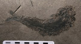
Palaeoniscum, a fossil fish from the Upper Permian of County Durham
By Hilary Ketchum, Collections Manager, Earth Collections
A few weeks ago Eliza Howlett (Collections Manager, Earth Collections) and I travelled to Wales to pick up an exciting collection of fossils that had been given to the museum.
The collection was kindly donated by Mr Phil Bennett, who has been finding fossils for over 20 years. In 2004, Phil won the Mary Anning Award for his outstanding contribution to palaeontology by making his collection available for researchers to study. He has an excellent eye for spotting new and interesting things, and thanks to this has a species of trigonotarbid (a spider-like animal) and crustacean named after him.
We had a fantastic day in Wales. After a delicious lunch we looked through the collection, and Phil told us all about the different specimens, pointing out some of his best finds.
One of his favourite specimens is a beautiful fossil fish called Palaeoniscum, from the Upper Permian of County Durham, which is approximately 270-250 million years old.
While Palaeoniscum is instantly recognisable as a fish, some of the older vertebrate fossils in Phil’s collection would look a bit out of place in a modern ecosystem. These fossils, from the Old Red Sandstone in Wales, date from the Lower Devonian period, approximately 410-420 million years ago.
An osteostracan from the Lower Devonian of Wales with a semi-circular head shield (left). The specimen is about 6 cm long from head to tail.
Featuring heavily in Phil’s Old Red Sandstone collection are fossils of strange, fish-like vertebrates (animals with backbones) called osteostracans. Their bodies were covered in large scales and they had massive bony head shields, but they didn’t have jaws or teeth.
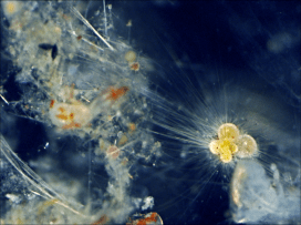
The head shields of osteostracans have a mysterious structure called a ‘cephalic field’ (shown in red in the image below). Palaeontologists do not know for sure what the cephalic field was for. Some think that it was a sensory organ that was used to pick up vibrations in the water or changes in electric fields, helping them detect prey or predators.
Image credit: Philippe Janvier
Phil had very carefully packed all the fossils into cardboard boxes before we arrived, so it didn’t take long for us to load the specimens into the back of the car and drive them safely back to their new home. We have now begun the process of incorporating the fossils into the museum’s permanent collections. The specimens will be taken out of their boxes and put into museum trays, ensuring that all of Phil’s labels are kept so that no information is lost. Over the next few months, the specimens will be catalogued on the museum’s electronic collections database. Each specimen will be given a unique museum number so it can always be easily identified.
It’s a fantastic collection, and we are really excited to have it in the museum. They can be used for display and teaching, and will be available for researchers to study for years to come.
They say a picture is worth a thousand words, and at the Museum we make thousands of pictures: pictures to document, pictures to investigate, and pictures to wow. We use a lot of different imaging techniques too, from standard close-up photography to scanning electron microscopy, which reveals the most minute details.
To coincide with the final week of the Wildlife Photographer of the Year exhibition here, on Saturday 20 September we held a new adult workshop to give people some hands-on experience of some of these processes. Imaging Techniques in Modern Natural History gave participants the chance to get up close to some wonderful specimens and make their own images to take home.
I had planned to review the day here, but Rose Parkin, who took part in the workshops, very helpfully sent in her own write-up of the sessions. So here’s a special guest post from Rose, along with some pictures taken by people on the day.
*
By Rose Parkin
When I signed up for the digital imaging course I expected a fairly dry, tech-heavy day. Instead, the experience was really exciting. Not only did it provide hands-on experience of viewing and recording images with new technology, it also gave me a brief glimpse behind the scenes of my favourite museum.
Laser Scanning and Digital Modelling For our first session our small group was led through a maze of corridors by Sarah Joomun, the Documentation Officer, to the laser scanning lab. It sounded a bit futuristic, and it turned out that it looks that way too. Sarah popped a fossil onto a mount, clicked a few buttons and red lasers appeared, scanning the fossil’s surface while it rotated. After ten minutes the first 3D image of the fossil was produced – a beautiful net of triangles, which looked like a teleporting object in a science fiction film.
Laser scanning Image copyright Tom Nicholson-Lailey
Sarah turned the fossil and scanned it again. The challenge was then to fit these two images together to make a complete 3D model. Amazingly, this technique enables other palaeontologists around the world to see and replicate, with the use of a 3D printer, the exact size and shape of a fossil without it ever leaving the museum.
Multi-plane Microscope Photography Our next session was upstairs, with artist-in-residence and photographer Katherine Child. Even though we were close to the main corridor of the museum it felt like a real working space, crammed full of equipment and insect specimens. Katherine had chosen the tiniest of insects for us to photograph with the multi-plane microscope. It looked like a small seed with some barely visible limb-like protrusions.
Multiplane photography. Image copyright Rose Parkin
But under the microscope a wonderfully strange insect became visible, with the most bizarre appendages and bright orange legs. While the microscope already showed a great deal of detail the multi-plane photography captured an incredibly crisp image. The microscope takes large numbers of photos of the specimen, using different focal planes each time, then the focussed elements are all stacked together to produce a crystal clear photograph.
Once we’d chosen and photographed some other insects from the collection and poked around the room a bit (finding a disturbing collection of large pickled spiders), we were taken on a tour of the entomology department. Katherine led us through corridors of offices and labs, up to a stunning store room that felt almost church-like, with rows and rows of cabinets full of fascinating insects.
Scanning Electron Microscopy After lunch we had a laboratory session with museum director Paul Smith to look at sand under an electron microscope. Luckily, that was much more exciting than it sounds! The sand was taken from Dog’s Bay on the west coast of Ireland and is rich with a wide range of tiny fossilized organisms. Paul showed us how to carefully select individual microfossils from a tray using just a microscope and a paint brush.
Professor Paul Smith demonstrates the scanning electron microscope. Image copyright Rose Parkin
We then viewed some of the microfossils using a scanning electron microscope. This allowed us to see an incredible level of detail. The microscope was so powerful that we could see hair holes in a fossil the size of a grain of sand.
DSLR Macrophotography My final session was a crash course in macrophotography. Held in the seminar room, the low lighting and floor-to-ceiling collection of specimens lent an almost eerie feeling to the set-up.
Macrophotography. Image copyright Keith BarnesBearded dragon. Image copyright: Rose Parkin
Once prepped, we were let loose on four separate camera setups. Being able to choose and shoot at our own pace made this feel like a really creative experience. The help given by professional photographer Keith Barnes and public engagement officer Scott Billings was perfect – very hands on but not patronizing (despite my lack of DSLR experience).
With this digital imaging course the museum has created a really exciting snapshot of the work that goes on behind the scenes, reinforcing the fact that this impressive place is much more than just an ordinary museum.
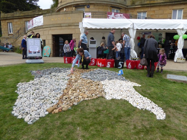
In rush hour traffic, carrying a precious cargo, the Museum’s Director, Professor Paul Smith and Head of Archival Collections, Kate Santry, headed north. They took the William Smith archive on tour to the Yorkshire Fossil Festival, in lovely Scarborough. Hosted by the Scarborough Museums Trust, in partnership with the Paleontological Association, the Yorkshire Fossil Festival had a wide array of exhibitors, lectures and events all celebrating fossils over the course of three days.
Festival-goers constructing a geological map of Yorkshire using stones. Smith would have been proud!
Despite some chilly and cloudy weather the festival saw a great turn-out. On Friday 12th September, a number of local primary and secondary schools made a visit, participating in activities that gave hands-on experience in understanding more about fossils. The school groups who visited our stall had the opportunity to act out a play exploring how fossils are made with our Director, Paul Smith, as the narrator!
Horace the travelling Pliosaur cinema
The crowds visiting the festival over Saturday and Sunday got a rare look at original material from the William Smith archive and were asked to help us transcribe the collection, which has recently been digitised and catalogued. Although he is ‘the father of English geology’, William Smith is not a universally known figure in the history of science. But it was a very different matter with the Scarborough crowd.
William Smith, ‘Father of English geology’ and Scarborough resident
Born here in Oxfordshire, Smith lived in Scarborough at the time he died in 1839 and was an active and important figure in the town. In addition to being an early member of the Philosophical Society, he was also consulted to solve the town’s water supply issues, select stone for the bridge between the town and its newly discovered spa, and most notably in helping to design the Rotunda Museum that was our base for the three days.
The biggest hit at our table over the weekend was the Geological Map of Yorkshire, published by Smith and Cary in 1820 as part of his County Map series. While approximately 400 people spent time looking closely at, and talking with us about this important map, its popularity was followed closely by a copy of Smith’s wine merchant’s bill from Scarborough dated 1839. It certainly appears that Smith was a fan of gin and marsala…
If you’re a regular reader of this blog you might have heard of Dr Tracy Aze already, and may even recognise the strangely-shaped specimen above as an example of planktonic foraminifera, the single-celled marine organisms that Tracy has been researching. This morning we have issued a press release about Tracy’s research which offers a warning from history about carbon emissions and global warming.
Surprisingly enough, the study shows how the fossils of these creatures hold clues to the impact on our oceans of man-made global warming. Around 56 million years ago, in a period known as the Palaeocene-Eocene Thermal Maximum (PETM), a rapid rise in greenhouse gases caused sea surface temperatures to rise as high as 40°C, with significant impacts on marine life.
Worryingly, the PETM – which lasted for around 170,000 years – saw the release of roughly the same volume of CO2 as expected from modern fossil fuel consumption. Tracy explains:
The amount of CO2 that is predicted to be released from the Industrial Revolution to around 100 years from now is roughly equivalent to what happened in the PETM. But the big difference is the rate of release: today we are releasing greenhouse gases at a far faster rate than 56 million years ago.
Although the research was conducted by Tracy, the project was led by Professor Paul Pearson of Cardiff University and funded by the UK Ocean Acidification research programme.
Tracy and her team used newly-extracted planktonic foraminifera fossils from Tanzania, dating from the PETM period. The tiny shells of these organisms contain different proportions of oxygen isotopes and these proportions are largely determined by the sea temperatures at the time. So the fossil shells offer a glimpse of the way sea temperatures were rising alongside the release of greenhouse gases, as well as a record of the relative abundance of this planktonic life in the oceans.
The PETM shows us that rapid increases in CO2 in the atmosphere have significant impacts on global temperatures, with the new information from our study site showing that tropical sea surface temperatures may have exceeded 40°C with an associated local disappearance of marine life.
The research paper, Extreme warming of tropical waters during the Paleocene–Eocene Thermal Maximum, was published in the September issue of Geologyand is available as open-access.
The tooth seen from different angles. Scale is 50mm
A few weeks ago, Dr David Martill from the University of Portsmouth visited the Museum’s collections to look at pterosaur fossils. While he was carrying out his research, he stumbled upon a tiny little tooth, about 1 cm in height, which looked like it might belong to a dinosaur.
Megalosaurus was a theropod, like the owner of the mysterious tooth
We showed the tooth to Dr Roger Benson, an expert on dinosaurs at the Department of Earth Sciences in Oxford. He confirmed that it belonged to a type of dinosaur called a theropod, because of its recurved shape and the presence of a series of serrations, called denticals, along one of its edges. The serrations are worn, making them difficult to see, which might explain why the tooth had not been identified as coming from a dinosaur before now. Theropods are the group of meat-eating dinosaurs that gave rise to birds, and include T. rex, Velociraptor and the UK’s own Megalosaurus. The tooth would have been from a small animal, probably less than a metre long. We can’t tell if it was from a small species or just a young dinosaur.
So why is such a diminutive dinosaur tooth potentially so exciting? The reason is that it might come from the Lhwyd Collection. If it does, it would make it the oldest surviving documented dinosaur tooth in the world.
Specimens from the collection were described by Lhywd in 1699 in his book Lithophylacii Britannici ichnographia, a catalogue of British fossils and minerals in the Ashmolean Museum, where he was Keeper from 1691 until his death in 1709. The collection was transferred to the Museum of Natural History after it opened in 1860. Sadly the collection became neglected, and at one stage the number of known Lhywd specimens in the collections was down to just two. However, thanks to the painstaking work of James Edmonds, one of the curators, in the late 1940s, the collection was brought back together, and the Museum now has around 80 of the specimens from the catalogue.
Lhwyd 92 catalogue entry
The problem is we can’t be completely sure that the tooth Dr Martill found is from Lhywd’s collection. Lhwyd wrote numbers on all his specimens, which correspond to the numbers in his catalogue (shown here), and this tooth is indeed labelled with the number 92. The handwriting on the dinosaur tooth, and the pen used to write the number are extremely similar to those used on other specimens from the Lhywd collection. It was also found with other numbered specimens from the Lhywd Collection. However, when we checked the catalogue entry, we found that the number 92 doesn’t correspond to a tooth, but to a piece of fossil coral.
Our next thought was that maybe the tooth was originally numbered 1292, but the 12 at the beginning had become worn away through handling. Examination under UV light, to increase the contrast between the writing and the tooth, revealed no sign of other numbers, and in any case the number 92 was written centrally on the tooth, rather than to the right as you would have expected if it had been the end of a longer number.
Here you can see 92 written on the tooth
We know that Lhwyd’s collection sometimes included several specimens under the same number, and there is another tooth in the collection labelled 1292. This specimen, a shark tooth, is slightly larger than the theropod tooth, so is it possible that Lhwyd wrote the full number on the larger specimens but abbreviated it to 92 for the smaller ones? Hard to say. It seems we have come tantalisingly close to a very exciting discovery, but for now it remains a mystery still to be solved.
Hilary Ketchum & Eliza Howlett, Collections Managers, Earth Collections


 In rush hour traffic, carrying a precious cargo, the Museum’s Director, Professor Paul Smith and Head of Archival Collections, Kate Santry, headed north. They took the William Smith archive on tour to the Yorkshire Fossil Festival, in lovely Scarborough. Hosted by the