Elsa Panciroli recently joined the Museum research team as an Early Career Leverhulme Research Fellow. Elsa is a Scottish palaeontologist, whose studies focus on the early evolutionary origins of mammals, working extensively on fossils from the Isle of Skye. Here she tells us how her work will combine studies of mammal evolution with stunning new fossil finds from Scotland.
We are mammals. This means we share a common ancestor with creatures as different as hippos, opossums and platypuses. All of us are united in one taxonomic group by a suite of characteristics in our bodies, but principally, that we feed our young on milk. Every mammal from a baboon to a blue whale produces milk for their offspring, and this makes us unique among animals alive on Earth today.
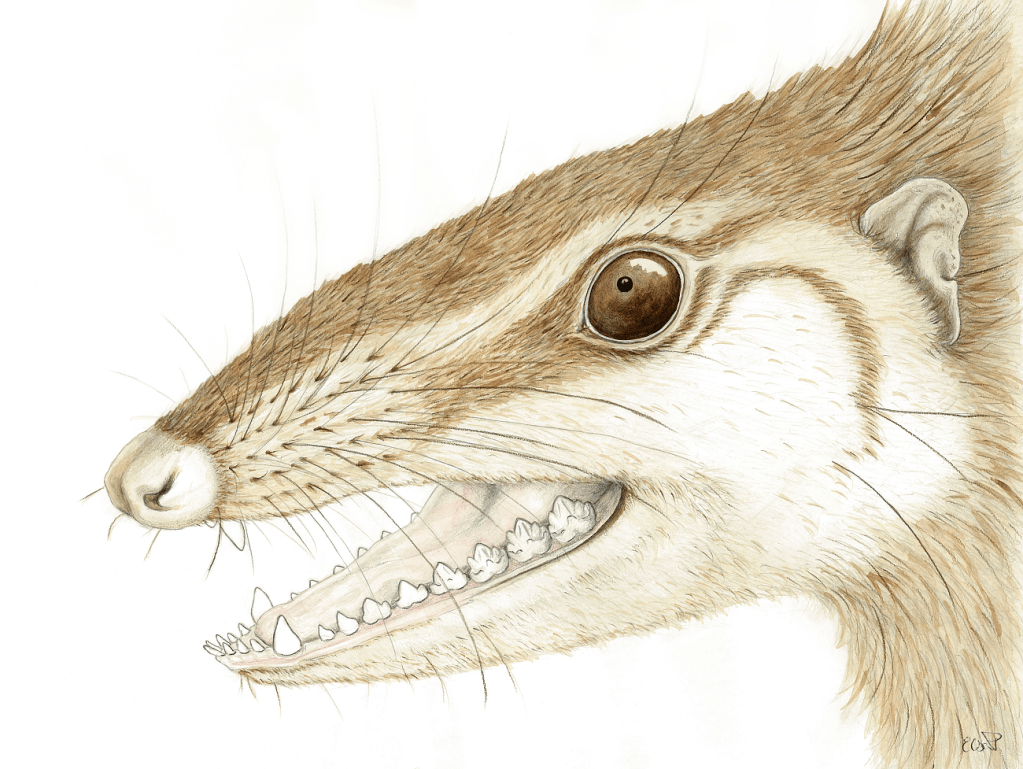
Wareolestes rex is a Middle Jurassic mammal, illustrated here by Elsa Panciroli
But not all mammals bring their young up in the same way; raising a kitten is nothing like raising a kangaroo or a platypus. Kittens are born stumbling around with their eyes closed, while platypus babies are laid in eggs – yes eggs – and when they hatch they look like little scampi. Both are underdeveloped at birth or hatching, but that’s nothing compared to kangaroos. They leave the womb only millimetres in length, and wriggle their way like living jellybeans toward a teat in the marsupial pouch, where they latch on. Only after two months of milk-drinking are they able to hop for themselves and leave the pouch.
The different ways that mammals are born and grow is a huge area of scientific research. But there are still some major questions to answer about the evolution of these growth patterns. When did the ancestors of mammals stop laying eggs? Were they born defenceless, or able to fend for themselves? How quickly did they grow up and how long did they live?
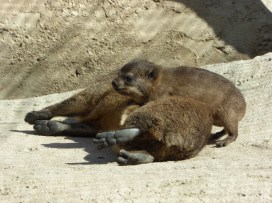
The Rock Hyrax (Procavia capensis) is a terrestrial mammal native to Africa and the Middle East
Over the next three years at the Museum, I’ll be looking for evidence in the fossil record to help us try and answer some of these questions. I’ll study living mammals to understand how they are born and grow, combining this information with data from some of the amazing fossils being found on the Isle of Skye. With collaborators in South Africa I’ll try and work out how the ancestors of mammals developed, and what this means for the bigger picture of the origin of mammals as a group.
Alongside my main research I hope to share lots of stories about our fossil past through the museum’s fantastic public engagement programme. I’m also very active on social media, and I write about science for online and in print publications. So if you see me on your next visit to the building, or find me online, feel free to ask about my research! I look forward to seeing you, and sharing more about the elusive and exciting origins of mammals – and ourselves.
Worms, fish and … Greenland? Hugely different topics which all have one thing in common – the Museum’s First Animals exhibition online lecture series. Running every other Wednesday from May until September 2020, this series provided a fantastic insight into a wide range of topics about how the first animals lived, died, and are studied. And illustrator Rachel Simpson tells us how she drew her way through them all…
I came across this lecture series just before the first talk and I knew I had to sign up. Drawing along to lectures is a hobby I seem to have developed in the past few months as we went into lockdown and didn’t have much to do. It’s the perfect combination for me – an opportunity to listen to interesting topics and brush up on my live drawing skills at the same time. There’s no pause button, there’s no asking the webinar speaker to just go back a few slides and hold on a minute whilst I draw; it’s fast paced, it’s inspiring and it’s a great way to just create art.
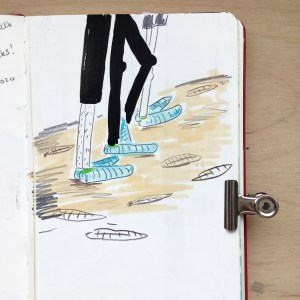
Barma Booties used on the rocks at Mistaken Point, and my first drawing of the series.
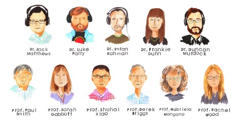
I’ve done some illustration work with the Museum before so I knew that it was going to be fun. In 2018, I worked with Dr Jack Matthews illustrating Ediacaran Fossils as part of a collaborative university project between the University of Plymouth and the Museum. I was also lucky enough to be able to go to Newfoundland and see some of the fossils myself, again with Jack. This was such an incredible opportunity and opened up a whole new world of science/art collaborative work which I didn’t know about before.
The First Animals series kicked off with Jack’s talk titled Don’t walk on the rocks! – an interesting insight into how protective “Barma Booties” (some rather funky socks worn to protect fossil sites such as Mistaken Point, Newfoundland) might actually be damaging to the fossils they’re meant to be protecting. Having been to Mistaken Point myself and worn these socks, it was interesting to hear about their possible impact and to learn about the experiments conducted to prove this fact.
Of course, at the same time as Jack was talking, I was scribbling away in my sketchbook trying to form some sort of visual response to the talk. At the end of the hour I’d managed a portrait of Jack and a family of Barma-Booted tourists trampling on the fossil site. It was a start. The beginning of my lecture drawings and a point at which I can retrospectively say started a new hobby.
Annelid worms drawn with Tombow brush pens.
Over the following weeks we heard about worms from Dr Luke Parry; 3D reconstruction from Dr Imran Rahman; The Chronicles of Charnia by Dr Frankie Dunn; and the first animal skeletons from Dr Duncan Murdock. Luckily for me, all the speakers kindly included photos and descriptions of the topics they were discussing which meant that I was never short of visual inspiration for my drawings. After all, it’s hard to try and draw an annelid worm if you’ve never seen one before.
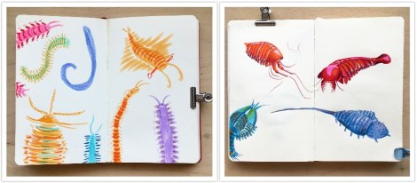
I love to look at the fossils being discussed and then try to draw a little character or creature inspired by them. They’re not scientifically accurate, nor are they always anatomically correct, but they have character and begin to bring to life the essence of something that’s been dead for many millennia. The fossils are obviously stone-coloured so I take as many liberties as possible when it comes to colour. I like to make them as vibrant and colourful as I can, so although they probably didn’t look like that, that’s how I like to think they looked.
Some fun little beasties from Dr. Imran Rahman’s talk.
Charnias galore! They come in all different shapes and sizes.
Small filaments which could have joined all those Charnia together.
Shells, bones and teeth from Dr. Duncan Murdock’s talk drawn in Tombow brush pen and Posca Pen.
Within my wider practice I like to use stamps as the basis of my illustrations. These however, are time consuming to make and therefore not very suitable for when I’m drawing along to lectures. As a result I’ve found myself using brush pens and pencils to make my lecture illustrations. If you’re interested in art, or thinking about getting into art, brush pens will be your best purchase. They create a wonderful quality of line and are quick and easy to use. Whereas a ballpoint pen will give you one line of a certain weight and thickness, brush pens are versatile and depending on the pressure applied, the line quality will change.
For the first few lectures I only used brush pens, but later on I decided to use coloured pencils as well, to add depth to the drawings. As I got more used to drawing in lectures I found that I was making more illustrations per talk. Early on, I managed to finish maybe a double page in my sketchbook but towards the end of the series I was filling four double pages! It’s amazing what a little bit of practice can do.
As the weeks went by the talks continued and we heard about the evolutionary origin of animals from Museum director Professor Paul Smith; an introduction to taphonomy, the study of fossilisation, by Professor Sarah Gabbott; and how the first animals moved by Professor Shuhai Xiao.
During this time I became a lot more confident drawing the specimens; looking back I can see that this was the period in which my work developed the most. My drawings began to have more character and life. The landscape drawings were slowly becoming more realistic and detailed. This was great news for me as this whole endeavour began as a way to practice my drawing skills in a timed environment.
Paul Smith’s lecture has to be my favourite of them all. He gave a wonderful talk all about the Evolutionary Origin of Animals and talked us through his fieldwork expedition to Greenland. How I would have loved to have been on that trip!
How I would have loved to have been on this trip! Drawings of Professor Paul Smith’s fieldwork to Greenland.
Some of the weird and wonderful fossils Professor Paul Smith found on his trip.
One of my favourite drawing from the lecture series! Drawn with Tombow brush pens and Polychromo pencils.
It was during Paul’s talk that I made one of my favourite drawings from the series – the plane –and coincidentally it was also at this point that I bought myself some new polychromo pencils. I started using these pencils in my illustrations on top of the Tombow brush pens. The pencils added a softer layer on top of the solid base colour from the brush pens and meant that I could add more details, shading and most importantly, the characterful eyes I love to add to my drawings.
Fish and animal studies from Professor Sarah Gabbott’s introduction to taphonomy, the study of the processes of fossilisation.
Imagine being the owner of this house and being told there were found fossils on your roof! Drawing from Professor Shuhai Xiao’s talk.
Buoyed by this development in my drawings, and some lovely responses to my work on Instagram and Twitter, I raced through the next few weeks of talks and made twelve pages of drawings over the next four talks. Professor Derek Briggs told us all about extraordinary soft-bodied fossils; Professor Gabriela Mángano told us about the trace fossil record; and Professor Rachel Wood gave us her thoughts about what triggered the Cambrian Explosion.
Another favourite drawings from the series, drawn from Professor Derek Briggs’ talk.
Close up of drawing from Professor Derek Briggs’ talk.
Trace fossil studies drawn in Tombow brush pens and Polychromo pencils.
The last drawings from the series from Professor Rachel Wood’s talk.
Another of my favourite drawings from the series was from Derek Briggs talk about extraordinary soft-bodied fossils. Here, I made a small series of drawings based on some of the animals mentioned in the talk and as soon as I’d finished drawing them I wished that they were real and that I could pop them in a fish tank and keep them as pets. These drawings got the best response on social media too and it’s wonderful now to look back and compare these drawings to the work I was creating at the beginning of the series.
Comparison between week 2, Luke Parry’s talk (left), and Week 9, Derek Briggs’ talk (right): What a difference 16 weeks of drawing practice makes!
The First Animals series may be over but keep your Wednesday evenings free because there are more talks to come! The next series, “Visions of Nature”, starts on 8 October so make sure you join us then! A huge thank you to all the speakers, to Jack for hosting and to the Museum for running the events.
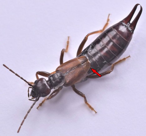
Earwigs are fascinating creatures. Belonging to the order Dermaptera, these insects can be easily recognised by their rear pincers, which are used for hunting, defence, or mating. But perhaps the most striking feature of earwigs is usually hidden – most can fly with wings that are folded to become 15 times smaller than their original surface area, and tucked away under small leathery forewings.
With protected wings and fully mobile abdomens, these insects can wriggle into the soil and other narrow spaces while maintaining the ability to fly. This is a combination very few insects achieve.
I have been working on research led by Dr Kazuya Saito from Kyushu University in Japan, which presents a geometrical method to design earwig wing-inspired fans. These fans could be used in many practical applications, from daily use articles such as fans or umbrellas, to mechanical engineering or aerospace structures such as drone wings, antennae reflectors or energy-absorbing panels!
Dr Saito came to Oxford last year for a six-month research stay at Prof Zhong You’s lab, in the Department of Engineering Science at the University of Oxford. He introduced me to biomimetics, an ever-growing field aiming to replicate nature for a wide range of applications.
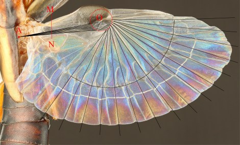
Biological structures have been optimised by the pressures of natural selection over tens of millions of years, so there is much to learn from them. Dr Saito had previously worked on the wing folding of beetles, but now he wanted to tackle the insect group that folds its wings most compactly – the earwigs.
He was developing a design method and an associated software to re-create and customise the wing folding of the earwig hind wing, in order to use it in highly compact structures which can be efficiently transported and deployed. Earwigs were required!
Here at the Museum we provided access to our insect collections, including earwig specimens from different species having their hind wings pinned unfolded. These were useful to inform the geometrical method that Saito had been devising.
Dr Saito was also interested in learning about the evolution of earwigs and finding out when in deep time their characteristic crease pattern established. Some fossils of Jurassic earwigs show hints of possessing the same wing structure and folding pattern of their relatives today.
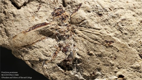
However, distant earwig relatives that lived about 280 million years ago during the Permian, the protelytropterans, possessed a different – yet related – wing shape and folding pattern. That provided the chance to test the potential and reliability of Saito’s geometrical method, as all earwigs have very similar wings due to their specialised function.
The geometrical method turned out to be successful at reconstructing the wing folding pattern of protelytropterans as well, revealing that both this extinct group and today’s earwigs have been constrained during evolution by the same geometrical rules that underpin the new geometrical design method devised by Dr Saito. In other words, the fossils were able to inform state-of-the-art applications: palaeontology is not only the science of the past, but can also be a science of the future!
We were also able to hypothesise intermediate extinct forms – somewhere between protelytropterans and living earwigs – assuming that earwigs evolved from a form closely resembling the protelytropterans.
As a collaboration between engineers and palaeobiologists, this research is a great example of the benefits of a multidisciplinary approach in science and technology. It also demonstrates how even a minute portion of the wealth of data held in natural history collections can be used for cutting-edge research, and why it is so important to keep preserving it for future generations.
Soon these earwig-inspired deployable structures might be inside your backpacks or used in satellites orbiting around the Earth. Nature continues to be our greatest source of inspiration.
Whether you’re a great white shark with a deadly conveyor belt of teeth, a deep sea snail with a coat of armour or a coral building the Great Barrier Reef one polyp at a time, mineralized skeletons are a crucial part of many animals’ way of life. These hard skeletons – shells, teeth, spines, plates and bones – are all around us.
The fossil record is full of the remains of the skeletons of long-extinct critters, so much so that entire layers of rocks can be composed almost completely of them. But this has not always been the case…
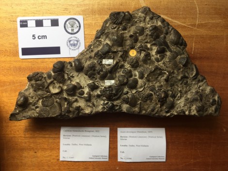
A piece of 425 million year old sea floor containing the skeletons of trilobites, brachiopods, bryozons, corals and gastropods preserved as limestone
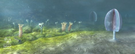
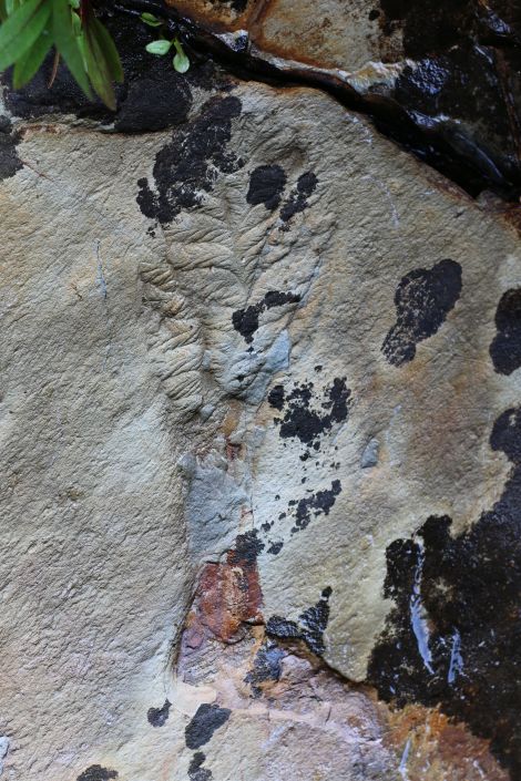
Travel back some 570 million years to a time known as the Ediacaran and the picture is very different. Although there were large-bodied creatures that were possibly animals, they were entirely soft-bodied. Then, right at the end of the Ediacaran Period, the first animals with hard skeletons evolved, creating strange tubes, stacked cones, and other bizarre forms such as Namacalathus, which resembles a baby’s rattle!
Some of the first animals with skeletons, Cloudina and Namacalathus alongside the soft-bodied Ediacaran fauna. Reconstruction based on rocks from Namibia, Southwest Africa, from 543 million years ago. Image: Mighty Fossils.
In the following few tens of millions of years, in the early part of the Cambrian Period, a whole host of animals burst onto the scene baring their ‘teeth’, hiding in their shells, and bristling their spines. In fact, we can trace the origin of almost every kind of animal skeleton to this relatively short window of the Earth’s past.
In my research, I have compiled the evidence for how and when these skeletons first appear. Three key observations have emerged. First, skeletons evolved independently many times in different animal groups. Second, there is both direct and indirect evidence, such as exceptionally preserved fossils and trace fossils, for entirely soft-bodied examples of animal groups that later evolved skeletons. And lastly, the first animal skeletons are less complex and more variable than later examples.
Added to what we know about how living animals build their skeletons, this all points to one explanation: Animal skeletons evolved independently in different groups by utilising a common ‘toolkit’ of genes, inherited from their common ancestor but used in different ways in different skeletons.
In other words, the soft-bodied ancestors of animals with hard parts had inherited all they needed to build simple skeletons that were then honed into the array of shells, teeth, spines, plates and bones we see today. For these skeletal pioneers, armed with their genetic ‘toolkit’, the environmental and ecological pressures of the early Cambrian prompted the evolution of similar, but independent, responses to their changing world – when life got hard.
Murdock, DJE. 2020. The ‘biomineralization toolkit’ and the origin of animal skeletons, Biological Reviews, is available for free here.
Top image: Tiny fragments of early skeletons, shells and spines, from around 510-515 million years ago.
With our Life, As We Know It redisplay project now underway, our Senior Archives and Library Assistant Danielle Czerkaszyn takes a behind-the-scenes look at how we captured the contents of the current displays for the Museum’s archive.
The archive here holds a unique collection of natural history books, journals and documents covering a wide range of subjects related to the Museum’s collections and research. It also contains papers and objects on the history of the building, providing an institutional memory of Oxford’s ‘University Museum’ since its foundation in 1860.
From an archive perspective it was really important to document the current layout of the cases, their specimens and text before they were removed from the court to make way for the new showcases in the first phase of our redisplay work.
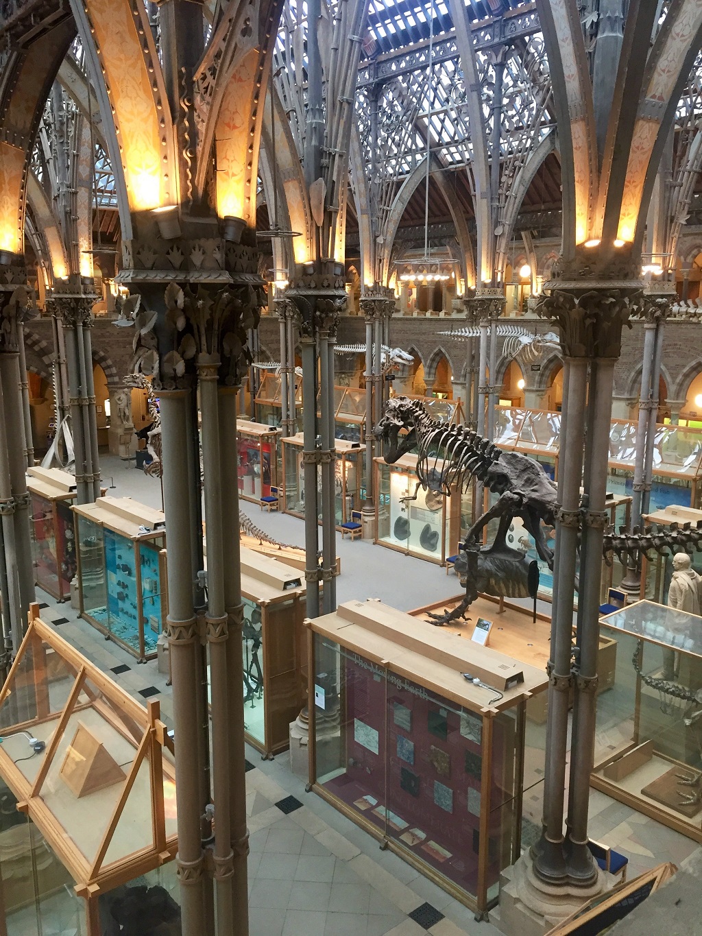
The museum in late 2019
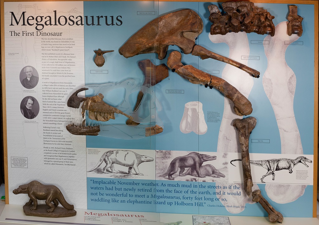
The displays as we know them – with exhibitions on the Oxfordshire dinosaurs, Alice in Wonderland, the Oxford Dodo, and more – were last changed in 2000. For the last 20 years visitors to the Museum would remember their first time being wowed by the Megalosaurus jaw – the world’s first scientifically-described dinosaur – or charmed by the Dodo made famous in Lewis Carroll’s Alice Adventures in Wonderland.
Although after 20 years it is time for a change, the stories and information in the displays are too good to be forgotten. So before anything was removed we began to build the archive for the future.
The previous display on Megalosaurus: The First Dinosaur
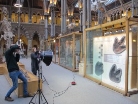
The best way to capture all the information of the displays was through high resolution photography, but this was not as straightforward as we hoped.
The first two obstacles to good photographs are pretty obvious to anyone looking at the cases: glass causes huge amounts of glare; and each case has a big dividing line down the centre where the two sliding glass doors meet, cutting what should be a lovely seamless image into two halves.
To avoid glare and the solve the problem of the dividing line, our photographer Scott opened each individual side of the case, photographed two or three images of the display, and then stitched the separate photos together using Photoshop.
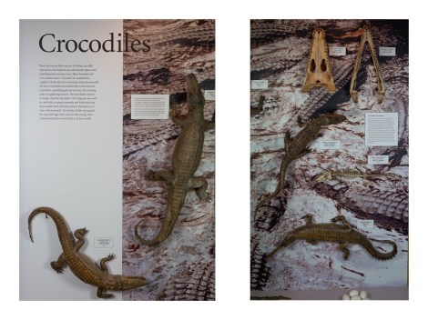
Each case was photographed in two or three segments
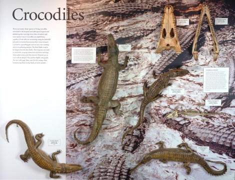
The segments were then stitched back together and adjusted for exposure and colour balance to create the final image
Another obstacle to taking good photographs of the displays came from the Museum itself. Some of our larger display furniture, such as the glass case for the Atlantic Bluefin Tuna or the huge T. rex plinth – got in the way of a nice straight shot. Because these items are so large and heavy they were impossible to move, so we had to improvise and do our best.
Capturing the displays before the current cases were removed allowed us to keep an archival record of their contents
Thankfully, we managed to get shots of all 24 displays before they were removed and so a record of each case now rests with the Museum’s archive. If anyone wants to know what the display cases in the court looked like from 2000 to 2020, they will now be able to look back at the images in the archive and recall the magic of the Oxford Dodo exhibit that perhaps first made them fall in love with the Museum.
Our new displays are now in development, and will include some beautiful presentations of the diversity of life, looking at the importance and fragility of biodiversity and human impact on the environment. These new exhibits will show how the biological processes of evolution combine with the geological processes of our dynamic Earth to give rise to the immense, interconnected variety of the natural world.
We look forward to telling you more about that here as the project progresses.
The Life, As We Know It redisplay project is supported by a generous grant from FCC Communities Foundation.
Some of the very oldest complex, macroscopic communities on Earth appear in the fossil record about 570 million years ago and record the presence of a group of organisms – the rangeomorphs – with an unfamiliar body plan that, at their ultimate extinction, was lost from life’s repertoire.
Rangeomorphs are characterised by a strange frondose branching anatomy, where large primary branches host smaller branches which themselves host smaller branches again. This arrangement appears to maximise the surface-area to volume ratio of the organism, rather like a lung or a gill would today.
The smallest known rangeomorphs are less than a centimetre in length, but they grew huge and the largest records indicate they could stand more than two metres tall. There is no evidence to suggest that rangeomorphs were able to move around, rather, they lived stuck to the sea floor in the deep ocean, far below the reach of light.
Despite this strange set of characters, there is growing consensus that rangeomorphs likely represent very ancient records of animal life. However, they lived at such a remote time in Earth’s history that they do not possess any direct living descendants. Given all this, it may not be a surprise to hear that we know relatively little about how these organisms made their living and came to dominate the ancient seafloors.
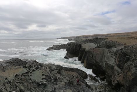
The UNESCO world heritage site Mistaken Point in Newfoundland, Canada, is one of the sites on which we find exceptionally preserved rangeomorph fossils. Photo: Alex Liu.
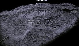
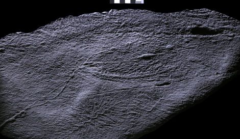
In order to better understand them, my co-author Alex Liu and I travelled to Newfoundland, Canada to explore the rocks which host these remarkable fossils and over the past few years we have made an unexpected discovery. We found that fine filamentous threads connect rangeomorph fronds of the same species, in some cases over many meters, though they are typically between two and 40 centimetres long.
An undescribed rangeomorph fossil with filamentous connections at the base of the frond. We find that this species of rangeomorph can be connected to each other over meters! Photo: Alex Liu.
It is possible that these filaments were involved in clonal reproduction, like strawberry plants today, but they may have had additional functions such as sharing nutrients or providing stability in strong ocean currents.
The discovery of the filaments means that we have to reconsider how we define an individual rangeomorph, and may help us understand how rangeomorphs (seemingly) rapidly colonised deep-sea environments. Either way, some reassessment of the palaeobiology of these unique organisms is certainly required!


")
")

")
")
")


")
")