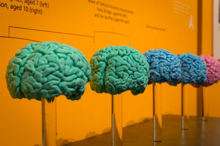
Can your brain rewire itself? How is the brain built and what can go wrong? And should the secondary school day start later to compensate for teenage sleep patterns?
Here we present a selection of videos from the exhibition. The full set is available on our YouTube channel now. And if you’re not able to visit the exhibition itself, we’ve built a special Brain Diaries website which contains all that neuroscience goodness.
Is brain-building a tricky business? Professor Zoltán Molnár of the University of Oxford specialises in the development of the brain. In this video he talks about the complex processes at play during the brain’s early development, including how things can sometimes go wrong.
Can my brain rewire itself? Associate Professor Holly Bridge works in the Nuffield Department of Clinical Neurosciences in the John Radcliffe Hospital in Oxford. Her research focuses on using MRI scans of the human brain to understand the organisation of the visual system in people with normal vision and in those with abnormal visual function. Here she talks about how the brain can rewire itself to compensate for damage to certain sensory areas.
School’s out – should the school day start later? Dr Christopher-James Harvey works at the Sleep and Circadian Neuroscience Institute at the University of Oxford. As part of the Teensleep research project, he is investigating how changes in the natural rhythm of sleep in adolescents, and the effects of sleep education, might impact on academic, health and sleep outcomes. Here he talks about initiatives to trial a later starting time for the secondary school day.
To read more about Brain Diaries and see the full programme of public events see braindiaries.org.
Our next exhibition – Brain Diaries: Modern Neuroscience in Action – opens on 10 March and in preparation we have indulged in a little bit of brain-washing… This article contains an image of a preserved human brain.
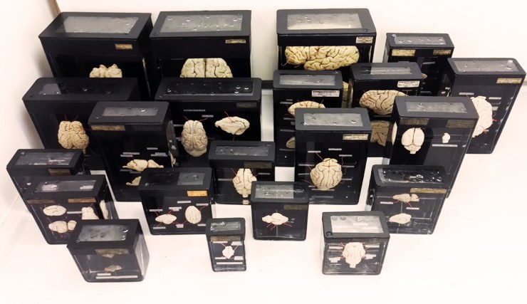
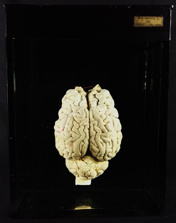
One of the first displays visitors will encounter is a ‘wall’ of 23 fluid-preserved mammal brains – from a Short-nosed Bandicoot to cow. The style of jar, with its black bitumen and paint backing, tells us that these were once used for display so it is exciting to put them in the public galleries again. Museum conservator, Jacqueline Chapman-Gray, runs us through the meticulous process she undertook to ensure these brains will look their best for their return to the limelight.
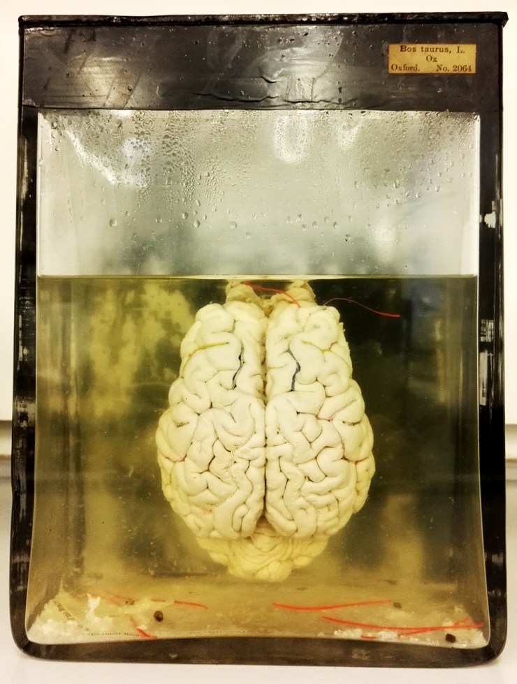
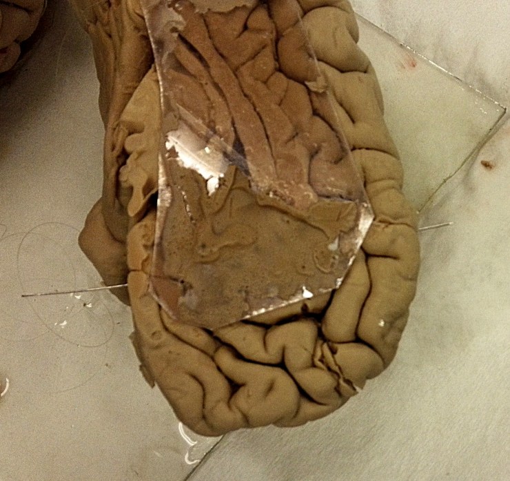
Cow brain before conservation treatmentA number of the brains had become dehydrated over time as the level of fluid – alcohol – had dropped. These needed to go through a rehydration programme to ensure their long-term preservation. This is more complex than simply adding more fluid to the jar. Instead the alcohol level needs to be increased gradually to avoid damaging the tissues.
Brains soaking in alcoholOthers had started to detach from their glass mounts, or anatomy labels that marked each of the different areas or sections of the brain had come loose. These were carefully remounted using specialist conservation-grade materials and a steady hand! Three brains had become completely detached and were repaired using a polyester monofilament thread, otherwise known as fishing line.
Repairing a human brain with a beading needle Labels found detached at the bottom of the jarFor the smallest of the brains a normal sewing needle was enough to pass through the tissues but for the larger two either a flexible 10cm beading needle or large 25cm mattress needle was needed. The original threading points were reused wherever possible though in one case this proved to be too difficult, as the tissue was soft and susceptible to breaking. With precision and patience I was able to gently stitch them back into place on the backing plate so they look as good as new.
All of the jars were given a thorough clean to ensure that seals were tight fitting and that the contents were shown off to their best. They were then filled with fluid to 4/5ths from the rim and the brains gently placed back inside.
Lids were sealed with clear silicone and each jar was topped up with a syringe through a small hole in the lid that is there for this very purpose – once full, this hole is also sealed.
Lastly, after the seals had dried, for the final finishing flourish black paint was reapplied to the backs and tops of the jars to provide a contrasting backdrop.
Ta-dah… the cow brain after conservation treatmentBrain Diaries opens on Friday 10 March and runs until Monday 1 January 2018. Take a look at the website to find out more about the exhibition and accompanying programme of events at braindiaries.org
The museum holds the only remaining soft tissue of the extinct dodo known anywhere in the world. The partially dissected skin of the head and scales from the feet of a single dodo represent one of natural history’s most iconic specimens. In fact, it is so tied to the museum’s identity and history that we use the dodo as our logo and it is even incorporated in the name of this blog.
Although the dodo head had been at Oxford University since the formation of the original Ashmolean Museum in the 17th century, it wasn’t really until the 19th century that the specimen really became celebrated.
Around this time, publications confirmed the extinction of the dodo from the island of Mauritius, where it was endemic. To capitalise on the rising interest in the animal, Ashmolean Museum Keeper John Duncan commissioned a number of casts of the Oxford Dodo head to give to, and exchange with, other museums.
One of the earliest of these casts was presented to the British Museum in 1828; later casts are recorded as being sent or exchanged with leading scientists of the time, as well as with Leiden Museum and the Royal College of Surgeons.
From these original and later casts further casts and models were presumably made, and eventually, dodo specimens spread to virtually every major natural history museum in the world. Today, many museums display casts of this head, all stemming from the single specimen held here in Oxford.
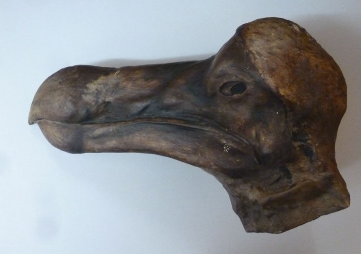
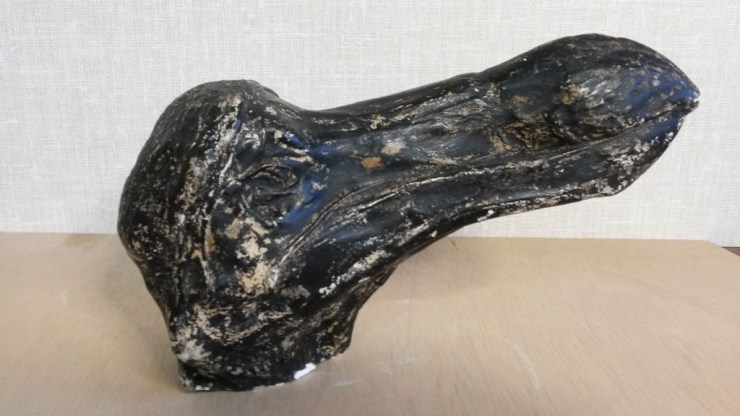
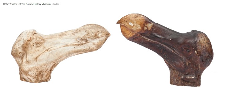
One of the many casts in the Oxford University Museum of Natural History, this one has been painted to match the original specimen
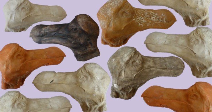
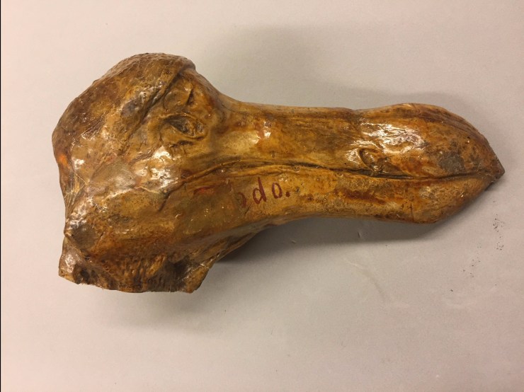
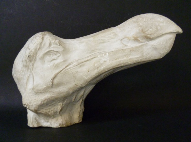
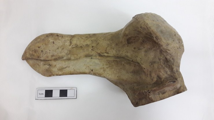
The Museum contains a number of models and casts of the head too; some are made from plaster and resin, some are painted to resemble the original specimen. The head of the dodo was actually dissected in 1847, by Henry Acland. He removed the skin from one side of the face so the early casts are a record of how the specimen would have looked originally.
In preparation for the Presenting display in the Museum I contacted natural history museums through the Natural Sciences Collections Association asking people to share information and photos about their casts and models of the dodo head. I wanted to try and construct a picture of how the dodo head was disseminated, as well as capture the diversity of quality and colours of representations of the original specimen. Here’s how far some of the dodos have flown:
Cast of the head labelled as coming from Cartwright Hall. Curator Gerry McGowan suspects this may have come via the Bradford Philosophical Society collections. The first curator of the society, Louis Compton Miall was friends with Thomas Henry Huxley and through him had contacts with many other geologists who may have gifted or exchanged this cast.
Bristol Museum & Art Gallery Bristol received a cast of the head directly from Oxford from Philip Duncan in 1834, keeper of the Ashmolean Museum between 1826 and 1855. Unfortunately, the head was likely destroyed in bombings of Bristol in 1940.
Canterbury Museum, New Zealand
Received a cast of a head from Professor Rolleston on 21 July 1871 in exchange for two kiwi skeletons which are still in the museum collections today.
Cast of head and foot presented to the museum by E.Ray Lankester in 1891/1892 just after leaving UCL and being appointed the Linacre Professor of Comparative Anatomy at Oxford.
The Great North Museum Hancock’s cast was presented by George Townsend Fox. This specimen had been presented to the Natural History Society of Newcastle in 1841 by Fox and had originally been presented by Philip Duncan.
Cast of the head that had quite a circuitous route to the Horniman Museum. The Horniman received the cast from the geology department of Queen Mary’s University of London in 1964 which received the cast from the Saffron Walden Museum in 1962.
Manchester Museum Cast of a head, presumed to have been presented by William Boyd Dawkins. The cast is currently on display in the Living Worlds gallery in Manchester Museum.
The National Geological Repository British Geological Survey
The Natural History Museum’s collections contain three casts of the Oxford Dodo head – two in the ornithology collections (pictured) and one in the palaeontology collections. The unpainted cast in the ornithology collections has the name ‘Johnson’ inscribed into the base.
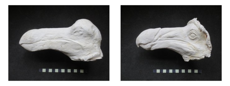
Colleague Adam Smith got in touch with some interesting specimens from Wollaton Hall. The first one looks like another cast in this series but the second cast is unlike any of the others gathered here. The cast shows an open eye, detail on the beak as well as a more defined hook to the end of the bill.
Unfortunately, there’s not much information about the origins of these two casts so it’s probable that the ‘open eye’ cast may be a cast of a model reconstruction or an in progress sculpt. There’s an extremely slight chance it’s a cast of an otherwise unknown dodo head… If you recognise this dodo head do get in touch so we can solve this mystery for colleagues in Nottingham (it’s not the model dodo that we have on display here!).
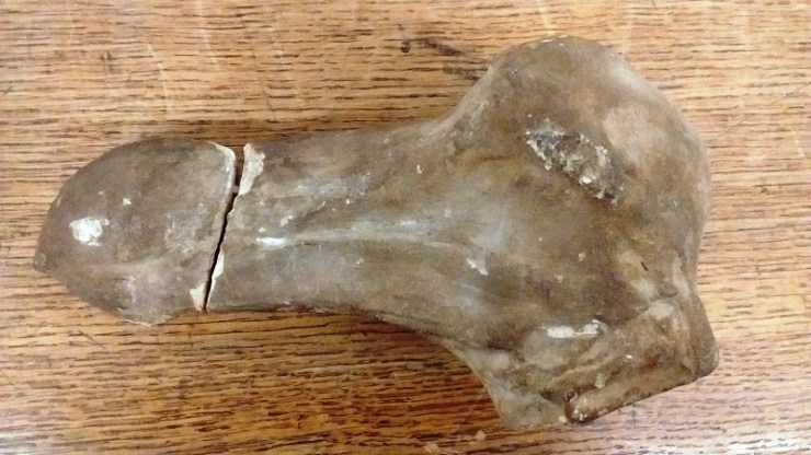
Cast of a head at Warwickshire Museum with damage to the beak. Donated to the museum by clergyman and naturalist Reverand Andrew Bloxham in the 19th century. As the museum is currently moving stores, further information about when this cast was acquired is inaccessible.
**
If you work at a museum and have a dodo head cast to share, please do get in touch and we’ll update this blog accordingly.
Last updated: 10/11/17
‘Presenting… The Flight of the Dodo’ was on display at the Museum of Natural History from the 25 January to 22 March 2017.
Acknowledgements
With many thanks to colleagues across the sector who helped with information and images about dodo specimens: Adam Smith, Alice Adams, Jack Ashby, Carol Davies, Bonnie Griffin, Dan Gordon, Yvette Harvey, Mike Howe, Emma-Lousie Nicholls, Laura McCoy, Gerry McGowan, Nigel Monaghan, Henry McGhie, Pat Morris, Paul Scofield, Paul Shepherd, and Paul Sweet.
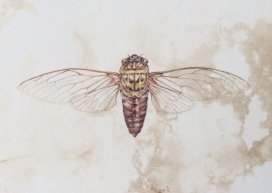
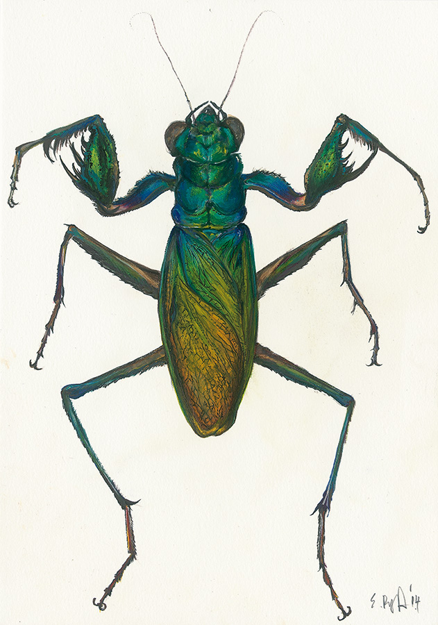
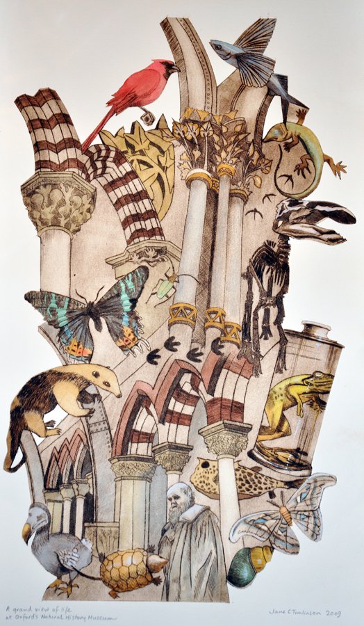
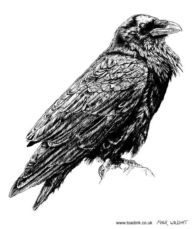
As part of our Visions of Nature year in 2016, we invited you to send in your own ‘visions of nature’. We didn’t know what to expect, but week after week we received pictures of beautiful paintings, drawings, photographs, and textile art, all inspired by the natural world or by the Museum and its collections.
All of these artworks can now be found in the online gallery, but we’ve picked out a few here to show you and to mark the end of the project. Thanks very much to everyone who took part.
Emma Reynard
Jake Spicer – Main court Oxford University Museum of Natural History
Jane Tomlinson – Oxford University Museum of Natural HistoryDeidre Bean – Cicada
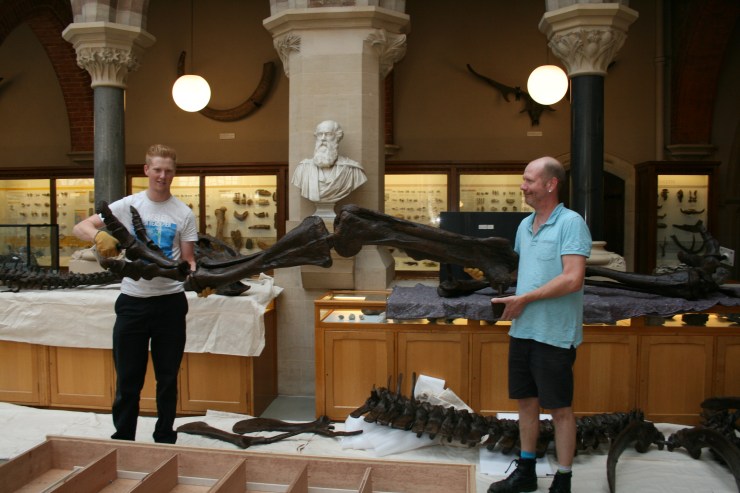
Last year we introduced you to Adam Fisk, our (then) new apprentice. He’s the slightly younger-looking one in the photo above; on the right is Pete Johnson, the Museum’s technician and Adam’s mentor and supervisor. Together, the two are a vital component that keeps the Museum machinery a-turning.
Over the past twelve months or so Adam has busied himself with many vital tasks: installing and de-installing our exhibitions, including Kurt Jackson’s Bees (and the odd wasp) in my Bonnet and Microsculpture; reconstructing an Edmontosaurus; and, more recently, sporting an excellent Star Wars Christmas jumper.
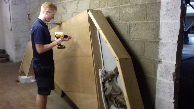
Packing an Attenborosaurus ready for transit
We’re therefore exceptionally pleased to announce that Adam has just been selected as a winner in the Annual University of Oxford Apprenticeships Awards. This calls for a small whoop, and a large ‘Well done Adam!’.
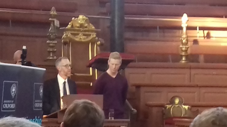
The awards, which celebrate the achievements of the University’s apprentices, supervisors and departments, were given out at a ceremony at the rather grand Sheldonian Theatre this afternoon.
Adam collecting his award at the Sheldonian Theatre in Oxford
When Adam joined the Museum last summer for his three-year apprenticeship he had just finished his GCSEs. Now he’s walking the stage at the Sheldonian… As Pete says:
Adam’s contribution to all aspects of work from maintenance and workshop demands to exhibition installation and Collections assistance has been outstanding. We have all benefitted greatly from his input, and he has helped us to achieve a high standard of public service.
Thanks for all the hard work Adam and we look forward to the next 18 months with you on the team.
The latest display in our changing Presenting… case showcases some pioneers of photography and features a recently-discovered original print of Charles Darwin by Julia Margaret Cameron…
In the early days of photography, in the mid-19th century, a number of photographic innovators had close links to the Museum and its collections. Perhaps the most famous of these is Charles Lutwidge Dodgson (1832-1898), better known as Lewis Carroll. Dodgson was an author and mathematician at Christ Church College in Oxford, and also an accomplished early photographer.
Dodgson’s friends were often the subjects of his photos. A well-known example is Alice Liddell who, as well as sitting for portraiture, provided inspiration for Dodgson’s book Alice’s Adventures in Wonderland, as did the Dodo in the Museum’s collections.
The image at the top of the article is titled The Anatomy Lesson with Dr George Rolleston and was taken by Dodgson around 1857. It shows Dr Rolleston, Professor of Medicine at the University of Oxford at the time, pictured with a selection of specimens from the Museum’s collections.
Two of Dodgson’s subjects were accomplished photographers in their own right, and perhaps the most famous female photographers of their time: Julia Margaret Cameron (1815-1879) and Sarah Angelina Acland (1849-1930).
Cameron’s innovative and distinctive style of portraiture raised photography to a true art form. Her intentional use of shifted focus and experiments with process and finish gave an ethereal look to her works resembling the artistic style of the contemporary Pre-Raphaelites.
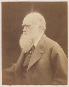
Many prominent scientists sat for Cameron, including naturalist Charles Darwin. She took the portrait above in 1868 at her home on the Isle of Wight. An original print of this photograph, made by Cameron, was recently discovered in the Museum Archive, uncatalogued. It features her signature on the back and the blind stamp from her printseller in London on the front.
‘Mr Ruskin & his old friend at Brantwood’ by Sarah Angelina Acland, 1893
Sarah Angelina Acland, inspired and taught by Cameron, is best known for her innovations in colour photography, but she also took many black and white photographs while learning the art form. The portrait above features her father Sir Henry Wentworth Acland and the artist John Ruskin at his home. Both Ruskin and Acland were instrumental in the founding of this Museum, and Sarah even helped to lay the founding stone in 1855.
In a 1904 Royal Photographic Society exhibition Acland was the first to exhibit work combining images taken with red, green and blue filters, three years before the launch of the colour photography process invented by the Lumière brothers.
These images, including the original print of Charles Darwin by Julia Margaret Cameron, are on display in our Presenting… case until 24 January.