Presenting… Christmas Island

By Eileen Westwig, Collections Manager in the Museum’s Life Collections.

About 320 km south of Java in the Indian Ocean lies Christmas Island. Although discovered and named on Christmas Day in 1643, the island remained unexplored until its first settlement in 1888, a development which had dire consequences for some of its native species.

Christmas Island is home to a variety of endemic animals such as rats, land crabs, butterflies and many birds. The accumulation of bird droppings over thousands of years made the island rich in phosphate, and the commercial potential of these deposits brought many expeditions to the island. With the ships’ cargo came black rats.

Two species of endemic rats, Maclear’s Rat (Rattus macleari) and the Bulldog Rat (Rattus nativitatis) went extinct within 20 years of settlement, despite having been previously very numerous on the island.

One of the skins of Maclear’s Rat (Rattus macleari) collected by H.E. Durham, and now held in the Life Collections of the Oxford University Museum of Natural History.

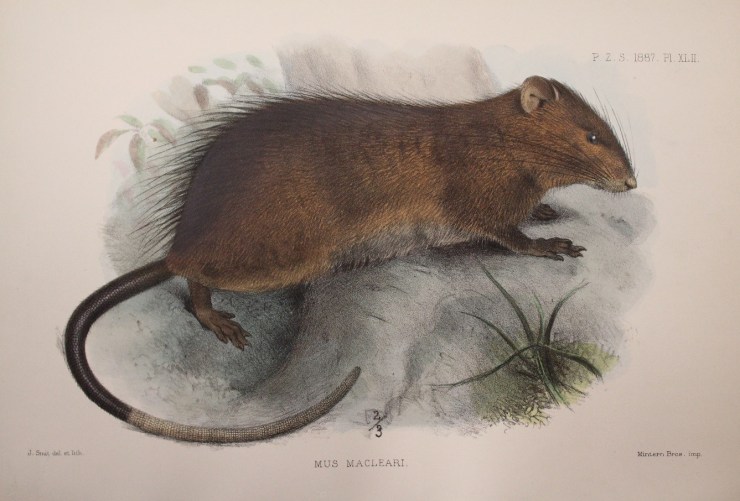
Maclear’s Rat, seen at the top of the page in an illustration from an 1887 publication, was described as chestnut brown above, with a partly white, long tail. It was once the most numerous mammal on the island ‘occurring in swarms’. The Bulldog Rat had a much shorter tail and a layer of subcutaneous fat up to 2 centimetres thick, the function of which is unknown to this day.

The likely cause of their extinction was the introduction of diseases by the ship rats, to which the Christmas Island rodents had no immunity. The disappearance of the native rats also had a knock-on effect: the parasitic Christmas Island Flea (Xenopsylla nesiotes) depended on the rats as hosts, and so the fleas became extinct with the rats’ demise.

In 1901 Dr. Herbert E. Durham, a British parasitologist investigating the origins of beriberi disease, led an expedition to Christmas Island. During his visit he collected several specimens of Maclear’s Rat, but was unable to find any Bulldog Rats, despite a lengthy search and the offer of a reward. Two of the nine Maclear’s Rats Durham obtained showed abundant parasites, trypanosomes, in their blood.

Christmas Island possesses quite a number of peculiar species in its fauna, and it is regrettable that observations were not made before animals had been imported to this isolated station, as well as that my own notes are so incomplete.

Dr. Herbert E. Durham

Durham also found blood parasites in the native fruit bats (Pteropus melanotus) but noted that these were unlikely to have been introduced, instead were “an old standing native occurrence.” These bats still inhabit various islands in the Indian Ocean, including Christmas Island, where they are critically endangered.

Original letter by H.E. Durham offering his Christmas Island specimens to the Museum in 1938.

The Museum holds a range of material from Christmas Island, including six skins and three skulls of Rattus macleari, which were collected by H. E. Durham in 1901-02, and donated in 1938.

Visit the Museum’s Presenting… case between now and 6 March to see Christmas Island specimens from the collections.
Precision antibiotics – the future treatment of infections?

by Hannah Behrens

In our Bacterial World Science Short event series, researchers present their latest findings related to themes in the exhibition. At a recent Science Short, Hannah Behrens, a University of Oxford PhD student, explained how bacteria become resistant to antibiotics and how the species-specific antibiotics she studies might reduce the worrying rise in antimicrobial resistance.

Bacteria that are resistant to antibiotics present a huge problem. I work on developing new antibiotics that will slow the development of bacterial resistance.

But let’s not get ahead of ourselves. Your body is full of bacteria. In fact, there are more bacteria than human cells in your body. Most of these bacteria are good for you; they help you digest food and protect you from diseases.

But once in a while a harmful bacterium causes an infection. This could be a lung, wound, or bladder infection, or something with a fancy name like, Black Death, tuberculosis, leprosy, syphilis or chlamydia. The doctor will then prescribe you antibiotics to kill the offending bacteria.

Hannah Behrens delivers her Science Short talk at the Museum

The development of antibiotics in the 20th century was a major breakthrough. For the first time bacterial infections could be effectively and rapidly treated. Since 1942, when antibiotics first became available, we have discovered many new antibiotics which have saved millions of lives.

However, in the last 30 years we have not managed to develop any new antibiotics. During the same time, many bacteria have adapted to become resistant to the antibiotics we do have. In 2017, a woman in the US died because she had an infection with bacteria that were resistant to all available antibiotics. It is estimated that already 700,000 people in Europe alone die because of resistant bacteria per year. What is happening?

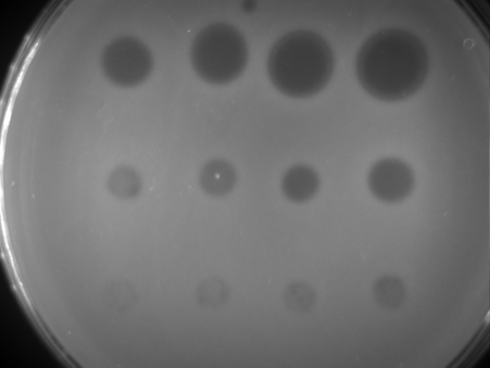
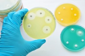
Bacteria are forming a lawn on this plate (light areas); where an antibiotic has been spotted on the bacteria they die and leave the surface blank (dark areas).

Every time we treat bacteria with antibiotics, most die, yet a few resistant bacterial cells survive. Like Rudolph the red nosed reindeer, the resistant bacteria are usually at a disadvantage until a special situation arises (a foggy night for Rudolph; treatment with antibiotics for resistant bacteria).

Under usual circumstances, producing a resistance mechanism is a disadvantage: it wastes energy and slows down growth, so very few bacteria are resistant. Only when all the non-resistant bacteria are killed by antibiotics do the resistant ones thrive. They have no more competition, and have all the resources, such as food and space, to themselves.

The more we use antibiotics, the more resistant bacteria we get. It is essential not to use antibiotics carelessly.

More antibiotics are used in animal farming than on humans. If we eat less meat, and so reduce the farming of livestock for food, we may reduce the growth of resistance bacteria. Another approach is to only take antibiotics when the doctor prescribes them. Antibiotics do not help against viral infections like colds. In many low and middle income countries, antibiotics are available in supermarkets and it is no coincidence that these countries have higher levels of resistant bacteria.

The precision antibiotics research group in the Department of Biochemistry at the University of Oxford

Apart from avoiding the unnecessary use of antibiotics, scientists – including me – are trying to develop better therapies against bacteria. I study precision antibiotics: drugs that specifically kill one species of bacteria. The advantage of this is that all good bacteria remain unharmed and only the disease-causing species is targeted. This also means that only resistant bacteria from this one species get an advantage to thrive.

I am interested in species-specific antibiotics against Pseudomonas aeruginosa. This bacterial species causes lung and wound infections and, according to the World Health Organization, is one of the three bacteria for which we most urgently need new antibiotics. Colleagues of mine tested different precision antibiotics against Pseudomonas and found one that is better than the others, called Pyocin S5.

Hannah’s painting of how researchers think pyocin antibiotics kill bacteria. The pink bacterium produces pyocins (pink balls), which enter the susceptible blue bacteria through pores (blue). The blue bacteria mistake the antibiotic for a nutrient and open the pore to let it in. Once inside the bacterium it forms a pore in the inner membrane which causes leakage of the cell contents and kills the cell.

I am now investigating how stable this antibiotic is, how it recognises this specific species of bacteria and how it enters the bacterial cells. This knowledge is important to decide on how to store, transport and administer the drug. I also hope that understanding why Pyocin S5 is more effective than the other antibiotics will allow us to design more effective, targeted antibiotics in the future.

My hope is that one day we will treat all bacterial infections with precision antibiotics and that antibiotic resistance will become a problem of the past.

Bacteria: captured and cultured

For our new exhibition, Bacterial World, we embarked an exciting science/art experiment to make visible the colonies of bacteria present on a wide range of our everyday items and belongings. Once cultured and photographed, eight of these colonies were captured by artist Elin Thomas as a set of crochet artworks that are on display in the exhibition. Our exhibitions officer Kelly Richards tells us more…

For every human cell in your body, a bacterial cell is also present. These bacteria are part of our microbiome, a vast array of microorganisms that use our body as a home and our food as a source of nutrients. In return, the bacteria help us to digest food, maintain our immune systems and keep dangerous bacteria at bay. In fact without these bacteria we would be very sick indeed.

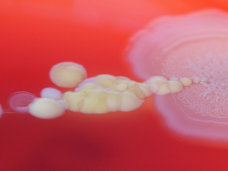
It’s hard to see our microbiome because individual bacteria can easily be as small as 0.2 microns; you could fit over a thousand of these smallest bacteria on one side of a red blood cell. But if we can select and artificially grow the bacteria, their colonies become living, breathing cities visible to the naked eye.

Click the images above to find out more about culturing bacterial colonies

Colonies, both natural and artificial, can contain billions of bacteria as well as the materials that they secrete such as slime, which helps them to move across surfaces, and antibiotics, which kill off other bacterial colonies that could compete for food and space. In their attempts to dominate the space and food available, as well as get enough oxygen to live, colonies can create beautiful, complex structures.

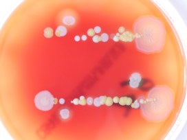
We had a go at visualising the bacteria that live invisibly alongside us by asking visitors to take part in a simple experiment. With the help of microbiologist Rachael Wilkinson, we took items such as coins, keys and jewellery and touched them lightly against agar plates – dishes containing a nutrient-rich jelly that aids bacterial growth. The agar plates were then given to Nicole Stoesser, a clinical microbiologist at the John Radcliffe Hospital, who grew them in the safe environment of the laboratory.

Many different types of colonies grew from the objects we printed. In the collage above, eight of these colonies have been represented as crocheted Petri dishes by artist Elin Thomas. These artworks are on display in the Bacterial World exhibition until 28 May 2019.

In the gallery below is a photograph of every participant’s plate, whether anything grew in it or not. Click on an image to see a larger version. If you took part in the experiment you will be able to identify your own plate from its number.

The results go to show that we really are living in a bacterial world!

Animating the extinct

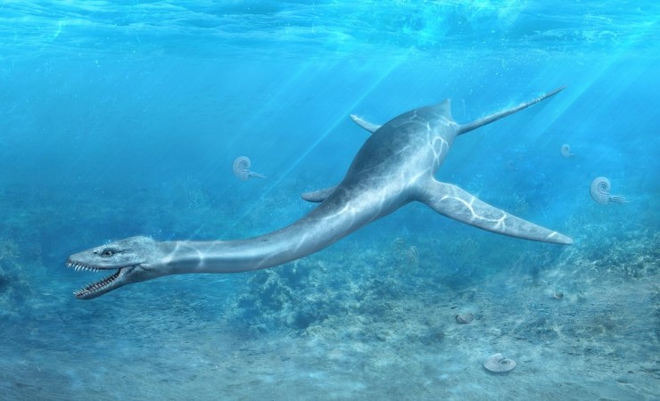
This sumptuous video features on our brand new Out of the Deep display and brings to life the two large marine reptile skeletons seen in the cases. The Museum exhibition team worked with Martin Lisec of Mighty Fossils, who specialise in palaeo reconstructions. Martin and his animators also created a longer video explaining how the long-necked plesiosaur became fossilised, as well as beautiful illustrations of life in the Jurassic seas. 
Martin explains the process of animating these long-extinct creatures:

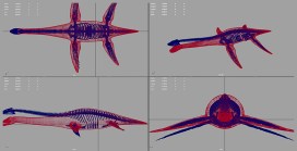
The first step was to make 3D models of all the animals that would appear in the films or illustrations. After discussion with the Museum team, it was clear that we would need two plesiosaurs (one short-necked, known as a pliosaur, one long-necked), ammonites, belemnites and other Jurassic sea life. Now we were able to define the scale of detail, size and texture quality of the model.

In consultation with Dr. Hilary Ketchum, the palaeontologist on the project, we gathered important data, including a detailed description of the discovered skeletons, photographs, 3D scans, and a few sketches.

This slideshow requires JavaScript.

We created the first version of the model to determine proportions and a body shape. After several discussions with Hilary, some improvements were made and the ‘primal model’ of the long-necked plesiosaur was ready for the final touches – adding details, mapping, and textures. We could then move on to create the other 3D models.

This slideshow requires JavaScript.

The longer animation was the most time-consuming. We prepared the short storyboard, which was then partly changed during the works, but that is a common part of a creative job. For example, when it was agreed during the process that the video would contain description texts, it affected the speed and length of the whole animation – obviously, it has to be slower so that people are able to watch and read all important information properly.

A certain problem appeared when creating the short, looped animation. The first picture had to precisely follow the last one – quite a difficult goal to reach in case of underwater scenery. Hopefully no-one can spot the join!

This slideshow requires JavaScript.

At this moment we had a rough animation to be finalised. We had to make colour corrections, add effects and sound – everything had to fit perfectly. After the first version, there were a few more with slight adjustments of animation, cut and text corrections. The final version of both animations was ready and then rendered in different quality and resolution for use in the display and online.

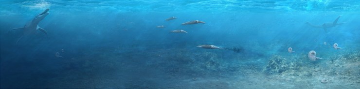
The last part of the project was creating a large illustration, 12,000 x 3,000 pixels, which would be used as a background for a large display panel. Text, diagrams and a screen showing the animations would be placed on this background, making the composition a little tricky. We agreed that the base of the illustration would be just the background. The underwater scene and creatures were placed in separate layers so that it would be easy to adjust them – move them, change their size, position etc.

This slideshow requires JavaScript.

In the first phase, we had to set the colour scale to achieve the proper look of the warm and shallow sea, then we made rough sketches of the scene including seabed and positions of individual creatures. We had to make continuous adjustments as the display design developed.

Then we finished the seabed with vegetation, gryphaea shells and plankton floating in the water. The final touch was to use lighting to create an illusion of depth for the Jurassic creatures to explore.

*

More Out of the Deep videos are available on the Museum website.

Pieces of a plesiosaur

We’ve just opened a brand new, permanent display called Out of the Deep, featuring two beautifully preserved plesiosaur skeletons. Remarkably, both of these marine reptile fossils have skulls, which is more unusual than you might think. Dr Hilary Ketchum, collections manager in the Museum’s Earth Collections and curator of Out of the Deep, describes how the skull of the long-necked plesiosaur made it safely from a quarry to a museum display.

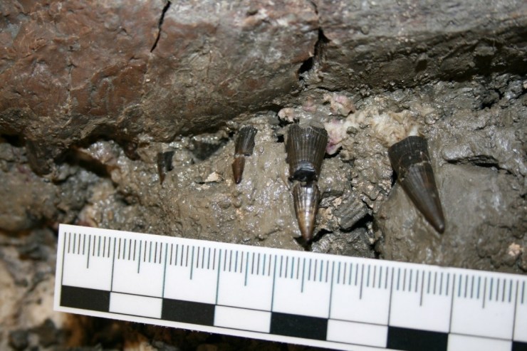
At the bottom of a clay pit in 2014, palaeontologists from the Oxford Clay Working Group discovered a 165-million-year-old fossil plesiosaur skeleton, and they knew they had found something special. Plesiosaur bones are fairly common in the quarry, but skeletons are rare. Skeletons with skulls are rarer still. Fantastically, at the end of their newly-found plesiosaur’s neck was a skull. Barely visible underneath the clay, only the tip of the snout and a few teeth were exposed.

Can you see the skull? Fossil hunting in the quarry takes time, patience and a good eye to distinguish between bones and clay. Image: Mark Wildman, Oxford Clay Working Group.

Plesiosaur skulls are usually made up of around 33 bones, not including the tiny bones from inside the eye sockets, called the sclerotic ring. The skull bones are among the smallest and most fragile in the entire skeleton. This means they are much less likely to be preserved, and less likely to be discovered, than the larger and more robust backbones and limb bones.

A plaster jacket was made around the skull while still in the quarry.
Image: Mark Wildman, Oxford Clay Working Group.

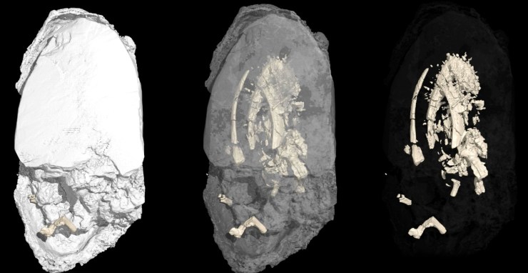
When the plesiosaur skeleton arrived in the Museum in 2015, the skull and some of the surrounding clay was encased in its protective plaster field jacket. As tempting as it was, instead of cracking open the jacket straight away, we decided on a more technological approach. Professor Roger Benson and Dr James Neenan took the specimen to the Royal Veterinary College to use their enormous CT scanner, normally used for scanning horses and other large animals, and took thousands of X-rays of the jacket. This allowed them to build up a 3D model of the fossil inside – our first tantalising glimpse of the whole skull!

The CT scan of the plaster jacket (left) revealed the location of the skull inside the jacket (middle). The jacket was then digitally removed (right) to reveal a 3D image of the skull.

Having the CT scan of the skull was like having a picture on a puzzle box
Juliet Hay, Earth Collections conservator and preparator

Although the CT scan was incredibly useful, we still had to proceed with the preparation with caution. It was possible that not all of the bones had not been detected by the scanner, especially the incredibly thin bones of the palate.

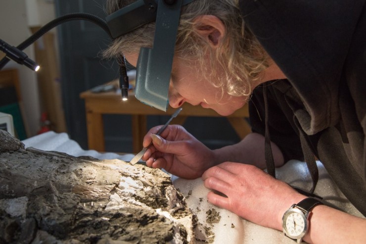
After opening the plaster jacket, Juliet began to carefully remove the clay from around the fossil bone.

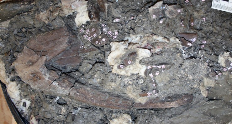
Slowly and carefully, Juliet and I removed the soft clay from around the skull. The weight of clay pressing on top of the skull for millions of years had crushed it, breaking some of the bones into a lot of smaller pieces. In order to keep track of them we attached a number to each piece of bone and photographed it from several different angles before removing it from the jacket.

Each individual bone was mapped using a numbering system. The numbers were attached with the conservation adhesive Paraloid B72 in acetone, so that they could be easily removed later.
The plesiosaur’s pointed teeth being revealed.

When all the bones had finally been removed from the clay, we had over 250 pieces. Next came the challenge of the three-dimensional jigsaw!

With knowledge about plesiosaur skulls from my PhD, and some extra expert help from Roger Benson and Dr Mark Evans, Curator of Natural Science and Archaeology, New Walk Museum and Art Gallery, I was able to build up the skull, piece by piece, until it was nearly whole again.

After many months of painstaking work, the beautifully preserved skull of this long-necked plesiosaur can finally be seen in the Out of the Deep display.

Amazingly, the skull is even more complete and more beautifully preserved than we could tell from the CT scan. The sutures between the individual bones can be seen in exquisite detail, and even though I work with fossils every day, I still find it amazing that it is 165 million years old.

*

With special thanks to:

Oxford Clay Working Group: Mark Wildman, Carl Harrington, Shona Tranter, Cliff Nicklin, Heather Middleton, and Mark Graham, who uncovered and excavated the long-necked plesiosaur.

Forterra, for generously donating the plesiosaur skeleton to the Museum, after it was discovered in a Forterra quarry. 

How to build a plesiosaur, in 836 easy steps

By Rachel Parle, Public Engagement Manager

Fitting together the remains of long-dead sea creatures is no easy job. A brand new permanent display, Out of the Deep, has brought together two Jurassic marine reptiles in the largest new display in the Museum for decades. But getting them out on show was a long, delicate process that took hundreds of careful steps.

The specimens are rare and remarkably complete, and on full display for the first time. The short-necked plesiosaur, known as a pliosaur, was discovered by a former Museum curator in the 1990s in Yarnton, Oxfordshire, just a few miles from the Museum.

The larger long-necked plesiosaur was found in a quarry in Cambridgeshire in 2014 by the Oxford Clay Working Group, and donated to the Museum’s collections by the quarry’s owner Forterra. You might remember this fossil from our excited article when the specimen first arrived at the Museum, back in 2016.

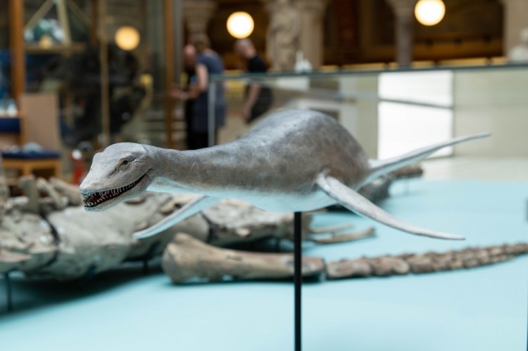
Model of the long-necked plesiosaur, made by Crawley Creatures

Before the fossils could take centre stage, their set had to be built, in the shape of two enormous display cases, designed and constructed to house these special specimens. Every day, for two weeks, the team from showcase specialists Click Netherfield pieced together slabs of glass, built walls, installed lights and fitted portholes. Yes, portholes!

A young visitor peers at the plesiosaur through a porthole

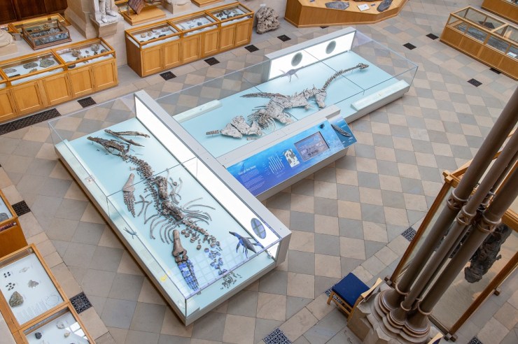
The display is positioned in the south aisle of the Museum – if you know the building, this is on the right as you come in, alongside large dinosaur skeletons. We’d measured and planned the space again and again, with our designers Calum Storrie and Pat O’Leary, but couldn’t quite picture this huge display sitting alongside the other cases. Each specimen is almost five metres in length, so the cases really are whoppers. Once the pieces started to come together, we could see that this was going to a really dramatic addition to the Museum. We chose a clean, contemporary look, with lots of glass and powder-coated steel. The colour of the metal case was carefully chosen to stand out (no oak-effect), but sit well in amongst the warm tones of the stone and brick work – RAL 7032 if you’re interested!

Here’s a time-lapse video to show you how the cases were built, piece by piece over two weeks.

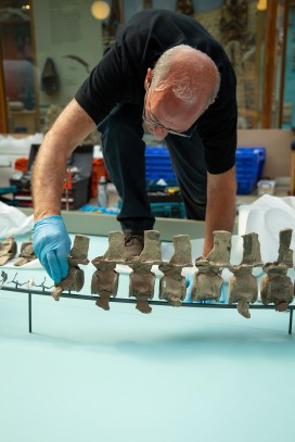
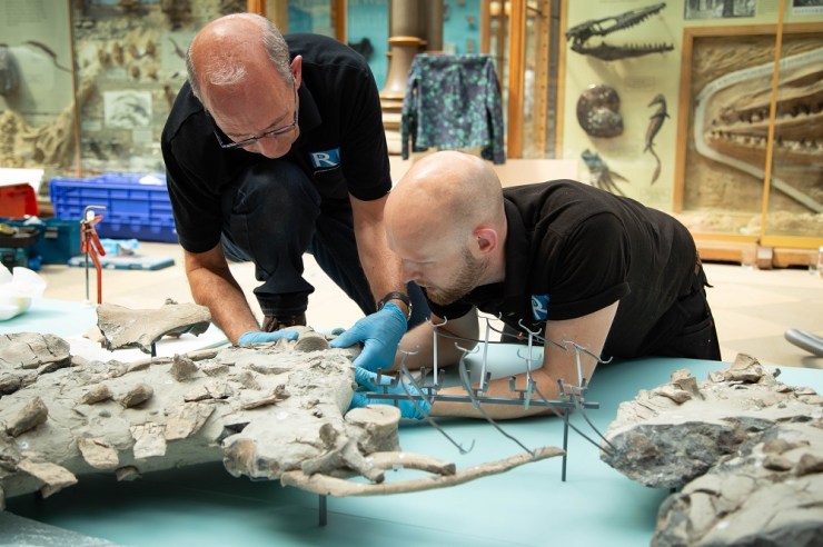
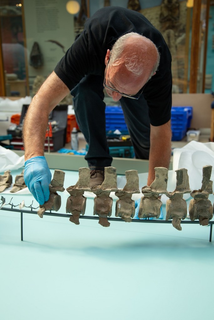
Once the enormous cases were in place, we could start installing the stars of the show – the two plesiosaur skeletons. Museum staff had worked closely with Richard Rogers Conservation to build an intricate web of steel that would hold each fragment of the skeleton in place, showing it off to its full potential. This mount was screwed into place on the acrylic base board then the individual bones were gently slotted into place.
Richard Rogers slots a vertebra into place on the carefully-crafted mount
Richard Rogers and James Dawson position plesiosaur ribs

Here’s a glimpse of the installation process in a handy time-lapse film:

After countless of hours of excavation, preparation, planning, measuring (measuring again) and installing, Out of the Deep is ready! It’s open now in the Museum and the specimens are accompanied by specially commissioned digital reconstructions (more about that soon), videos that share the stories behind the specimens’ discovery and even a touchable 3D print of a plesiosaur flipper. Come along and meet the Jurassic beasts that deserved all this care and attention.

The finished Out of the Deep display is open now

**

The project has been generously supported by grants from the DCMS/Wolfson Museums and Galleries Improvement Fund, and WREN’s FCC Community Action Fund.