On the trail of the Piltdown hoax

The latest display in our single-case Presenting… series takes a look at the famous Piltdown Man hoax, and Life Collections manager Mark Carnall tells us how the display came about…

Visiting researchers to the zoology collections at the Museum often give us an excuse to dig deeper into our own material, and one such recent enquiry led me into the intriguing story of the Piltdown Man hoax.

Professor Andrew Shortland from Cranfield University contacted us to enquire about the Piltdown Man material in our collections, as part of research for a book on hoaxes and forgeries in anthropology that he is writing with Professor Patrick Degryse of KU Leuven.

I knew we had some Piltdown material here thanks to this page written by Malgosia Nowak-Kemp, but I hadn’t had an excuse to investigate any further. The enquiry was also timely as we’d just transferred a collection of palaeoanthropology casts, models and reconstructions from our Earth collections to bring our human collections into one place. I knew from our move project team that there was some Piltdown material awaiting processing – perfect.

For those who don’t know the Piltdown Man story, a short history is in order. In the early 20th century, amateur fossil hunter Charles Dawson brought a collection of human remains excavated from gravel pits in Sussex to the attention of Arthur Smith Woodward, then Keeper of Geology at the British Museum (Natural History). Woodward and Dawson collected further material and presented the remains as those of Eoanthropus dawsoni (‘Dawson’s dawn man’), an important fossil human from Britain.

Group portrait of the Piltdown skull being examined. Back row (from left): F. O. Barlow, G. Elliot Smith, Charles Dawson, Arthur Smith Woodward. Front row: A. S. Underwood, Arthur Keith, W. P. Pycraft, and E. Ray Lankester. Charles Darwin looks on from a portrait on the wall. Image via Wikipedia.
R.F. Damon-produced endocast and associated label recording the presentation of this specimen to the Museum by Arthur Smith Woodward

The discovery looked set to put Britain on the map when it came to evidence of human evolution, but suspicions were quickly raised about the authenticity of the material. Such was the skill of the forgery – meticulous breaking, abrading and staining of various archaeological and historic specimens – that it wasn’t until dating techniques, chemical analyses and some experimental palaeoanthropology in 1953 that the hoax was conclusively put to bed.

In turned out that the Piltdown ‘remains’ were a mix of medieval bone, an orangutan jaw, and chimpanzee teeth maltreated to look like an evolutionary intermediate between humans and other apes.

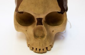
For 40 years or so the hoax refused to go away and numerous casts, models and reconstructions of Piltdown Man were made, sold, exchanged and gifted to museums and universities. These included casts of the original material as well as reconstructions of the skull and even reconstructions of the endocast – a cast of the inside of the skull.

The Museum has a selection of this material, but as Professor Shortland examined the collections, two specimens stood out.

The first is an R. F. Damon-produced endocast presented to the Museum by Arthur Smith Woodward himself. Smith Woodward was known as an expert on fossil fish but published widely on zoological topics. As a scientist of some repute there’s been long-standing speculation about his role in the hoax. Was he wholly duped by Dawson, or was he in on the hoax from the beginning? If it’s the former, then the presentation of this endocast shows Smith Woodward disseminating research he presumably took some pride in. If it’s the latter, perhaps it was a way of cementing the hoax as legitimate by spreading specimens far and wide.

Joseph Weiner’s experimental fake created by modifying an orangutan jaw, alongside a cast of the Piltdown jaw

The second significant specimen is a worked orangutan jaw produced by Joseph Weiner, one of the three authors who debunked the hoax in a 1953 Nature paper titled The Solution of The Piltdown Problem. Weiner modified the orangutan jaw to replicate the original hoax specimen. Thanks to Professor Shortland’s knowledge of the hoax, he sent through a copy of Weiner’s book on the Piltdown Man where this exact specimen is pictured.

The Piltdown Man hoax wasn’t the first and certainly won’t be the last hoax, fake or forgery in the history of science, but it remains one of the most well-known and stands as a warning of the dangers of hubris in the discovery and description of the natural world.

The Weiner jaw and Damon endocast will be on display alongside other Piltdown Man material in our Presenting… case from 9 January to 8 March 2020.

Birds of paradise

By Eileen Westwig, Life Collections Manager

The latest in our Presenting… series of single-case displays takes a look at one of the world’s most spectacular groups of birds – Paradisaeidae, or the birds of paradise.

A beautiful male Magnificent Riflebird (Lophorina magnifica)

The first bird of paradise to arrive in Europe was a skin that came to Spain in 1522. Many of these early skins were prepared by native hunters without wings or feet to better show off the bird’s spectacular plumage. Upon arrival in Europe, the apparent lack of wings and legs led to the myth that these birds originated from paradise and floated high in the skies, only to fall down to earth after their death.

Birds of paradise are members of the family Paradisaeidae, which contains more than 40 recognised species. Their closest relatives are crows and jays, of the Corvid family.

They inhabit the rainforests of Papua New Guinea, Eastern Indonesia and Eastern Australia and mainly feed on fruit and some insects. Hybridisation, when two birds of different species crossbreed, is quite common and can explain why many of the early described species were so “rare”.

Male Paradise Riflebird (Lophorina paradiseus) showing off iridescent plumage on its chest
Female Paradise Riflebird (Lophorina paradiseus) without colourful plumage, which helps to blend into the environment

Most species of birds of paradise are sexually dimorphic, meaning males exhibit the spectacular plumage these birds are best known for, whilst females have much less ornamentation and coloration. The male’s display feathers are highly specialised and have evolved from basic feathers. Like all feathers, they are shed and regrown every single year, which puts quite a strain on the males.

One of the first few Westerners to see these birds in their native habitat was naturalist and explorer Alfred Russel Wallace. He described the encounter, from a 19th-century Westerner’s point of view, in Narrative of Search after Birds of Paradise (1862) as:

Nature seems to have taken every precaution that these, her choicest treasures, may not lose value by being too easily obtained. […] In […] trackless wilds do they display that exquisite beauty and that marvellous development of plumage, calculated to excite admiration and astonishment among the most civilized and most intellectual races of man…

The Presenting… Birds of paradise case will be on display until 3 September 2019.

Happy 250th William Smith

Today is the 250th birthday of the remarkable English geologist William Smith, creator of the first geological map of England and Wales – ‘the map that changed the world’. Here Danielle Czerkaszyn, Senior Archives and Library Assistant, tells us more about Smith’s achievements and his relationship to the Museum.

William Smith (1769-1839)

William Smith (1769-1839) began his career as a land surveyor’s assistant in his home village of Churchill, Oxfordshire. He soon travelled the country working on mining, canal and irrigation projects. This gave him the opportunity to observe the patterns in layers of rock, known as strata, and to recognise that they could be identified by the fossils they contained. This would earn him the name ‘Strata Smith.’

Smith’s observations of strata over hundreds of miles led to the ground-breaking 1815 publication of his map A delineation of strata of England and Wales (pictured top) that ultimately bankrupted him.

Smith’s map set the style for modern geological maps and many of the names and colours he applied to the strata are still used today. While Smith’s accomplishment was undoubtedly remarkable, he was only officially recognised for his discoveries late in life. His lack of formal education and his family’s working class background made him an outcast to most of higher society at the time.

Geological Map of Bath, 1799. This map is considered to be one the earliest geological maps ever created. It demonstrates an early use of Smith’s ‘fading’ colouring technique which emphasised the outcrops of each stratum. The yellow tint represents the Bath Oolite, the blue marks the base of the Lias, and the red the base of the Trias.

It wasn’t until a few years before he passed away that Smith received any recognition for his contribution to the science of geology, receiving a number of awards, including the prestigious Wollaston Medal from the Geological Society of London in 1831, and an honorary degree from Trinity College Dublin in 1835.

A bust of William Smith is on display in the Museum’s court

His legacy lived on with his nephew John Phillips, one of our Museum’s founders and Professor of Geology at Oxford. Recognising its importance, Phillips left Smith’s archive to the Museum on his death in 1874. Thanks to generous funding from Arts Council England a few years ago, the Smith collection has been catalogued, digitised and is available online to the public.

Few men in the history of science contributed as much, but are as little known, as William Smith. He was a hardworking and determined man who dedicated his life to understanding the world beneath us. So here’s a big Happy 250th birthday to William Smith – the ‘Father of English Geology.’

A small display, Presenting… William Smith: ‘The Father of English Geology’ 250 years on, is running in the Museum until 2 May 2019.

Presenting… Christmas Island

By Eileen Westwig, Collections Manager in the Museum’s Life Collections.

About 320 km south of Java in the Indian Ocean lies Christmas Island. Although discovered and named on Christmas Day in 1643, the island remained unexplored until its first settlement in 1888, a development which had dire consequences for some of its native species.

Christmas Island is home to a variety of endemic animals such as rats, land crabs, butterflies and many birds. The accumulation of bird droppings over thousands of years made the island rich in phosphate, and the commercial potential of these deposits brought many expeditions to the island. With the ships’ cargo came black rats.

Two species of endemic rats, Maclear’s Rat (Rattus macleari) and the Bulldog Rat (Rattus nativitatis) went extinct within 20 years of settlement, despite having been previously very numerous on the island.

One of the skins of Maclear’s Rat (Rattus macleari) collected by H.E. Durham, and now held in the Life Collections of the Oxford University Museum of Natural History.

Maclear’s Rat, seen at the top of the page in an illustration from an 1887 publication, was described as chestnut brown above, with a partly white, long tail. It was once the most numerous mammal on the island ‘occurring in swarms’. The Bulldog Rat had a much shorter tail and a layer of subcutaneous fat up to 2 centimetres thick, the function of which is unknown to this day.

The likely cause of their extinction was the introduction of diseases by the ship rats, to which the Christmas Island rodents had no immunity. The disappearance of the native rats also had a knock-on effect: the parasitic Christmas Island Flea (Xenopsylla nesiotes) depended on the rats as hosts, and so the fleas became extinct with the rats’ demise.

In 1901 Dr. Herbert E. Durham, a British parasitologist investigating the origins of beriberi disease, led an expedition to Christmas Island. During his visit he collected several specimens of Maclear’s Rat, but was unable to find any Bulldog Rats, despite a lengthy search and the offer of a reward. Two of the nine Maclear’s Rats Durham obtained showed abundant parasites, trypanosomes, in their blood.

Christmas Island possesses quite a number of peculiar species in its fauna, and it is regrettable that observations were not made before animals had been imported to this isolated station, as well as that my own notes are so incomplete.

Dr. Herbert E. Durham

Durham also found blood parasites in the native fruit bats (Pteropus melanotus) but noted that these were unlikely to have been introduced, instead were “an old standing native occurrence.” These bats still inhabit various islands in the Indian Ocean, including Christmas Island, where they are critically endangered.

Original letter by H.E. Durham offering his Christmas Island specimens to the Museum in 1938.

The Museum holds a range of material from Christmas Island, including six skins and three skulls of Rattus macleari, which were collected by H. E. Durham in 1901-02, and donated in 1938.

Visit the Museum’s Presenting… case between now and 6 March to see Christmas Island specimens from the collections.

Marvellous Mantodea

In the latest display in our Presenting… series, collections manager Amoret Spooner takes a look at the wonderful and sometimes strange world of the praying mantis.

Praying mantis is the common name given to an order of insects called Mantodea, a word which derives from mantis meaning prophet, and eidos meaning form or type. The more familiar ‘praying mantis’ refers to the striking way that they hold their large forelimbs, in a ‘praying’ posture.

Display of different mantis species

There are over 2,400 species of mantis worldwide, split into 21 different families. The image above shows their incredible diversity of colour, shape and size. But while they may differ in appearance, their biology and many behavioural traits are the same.

Mantis are predators of insects, including other mantis, but larger species will eat small lizards and birds. But they are perhaps best known for being cannibalistic. This behaviour is most commonly seen in nymphs straight out of the egg case, or ootheca, but it can also occur when the female eats the male after mating. However, cannibalism is not required to mate, so when it happens it’s usually because the female is hungry!

The egg case, or ootheca, of mantis vary greatly depending on the size and behaviour of the species.
Revisio Insectorum Familiae Mantidarum was one of John Obadiah Westwood’s greatest works. Thankfully he kept all of his drawings, annotated pages and notes for the publication, allowing us an insight into the years of work he put into its production.

Praying mantis are ambush hunters, either camouflaging themselves while waiting for their prey to approach, or actively stalking prey. Their compound eyes are specialised in perceiving motion, and are widely spaced giving them a wide field of vision. Along with powerful front legs and an ability to move the head up to 180°, this makes them successful predators.

The Museum’s archive contains original drawings and annotations by John Obadiah Westwood (1805–1893), the first Hope Professor of Zoology. As a renowned scientist Westwood described many new mantis species, and he was also a talented artist.

The Presenting… Marvellous Mantodea case is on display at the Museum until 1 November 2018.

Argonauts: astronauts of the sea

by Mark Carnall, Life Collections manager

Cephalopods are a remarkable group of molluscs that includes nautilus, octopuses, cuttlefish and various groups of ‘squid’. The other major groups of molluscs includes more familiar shelled animals such as gastropods (snails and slugs), bivalves, and chitons, as well as some less familiar forms.

In natural history museums, molluscs are normally represented by shell collections because the hard shelly parts are easier to preserve and store than the soft tissue. This creates a bias against the soft-bodied cephalopods, such as squids, octopuses and cuttlefish, because aside from the cuttlebones of cuttlefish and the thin gladius in squids there aren’t many hard parts that can be preserved to represent these animals in dry collections. For octopuses it’s normally only the beak and microscopic radulae, a toothed tongue-like structure, that can be preserved. But there is one notable exception: the eggcases of argonauts.

Model of Argonauts argo. Image: Mark Carnall
Model of Argonauta argo. Image: Mark Carnall

Argonauts, four* species of octopuses in the genus Argonauta, are unusual in that they produce a paper-thin eggcase, sometimes referred to as a shell. Unlike a true shell it’s not attached to the body of the argonaut, but secreted by two specialised webbed arms. The eggcases themselves are sometimes called paper nautiluses because they resemble the spiral shells of nautiluses, but they are structurally and functionally very different.

External morphology of a female paper nautilus (Argonauta argo) with egg case. Poli, Giuseppe Saverio. Testacea utriusque Siciliae. (1791-1827).
External morphology of a female paper nautilus (Argonauta argo) with eggcase. Poli, Giuseppe Saverio. Testacea utriusque Siciliae. (1791-1827). http://biodiversitylibrary.org/page/44020354

Argonaut eggcases wash on up shorelines around the world and have been known for centuries. But it’s only comparatively recently that the origin and use of these cases has been described. When eggcases containing live argonauts were first encountered it was supposed that argonauts were reusing empty shells created by another animal, much like hermit crabs repurpose gastropod shells.

Pioneering research by marine biologist Jeaneatte Villepreux-Power in the 19th century led to observations of Octopus and Argonauta, confirming that the eggcases are made and repaired by female argonauts. It wasn’t until 2010 that we understood how argonauts use these cases to float in the ocean. It turns out that they ‘bob’ their shells to gulp a pocket of air. Then, using their second pair of arms, they trap the air in the top of the shell and dive releasing enough air to maintain the required buoyancy.

Only female argonauts make the eggcases, so the free-floating males are tiny in comparison. In addition to providing a home for female argonauts, these structures are used to brood embryos in. One eggcase was described with nearly 50,000 embryos attached to the inside of the shell.

Preparation showing series of argonaut egg cases of varying sizes.

Thanks to their oddity and beauty these eggcases are common in museum collections, but they represent one of the marvels of evolution. Unlike many bottom-dwelling octopuses, female argonauts have evolved this amazing structure to function as an underwater craft to allow them to leave the ocean floor and inhabit the open oceans: the true astronauts of the sea.

To celebrate the pioneering work of Jeaneatte Villepreux-Power, these amazing animals, their eggcases, and a selection of museum specimens are on display in the Museum’s Presenting… case until the 3 July 2018.

Mark writes more about these ‘astronauts of the sea’ on the Guardian’s Lost Worlds Revisited blog.

* Tens of species of living argonauts have been described, however four are currently recognised with a few dubious species.