Is it real? – Taxidermy

One of the most common questions asked about our specimens, from visitors of all ages, is ‘Is it real?’. This seemingly simple question is actually many questions in one and hides a complexity of answers. 

In this FAQ mini-series we’ll unpack the ‘Is it real?’ conundrum by looking at different types of natural history specimens in turn. We’ll ask ‘Is it a real animal?’, ‘Is it real biological remains?’, ‘Is it a model?’ and many more reality-check questions.

First up: Taxidermy, by Mark Carnall

Taxidermy
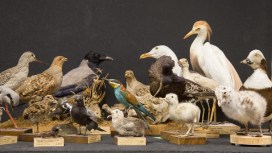
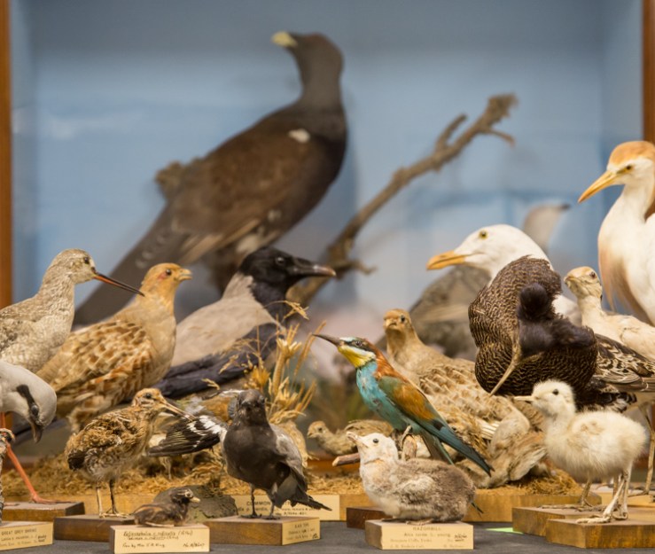
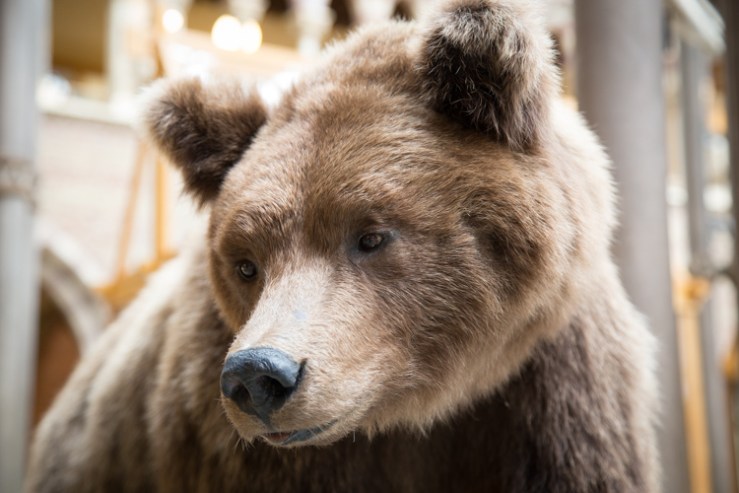
The Museum is well-known for its touchable taxidermy. As of today, we have two large bears, a Black Bear and a Brown Bear, greeting visitors as they enter the main court, as well as taxidermy specimens on our Sensing Evolution touch-tables. For children and adults alike, this close encounter with a taxidermy animal prompts the question – is it real?

Taxidermy, or ‘stuffed’ animals, are specimens that have been specially prepared, preserved and posed to show what the creature may have looked like in life, but real and not real here is tricky. The animal itself is, or was, a real animal – there are no taxidermy unicorns, for example. But the biologically real parts may only be the skin, the skull, and the skeleton inside the paws and feet, depending on the type of animal.

The touchable taxidermy Brown Bear greets visitors to the museum.

Inside taxidermy specimens there may be sculpted statues over which the skin is stretched; for older specimens, a wire and wood framework with paper, wood wool, straw and seeds may be used to fill out the skin. The animal’s squishy parts, which are not easy to preserve –such as eyes, lips and tongues – are normally made of glass or plaster.

Animals that have skins and skeletons that are relatively easy to preserve – including mammals, reptiles, and birds – are generally better suited to taxidermy. Marine mammals such as whales and dolphins, amphibians such as frogs and salamanders, and fish are all less common as taxidermy because their skins are harder to treat and keep stable.

Dogfish and piranha taxidermy which have been painted and varnished in an attempt to make them resemble the living animals. Note the comedic eyes on the shark.

The hard parts of skin, such as crests, wattles and skin patterns in reptiles, are susceptible to discolouring and fading in light, so these areas may be repainted to show what the animals look like in life. This introduces another ‘non-real’ element: paint.

So although there are certainly real parts used in taxidermy, there’s yet another complication in answering the question: the animals are usually posed by a human, so even their posture and appearance could be considered ‘subjective’ and perhaps therefore not quite ‘real’.

In fact, some of our older taxidermy may have been prepared by taxidermists who hadn’t ever even seen a living example of the animal they were working on. This can lead to inaccurate positioning and posing, as in the taxidermy kiwi on display in our main court.

So, is it real? You decide.

Next time… Skeletons and bones


The Big Brain Competition

What happens in your brain when you receive compliments? And what’s going on in your mind when you watch your football team win a match? Does the brain respond differently when recalling music, compared to listening to it? All these questions, and more, have been posed in our Big Brain Competition

Coinciding with the Museum’s Brain Diaries exhibition, the Wellcome Centre for Integrative Neuroimaging is inviting you to ask your own question about the brain to be in with a chance to have it tested by neuroscientists using Oxford’s state-of-the art Magnetic Resonance Imaging (MRI) scanner.

The advanced MRI scanner at the John Radcliffe Hospital in Oxford is one of the strongest in the world. It allows scientists to carry out functional MRI (fMRI) scans to see the brain in action. This mind-blowing procedure can reveal how the brain changes when learning a new skill or how it compensates when someone recovers from brain damage. It can also reveal which areas are used when people speak, move or laugh, to give just a few examples.

This fMRI scan shows how blood flows to the visual cortex region at the back of the brain when viewing a visually-stimulating checkerboard pattern
layouttile
Dr Stuart Clare of the Nuffield Department of Clinical Neurosciences is asking you for questions about the brain

Functional MRI shows when a brain area is more active by detecting the changes in blood oxygen levels and blood flow that happen in response to neural activity. The technique can be used to produce activation maps showing which parts of the brain are involved in a particular mental process.

The scientist behind the Big Brain Competition is Dr Stuart Clare, whose research involves pushing the technological boundaries of the fMRI technique to reveal new insights about how the brain functions normally and how it is affected by disease. There is still so much that the fMRI scans can bring to light, so Stuart is asking you for ideas!

Over several years of inviting people in to see the beautiful pictures that our MRI scanner can produce, I’ve been fascinated by the questions they have about the brain and whether you can see this thing or that thing in our fMRI scans.  With this competition we want to give people the unique access to our scanner and the chance to try an idea out for themselves.

When coming up with an idea for investigation there are a few practical things to bear in mind. Any activity has to be something people can do when lying down in the scanner and it has to be clear when they start and stop doing the activity. But Stuart is very open to ideas for experiments that they haven’t come across before – something that scientists really don’t already know the answer to.

The animation below explains how fMRI works and what it can do. So take a look, think up an experiment of your own and enter your idea via this form. The best one will be put into action by the research team and you will be able to watch the scans take place at the John Radcliffe Hospital yourself!

Comedy rears its ugly head

Logo blob fish
Is the Blobfish the ultimate ugly animal?

Animals with big eyes, fluffy tails and cute noses are easy to love. But what about those with tentacles, slimy skin or a large throat pouch?! Luckily the Ugly Animal Preservation Society is here to fight their corner. Education Officer Chris Jarvis shares some tales from a special event…

Comedy met conservation on Saturday night, as the Museum teamed up with Berkshire, Buckinghamshire & Oxfordshire Wildlife Trust, compere Simon Watt and the Ugly Animal Preservation Society. During the evening, six comedians put the case for their own neglected animals in need of conservation: the Scrotum Frog; Christmas Island Frigate Bird; Kaluga Sturgeon; Slimehead; Okapi, and Yak. The audience then voted to choose the official mascot for Oxford, but which of these lovely uglies did they choose?

The Scrotum Frog

Par Samuel Garman (1843-1927) [Public domain], via Wikimedia Commons
Image: Samuel Garman via Wikimedia Commons

Chosen by Iszi Lawrence, this unfortunately-named amphibian’s population in Lake Titicaca is now dwindling. It’s threatened by the introduction of alien salmon for the angling fraternity and is also blended to make a medicinal frog frappe by Andean locals with strong stomachs!

Christmas Island Frigate Bird

By Charlesjsharp (Own work) [CC BY-SA 3.0 (http://creativecommons.org/licenses/by-sa/3.0)], via Wikimedia Commons
Photo: Charlesjsharp via Wikimedia Commons
Some may find its inflatable throat pouch off-putting (in the same way the Scrotum Frog brings certain disturbing images to mind), but not advocate Eleanor Morton. This critically endangered seabird is one of a family of only five species that grace our oceanic skies and deserves our attention.

Kaluga Sturgeon

By User:Cacophony [GFDL (http://www.gnu.org/copyleft/fdl.html), CC-BY-SA-3.0 (http://creativecommons.org/licenses/by-sa/3.0/) or CC BY 2.5 (http://creativecommons.org/licenses/by/2.5)], via Wikimedia Commons
Photo: Cacophony via Wikimedia Commons
Hideous and aggressive, these formidable fish grow to a ton in weight and have suffered a population decline of 80% over the last 90 years: we humans like to eat their eggs and their spawning grounds are now severely polluted. Without action we may lose one of the largest fresh water fish in the world. Kaluga Sturgeon champion Paul Duncan McGarrity made the case for this fugly fish.

Slimehead

By Pengo (Own work) [CC BY-SA 3.0 (http://creativecommons.org/licenses/by-sa/3.0) or CC BY 3.0 (http://creativecommons.org/licenses/by/3.0)], via Wikimedia Commons
Photo: Pengo via Wikimedia Commons
Nigel Lovell chose this sociable, deep sea fish which has been severely overfished due to the remarkable success of a rebranding campaign. You might know it better as the ‘Orange Roughy’ seen in fishmongers since the 1970s. But just remember it’s a ‘Slimehead’ – your dinner guests might not want to see that on their menu!

Okapi

By Raul654 (Own work) [GFDL (http://www.gnu.org/copyleft/fdl.html) or CC-BY-SA-3.0 (http://creativecommons.org/licenses/by-sa/3.0/)], via Wikimedia Commons
Photo: Raul654 via Wikimedia Commons
A rather beautiful (to my mind) relative of the giraffe, which looks like it put on a zebra’s pyjama bottoms by mistake! Rosie Wilby’s case for the Okapi was strengthened by a wonderful demonstration of its courtship behaviour involving a member of the audience and an 18-inch purple tongue*!

*Photographs cannot be shared.

Yak

Photo: travelwayoflife via Wikimedia Commons

Chosen by Teiran Douib, and not to be confused with the domestic yak, the wild yak is listed as vulnerable in its native Himalayas due to poaching, cross-breeding and climate change. These wonderful creatures are blessed with electrically non-conductive fur to survive electrical storms. They are one of the largest of all cow species, reaching up to 2.2 metres at the shoulder. However, their udders and scrotums are particularly small and hairy as protection against the cold.

Once the comedians had made their pitch it was time to decide on an ugly animal mascot for Oxford. Voting was frenetic and passionate as the issues at stake clearly hit home. But finally a champion emerged…

The winner was…the Kaluga Sturgeon! Yes, it’s official, Oxford’s Ugly Animal Mascot is a one-ton, aggressive fish that occasionally upends boats. As Paul Duncan McGarrity pointed out, it’s a shame that Oxford doesn’t have a long-standing boating rivalry with another city where this might come in handy…

Skeletons in the closet

Once found in almost every museum and university in the western world, human anatomy collections have slowly disappeared over time, as medical practices have changed. These collections are historically important and medically relevant, providing direct evidence of patients’ lives and illustrating a pivotal period in the history of medicine.

Over the past seven months, Dr Kathryn Krakowka has been going through the Museum’s human remains specimens to create a new detailed database of the contents. Here, Kathryn talks about the fascinating material she is working on…

The Museum holds many specimens that are not on public display, and perhaps one of the largest collections hidden from view is the human remains. This collection was started in the first half of the 18th century by Dr Matthew Lee, and eventually passed to Dr Henry Acland, founder of the Museum. Acland added a lot to the collection, acquiring his own samples as well as purchasing specimens and collections from other physicians.

When Acland gathered material for the new museum building in 1860, he brought this anatomical and physiological material with him, including the human remains. The collection continued to develop throughout the 19th century and grew to contain over 1,400 specimens. It is now one of the few collections from this period still in existence in Britain.

As a specialist in human osteology, I am trained to assess human skeletonised remains for such characteristics as sex, age-at-death, and any pathologies or trauma that the individual was affected by in life.

The Museum’s collection revealed specimens that tell a tale of health and medicine at Oxford in the 18th and 19th centuries. Many of the specimens were used to teach medicine, so many different skeletal pathologies are represented, including bacterial infections, genetic deformities, nutritional deficiencies, and traumatic injuries.

A skull fragment showing a large growth. The four round marks are thought to have been made by a tool during investigations after death.

One example is the frontal bone of a possible child, pictured above. This presents a large porous growth affecting both the outer and inner table of the bone. The characteristics of the lesion suggest osteosarcoma (bone cancer). There are then what appear to be four tool markings on the outer table of bone that indicate some sort of drill may have been used to take samples of the bone after death, possibly in an attempt to determine what was ailing the individual during life.

As this growth is quite large and just above the forehead, it would have been very noticeable while the individual was alive. This specimen provides an interesting example of how medical practices were often developed through exploratory postmortem analysis.

Now that the collection has been fully catalogued and digitised, it is hoped that further research into specimens like this one can help shed light on historical pathologies and the development of medicine as a discipline.

The database and digital images of the specimens will soon be available on the Museum’s website, thanks to funding by the John Fell Fund.

Delightful dung beetles

The latest display in our changing Presenting… case showcases a wonderful array of dung beetles. Darren Mann, head of our Life Collections, tells us why they are so important.

Worshipped during ancient Egyptian times, dung beetles have a long history of human appreciation. Jean-Henri Fabre (1823-1915), one of the first to popularise insects in his writings, began his Souvenirs entomologiques series with the Sacred Scarab, and even Charles Darwin appreciated the weaponry adorning many dung beetles.

Dung beetles can be divided into three main groups based on their nesting behaviour. The rollers, often seen on television wildlife documentaries, make a ball out of dung and roll it some distance before burying it. The tunnellers dig directly below the dung pile and bury as much as needed for nest construction. Finally, the dwellers nest within the dung pile.

The South American Phanaeinae is one of most colourful groups of beetles. They are often referred to as Rainbow Scarabs due to their bright metallic bodies. We don’t fully understand why these beetles are quite so colourful.

Dung beetles are one of the more popular groups of insects used in ecological and evolutionary research today. They can help us to understand questions about how biodiversity loss impacts on ecosystems, or act as model organisms in the field of evolutionary development.

Unlike the much-publicised importance of bees and their pollination services, dung beetles are relatively unknown, despite their huge ecological and economic value. Their feeding and nesting behaviours provide many useful ecosystem services such as dung removal, pest fly control, parasite suppression, nutrient cycling, plant growth enhancement, improvement of soil structure, secondary seed dispersal, and a possible reduction of greenhouse gas emissions.

Through these activities, one study calculated that dung beetles are worth around £367 million a year to the UK cattle industry alone.

The largest dung beetles belong to the genus Heliocopris, which can reach up to 69 mm (pictured is Heliocopris dominus). These large beetles specialise on elephant and rhino dung. From around 2 mm in length is the oriental genus Panelus. These small beetles probably feed on the ‘dung’ of other insects and fungi.

Ancient Egyptians believed that the dung beetle kept the Sun moving across the sky like a giant ball of dung, linking the insect to the god of the rising sun Khepri. Some historians believe that it was through observing dung beetle behaviour and biology that Egyptians developed ideas about life after death.

The two most widely depicted species in Egyptian art are Kheper aegyptiorum and Scarabaeus sacer. Nowadays, only Scarabaeus occurs in this region of Africa; Kheper is now a more southern species, possibly indicating climatic changes since Ancient Egyptian civilization.

Kheper aegyptiorum on display in the museum’s Presenting… case

The UK has about 60 species of dung beetle and most of these belong to the ‘lesser dung beetle’ subfamily Aphodiinae. The largest of our dung beetles are the Dor Beetles which can reach 28 mm. Our smallest, Plagionus arenarius, is a meagre 2.5 mm. Sadly, over 50 per cent of our dung beetles are in decline due to agricultural intensification, pesticides and habitat loss.


Imitation game

Last month we had the pleasure of hosting artist and scientist Dr Immy Smith as part of her week-long takeover of @IAmSciArt on Twitter. Drawing inspiration from the Museum’s collections, Immy has created some beautiful paintings. Here she tells us a little more about her interests and work…

My current artwork is focused on crypsis and mimicry – the ways that animals and plants disguise themselves or pretend to be something they’re not. Cryptic camouflage helps animals to avoid being seen, often to help them catch prey – or to avoid becoming prey themselves! Mimicry is also often about trying not to get eaten: the harmless hornet moth, for example, mimics a stinging insect to deter predators. I use these themes to develop print art projects, and also public workshops to help people learn more about the ecology of cryptic animals.

Cryptic Cards by Immy Smith

In my arts practice I try to imagine how animals and plants might evolve to camouflage themselves on human-made materials, and what they might look like. Will we one day find moths adapted to hide on advertising hoardings, or beetles mimicking litter? I made an entire deck of Cryptic Cards as a response to this kind of question.

Another project I’m working on at the moment is called Emergent Crypsis. This is a collaboration with Norweigan generative artist Anders Hoff who makes art using algorithms executed by a computer. I’m imagining how creatures might adapt to an extreme example of human-made patterns – computer generated abstract images.

Violin Beetle (Mormolyce phyllodes) by Immy Smith

My work requires me to closely study many animals and plants, but how do I learn about all these species in order to draw their imaginary relatives? How do I make my art a convincing representation of how life might find ways to hide on human-made art?

One answer is of course, the internet. I’ve been lucky enough to find many wildlife photographers online who are kind enough to let me use their images as reference. But photographs alone are not always enough to get to know the fine details and defining characteristics of a species: the joints and articulations of small insects, for example, are best studied from specimens. And some species are rare, or even extinct, and it can be hard to find photographic a reference.

Leaf-footed Bug (Diactor bilineatus) by Immy Smith

This is where scientific collections come into the picture. The collections held in museums and other institutions are not only essential for scientists and scientific illustrators, they are also an invaluable resource for artists of many disciplines, science communicators, and educators of many kinds. In the collections at the Oxford University Museum of Natural History I can photograph and sketch leaf-mimicking insects, for example, that are native to the forests of South America which I may never visit. I can study in minute detail the articulation of beetles that are rarely seen, and which might be difficult to find – and irresponsible to collect – myself.

A display of terrestrial bugs (Heteroptera) in the Museum, including the Leaf-footed Bug painted by Immy Smith

Not only do I find specific species that I want to study in natural history collections, I often see new ones – animals I didn’t know about or hadn’t thought of drawing before. In the same week that I visited Oxford, I also made a trip to Herbarium RNG in Reading to study plant mimicry, and found similar inspiration there. I can channel all this into both aesthetic art destined for print and sciart workshops that communicate the wonders of insects or plants with the wider community.

Working on sciart projects and educational workshops helps me appreciate the multitude of ways in which collections benefit research and education. We must try to communicate the plethora of roles they play, and the host of ways they cross into our lives – whether through scientific research on insects pollinators of the crops we eat, or via a deck of cards made by someone like me for mainly recreational purposes. We must fight to protect scientific collections because they are a resource that benefits all of us as a society.