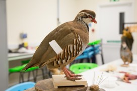
Partridge in a laboratory

IMG_6669

We recently ran a second series of taxidermy workshops here at the Museum, run under the expert guidance of professional taxidermist Derek Frampton. Once again they proved very popular with participants, so we asked one of those budding taxidermists, Kit Collins, to give us a short write-up of the day…

As a child I was always fascinated by nature, finding adders, baby hares, grass snakes, slow worms, and watching dolphins, buzzards, and Red Kites, when they were much rarer. I even once skinned a mouse that had been caught in our mouse trap.

I have always wanted to try taxidermy and I now work at an auctioneers where I regularly see all sorts of taxidermy – skins, horns, and skulls, including a hippopotamus skull. So I was keen to know more about the process. This was the first taxidermy course I’ve seen so I jumped at the chance to try something new and learn from an expert taxidermist.

IMG_6677
Derek Frampton puts some finishing touches to his partridge

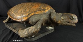
During the workshop we were taken through each step of the process, first watching Derek demonstrating on his bird then copying these steps on our own Red-Legged Partridges.

Kit and partridge
Kit and his finished specimen regard one another

We could see the finished article that had been made in the previous day’s workshop, sitting watching us on a nearby windowsill. Unfortunately, our specimens looked nothing at all like this at the start and as the morning went by it looked less and less likely that our piece of wet skin and feathers with a few bones attached would end up looking anything like a real bird again…

However, with the help of a blow dryer the feathers regained their soft, striking plumage. We then spent the afternoon piecing the bird back together using a kind of packing straw to recreate the shape of the body, and wire, clay, false eyes, and car body filler to do the rest.

We each ended up with a beautiful bird to take home, as well as the memories of a fun and unusual day out (and anatomy lesson) at the Museum. I would love to do it again.

Kit Collins

More than mimicry

figure_3
Hunting for Bates specimens. The labels with pink strips tell us they were part of Bates’ collection.

A small exhibition has popped up on the upper gallery of the Museum, showcasing natural historian Henry Walter Bates. He’s famous for his theory on mimicry, but, as exhibition curator Gina Allnatt explains, there’s a lot more to discover about Bates.

**

Charles Darwin and Alfred Russel Wallace are now widely recognised as the co-discoverers of the theory of evolution. They are both established figures in the natural history world, but it was a lesser known contemporary who gave them a missing piece of the puzzle.

A panel from the display is put together behind the scenes

Henry Walter Bates was born in Leicester on 8th February 1825. He was originally apprenticed to a hosiery manufacturer, but his passion for insects sent him on a completely different path in life. In 1844, Bates encountered Wallace in a library, and the two men found they shared a mutual love of nature. Bates introduced Wallace to the field of entomology (the study of insects) and it wasn’t long before the two were planning a joint expedition to the Amazon. They funded the expedition almost entirely through the sale of specimens they collected. Wallace returned to England after four years, but Bates remained in the Amazon for a further seven years. When he finally returned to England he had amassed a collection of over 14,000 insects. 8,000 of these were new to science.

LEPI4132b_heraldica_Bates_dorsal
Bates Type specimen from the Godman-Salvin collection

Bates is most famous for the work which bears his name: Batesian mimicry. Batesian mimicry is when a harmless species mimics the warning colours or behaviour of a harmful species.

The mimic  then benefits from the protection of the model. For example, the Hornet Moth (Sesia apiformis) is completely harmless, but looks like a wasp and benefits from the protection of the wasp’s warning colours. The moth is the mimic and the wasp is the model.

figure_2
Net cocoon (Urodidae) from the moth collections

This Museum’s historical butterfly collection contains over 200 specimens collected by Bates, and thousands of other insects collected by him in the overall Entomology collections.

The specimens came to the collections via different routes. Some were purchased directly from Bates by Professor J.O. Westwood, the Museum’s first Hope Professor of Zoology. Bates material also arrived through acquired collections, such as those purchased from natural history specimen dealer Samuel Stevens. Other specimens of interest in the Lepidoptera collections purchased from Bates include an unusual net cocoon from a rare moth in the family Urodidae. The Urodidae are an unusual form of moth that build cocoons resembled a mesh bag with an opening at the bottom. One theory for this unusual structure is  to allow rainwater to flush through it easily, without drowning the pupa inside. In the Amazon rainforest, which is prone to heavy rainfall and flooding, this is a huge advantage.

The exhibition, which can be seen until 26th February 2016, will reveal much more about Bates and his contribution to modern-day science.

Gina Allnatt, Curatorial assistant (Lepidoptera)

#Dungisfun

Onthophagus nuchicornis male

Dung doesn’t always get a lot of attention, but his week a new project, known as DUMP, has been all over the news. The project team are here to try and convince you that dung really is fun.

**

How often do you think about dung? Possibly not at all, yet without Dung Beetles we would literally be up to our necks in it.

There’s no doubt, Dung Beetles are an important group of insects, particularly for the agricultural environment. Recent research estimated that dung beetles save the UK cattle industry £367 million per year (Beynon et al. 2015). They provide all sorts of ecosytem services, including their famous ‘dung removal’ and others you may not be aware of. For instance, they reduce gastrointestinal parasites of livestock, nuisance flies, and play a key role in improving soil condition through aeration and nutrient recycling.

Aphodius subterraneus
Aphodius subterraneus – possibly extinct in the UK

In the UK there are 100 species of Scarabaeoidea, which includes the Dung Beetles, Chafer and Stag Beetles. Over half of these are dependent on dung. As part of the on-going Species Status Assessment Project with Natural England in collaboration with Buglife , a review of the scarce and threatened Dung Beetles and Chafers is currently in progress (S.A. Lane & D.J. Mann, in prep.). The preliminary results indicate an alarming decline in our Dung Beetle fauna.

Just over 25% of UK Dung Beetles are ‘Nationally Rare’ and four species may even have become extinct in the past 50 years. This project also highlighted the lack of modern records for many of the rare species and that many areas of the UK are severely under recorded.

All this prompted us (Darren J. Mann, Steve Lane, Sally-Ann Spence & Ceri Watkins) to go out and look for beetles and to re-survey sites where rare species were previously known. DUMP was born.

The Dung Beetle UK Mapping Project (DUMP) aims to record Dung Beetles across the UK, provide distributional records and gather information on habitat requirements and ecology. The DUMP team will also engage with landowners, farmers and the general public on the benefits and value of dung beetles.

Ceri Watkins making new friends during DUMP fieldwork
Aphodius lividus
Aphodius lividus

Over the past few years the team has travelled across the country from the Orkneys to the Channel Islands sampling across a range of habitats. A targeted survey for Onthophagus nuchicornis discovered healthy populations in North Devon and South Wales, but highlighted the dramatic decline of this species in its previous strongholds in Norfolk and Suffolk.

We also made some positive new discoveries including finding the rare Aphodius lividus and Aphodius sordidus on the Norfolk–Suffolk border, and Aphodius porcus at a new site in South Wales.

The DUMP project is in its early stages and comprised of a small team of volunteers. In the near future we hope to provide further information, distributions maps, online recording, and advice for management plans to help conserve our dung-inhabiting fauna. You can help out on social media too – tell the world why Dung Beetles matter and why #dungisfun!

Ceri Watkins, TCV Natural Talent Trainee

TLC (turtle loving care)

Turtle post-treatment

The Green Turtle (Chelonia mydas) above has been receiving some much-needed TLC from Abby Duckor, our first conservation intern from UCL’s MSc in Conservation for Archaeology and Museums. Here Abby explains what she’s been up to…

During my time at the Museum I have been lucky enough to spend a fair amount of time working on this taxidermy turtle. There was plenty to do: the specimen was covered in a dark layer of dust; there was a large tear in the neck, perhaps from a knock; and the taxidermy was generally overstuffed, noticeably on the stomach plate which had become completely detached, revealing the inner filling material.

The turtle shell (carapace) in the middle of treatment. The carapace was cleaned with detergent and de-ionized water. Toothbrushes were used to scrub the hard shell and to help remove any ingrained dirt.
Most of the filling was wheat with a small grain size, dating it to pre-1950, according to Dr Stephen Harris: after 1950 wheat grains were cultivated to be fatter and the stalks shorter. The wheat and other plant materials in the filling suggest an English location for the taxidermy.

The specimen itself is labelled as part of Rev. Buckland’s collection, dating it to the early 1800s, if not earlier. William Buckland, an important early geologist and palaeontologist, was quite a character. During his life he amassed a large collection of living and mounted animals. He claimed to have eaten his way through the animal kingdom, and you have to wonder if he ever tasted this fellow… Green Turtles were a popular food for sailors and locals, reducing their population size. Today they are listed as endangered.

Abby working on the turtle in the conservation lab.
Conservation treatment of this specimen involved cleaning the shell with detergent and deionized water, revealing a colourful shell underneath. You may have noticed that this Green Turtle is not actually green. In fact, the Green Turtle is named after the colour of its fat, not the colour of the skin. Ours had been painted a dark greenish-brown colour, because they lose their skin colour after they die, but I removed the top layer of paint to better reveal the yellow scales on the turtle’s head, tail and limbs.

The final touch was the replacement of the stomach plate, or plastron, which is now held in position with epoxy putty wedges attached to the metal stakes that hold up the specimen. Cleaned, and with everything in its right place, our Green Turtle is now in much better shape, as you can see in the photo at the start of the article.


Twice in a whale

natural-history-museum-oxfordsmall-41

By Mark Carnall, Life Collections

Followers of our Once in a Whale blog a while back may be aware of the huge task that faced our Life Collections conservator Bethany Palumbo and her team as they set out to clean, restore and repair the whale skeletons that hang from the Museum roof.

In my first week here, I received an enquiry about the history of these specimens, and digging through the archives I was pleased to find that they are not just ‘prop’ skeletons acquired for the purpose of display – they are important in the history of whale biology too. So this article is something of a postscript to the Once in a Whale project.

Surprisingly, it wasn’t until the mid-19th century that much was understood about the science of the largest animals to have lived on Earth. Some species were known from strandings; others from accounts – varying in reliability – from fishermen.

img_5415small-copy
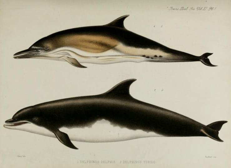
Yet difficulties in preserving and transporting such large creatures (as well as the penchant for eating stranded whales at community festivals) meant that the biology and behaviour of whales was poorly-described and documented until fairly recently. So much so, that in early scientific literature just a few scientists are singled out as having actually seen the animals they were studying.

In the Museum there are five whale skeletons suspended from the roof, along with the skull of a Humpback Whale and the mandible of a Sperm Whale. Some of the earliest ‘whaleologists’ made the trip to Oxford to see these specimens in a race to formally describe new species or new aspects of whale biology.

In particular, two 19th-century anatomists competed to make new discoveries about whales. Dr John Edward Gray, keeper of zoology at the British Museum (Natural History), and Professor Daniel Frederick Eschricht, a Danish comparative anatomist, were so competitive that Gray made sniping comments in formal papers, questioning Eschricht’s observations.

John Edward Gray ‘destroying’ Eschricht’s observations. From Gray, J. E. 1864. On the Cetacea which have been observed in the seas surrounding the British islands.
John Edward Gray ‘destroying’ Eschricht’s observations. From Gray, J. E. 1864, On the Cetacea which have been observed in the seas surrounding the British islands

But perhaps this competition was more sporting than malicious: Gray did name the gray whale, Eschrichtius robustus, in Daniel Eschricht’s honour.

Eschricht actually presented the Museum with two of the specimens now on display: the Humpback Whale skull at the entrance and the suspended Minke Whale skeleton. Of the others, the Bottlenose Dolphin skeleton was caught near Holyhead in 1868 and was drawn by another notable natural historian, William Henry Flower, before being skeletonised for the Museum.

The Orca skeleton is from an individual killed in the Bristol Channel by fishermen in 1872, and the Beluga Whale was collected from Spitsbergen, Norway in 1881 and presented by Alfred Henge Cocks, who donated a range of mammal specimens to the University of Oxford.

The female Northern Bottle-nosed Whale skeleton has been harder to track down. It’s possibly a specimen shot in Weston Super-Mare in 1860 mentioned by Gray, but it isn’t clear. Lastly, there’s the large Sperm Whale mandible that greets visitors at the entrance. It doesn’t have much of a recorded history, but is allegedly one of the largest specimens in the UK according to a ‘researcher’ whom I’ve yet to track down.

William Flower’s drawing of the bottlenose dolphin (lower) the skeleton from this individual is on display in the museum Flower, W. H. (1880), I. On the External Characters of two Species of British Dolphins (Delphinus delphis, Linn., and Delphinus tursio, Fabr.). The Transactions of the Zoological Society of London, 11: 1–6. doi: 10.1111/j.1096-3642.1980.tb00343.xWilliam Flower’s drawing of the bottlenose dolphin (lower) the skeleton from this individual is on display in the museum Flower, W. H. (1880), I. On the External Characters of two Species of British Dolphins (Delphinus delphis, Linn., and Delphinus tursio, Fabr.). The Transactions of the Zoological Society of London, 11: 1–6. doi: 10.1111/j.1096-3642.1980.tb00343.x
William Flower’s drawing of the Bottle-nosed Dolphin (lower); the skeleton from this individual is on display in the Museum

The next time you are in the Museum, do look up: the skeletons there are not simply representing ‘whaleness’ but are also individual animals and important specimens in the early discovery and description of whale biology.

Talkin’ ’bout taxidermy

IMG_1691 (1)

Here at the Museum we have many bird, mammal, fish and reptile taxidermy specimens on display. There are a fair few that you can touch too, on our Sensing Evolution tables in particular. Outside museums, taxidermy has seen a wider surge in popularity in recent years, featuring in artistic practice and being offered as a practical skill through taxidermy courses. It has also been the subject of ethical questions and debates.

In the 19th century, taxidermy was seen as one of the ways of bringing strange creatures from the around world to museum visitors. They were collected at a time when the natural world was thought to be an infinite resource. Many early biologists would combine scientific expeditions with collecting trips, gathering material to be studied, described and displayed in public museums.

IMG_1639 (1)
Unwrapping the American Black Bear

Today, it’s no longer possible to simply order an animal from a collector and expect to receive a crate containing a specimen prepared for display. There is a raft of national and international legislation designed to ensure that biological material is ethically, legally and safely collected and transported. It is also concerned with the impact on source communities, species conservation or ecosystems, and imposes checks on the transmission of disease or pests.

When Sensing Evolution opened here in May 2015 the museum obtained two larger pieces of taxidermy to greet visitors as they enter the building: a Red Deer purchased from a farm in Belgium, and an American Black Bear, which is a donation from the New Jersey Fish and Wildlife department in the USA.

Commissioning taxidermy is not straightforward and it’s important to know which permits are needed and whether you can legally qualify to receive them. There are many laws surrounding the import and use of animal parts that you need to be familiar with.

The Museum believes in the importance of wildlife conservation and we wanted to be sure that our taxidermy specimens are ethically obtained. Neither the Red Deer nor the American Black Bear were killed for the purpose of museum taxidermy, and both were sourced following a lot of research and communication with professional taxidermists and zoological societies.

IMG_1674 (1)
Team Bear and the bear itself, now installed in the court

The Red Deer skin was discarded from a farm where the specimen was raised for meat; the Black Bear was euthanized by the New Jersey Fish and Wildlife department as part of its standard control measures, although in this case rather than incinerating the carcass, the skin was removed, processed and donated to the Museum. We waited for many months for a suitable specimen to become available in this way.

The result is two touchable pelts which we hope visitors will find fascinating and beautiful, and which will contribute to our understanding of these incredible animals.

For more advice or information about taxidermy contact your local museum or the Guild of Taxidermists.