Sight without eyes

By Lauren Sumner-Rooney, Research Fellow

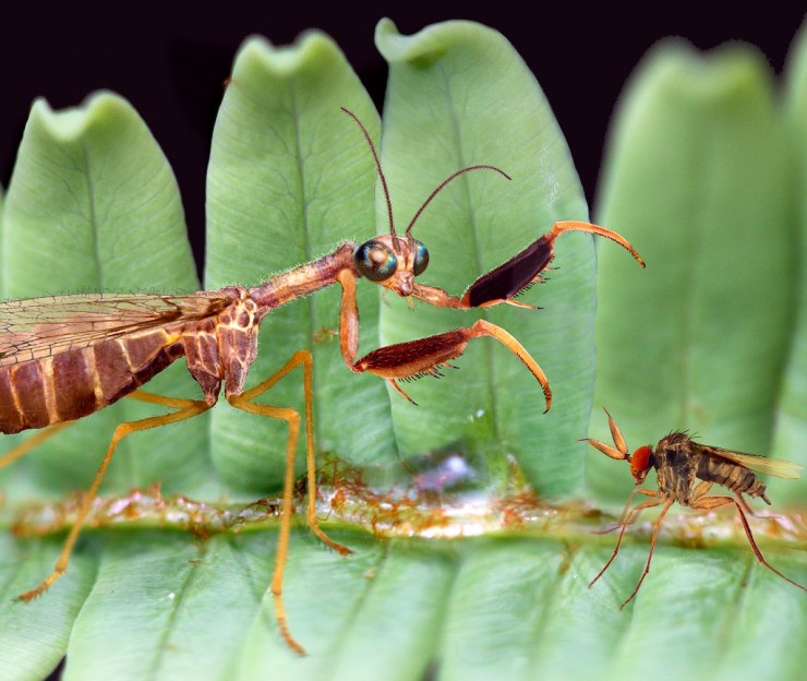
Vision is among the most important innovations in animal evolution. The ability to see predators, prey, mates, and the environment transformed the way animals interact with each other and the world around them. Eyes can take many different forms, but this month saw the description of a visual system unlike almost any other known to science, found in a brittle star called Ophiocoma wendtii.

Brittle stars are marine invertebrates related to starfish. They have long, slender arms connected to a central disk, but no head, no brain, and – so we thought – no eyes. But recent experiments have shown that some brittle stars are able to see the world around them.

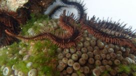
Ophiocoma wendtii is a common species found throughout the Caribbean Sea and the Gulf of Mexico. If you rummage around in coral rubble in shallow water, you’ll probably find Ophiocoma hiding underneath rocks and other debris, sheltering from their fishy predators. It has beautiful bright red tube feet (small, water-filled tentacles) and a neat party trick: it changes colour. During the day, the animals are a deep reddish-brown colour, but after dark they become beige with dark stripes.

The red brittle star, Ophiocoma wendtii

For more than thirty years, O. wendtii has been something of a mystery to scientists like myself who are interested in animal vision. It’s covered in light-sensing cells – thousands of them – and it hates being exposed to bright light, quickly dashing for cover if possible. However, it’s possible to head for dark, shadowy places without vision; you only need to be able to tell that one direction is brighter than the other. So, with a team of colleagues from Germany, Sweden and the USA, we set about giving the brittle stars an eye-test.

Lauren Sumner-Rooney, collecting specimens of Ophiocoma wendtii. Image: Jane Weinstock

We know that when they’re exposed to sunlight, O. wendtii try to hide underneath nearby rocks or other objects, so we designed a circular arena with a stimulus printed on one side – the idea is that the stimulus might resemble an object under which the animals can shelter, and the animal will move towards it.

We ran three experiments, changing the stimulus and background of the arena in each to test whether the brittle star can just see relative light or dark areas, or whether it can resolve finer points of contrast. To my surprise, O. wendtii moved towards the stimuli in all three experiments significantly more frequently than expected by random chance, as you can see in the video below. This was super exciting, as it represents not only the very first evidence of vision in these animals, but the second known example of any animal that can ‘see’ without having eyes (the first is a close relative, a sea urchin).

While O. wendtii is known to shelter during the day, we were also curious to test its behaviour at night. Running the same experiments again in natural darkness, we found that animals no longer moved towards any of the stimuli. There could be a whole number of reasons behind this, so we devised tests that eliminated several possibilities, and were left with a remaining explanation that the animal’s colour-change between night and day was somehow responsible.

Close-up of the arm plates of Ophiocoma wendtii

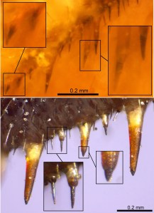
Colour-changing in the brittle star is controlled by the expansion and contraction of cells, called chromatophores, that are filled with pigment granules. These sit inside pores in the skeleton, alongside the light-sensing cells. During the day, the chromatophores expand, pushing up through the pores and spreading over the body surface. The pigment is spread over the outside of the animal, which looks dark brown as a result. During the night, the chromatophores contract, bringing all the pigment granules back inside the skeleton and giving a paler appearance.

The red brittle star, Ophiocoma wendtii. Image: Heather Stewart

We thought that during the day the pigment granules surrounding the light-sensing cells might block light reaching them from most directions. To test this, we constructed digital models of the visual system, creating 3D models of the light-sensing cells, the skeleton, and the pigment granules.

We found that in light-adapted systems, those with pigment, light could only reach the sensory cells from an angle of around 60° out of 360° which, though probably very coarse, could support vision. By removing the pigment from the models, vision was made impossible, as light could reach the sensory cells from too many different directions. It looked as though it was the chromatophores that made all the difference.

This is the first proposed example of whole-body colour change enabling and disabling vision in any animal, and raises many new questions about image formation and information processing. There are exciting parallels with the only other example of ‘extraocular’ (=without eyes) vision, the sea urchin we mentioned earlier: these sea urchins can also change colour in response to light levels, using similar chromatophores. Have they independently evolved a similar trick?

Top image: Heather Stewart

On the trail of the Piltdown hoax

The latest display in our single-case Presenting… series takes a look at the famous Piltdown Man hoax, and Life Collections manager Mark Carnall tells us how the display came about…

Visiting researchers to the zoology collections at the Museum often give us an excuse to dig deeper into our own material, and one such recent enquiry led me into the intriguing story of the Piltdown Man hoax.

Professor Andrew Shortland from Cranfield University contacted us to enquire about the Piltdown Man material in our collections, as part of research for a book on hoaxes and forgeries in anthropology that he is writing with Professor Patrick Degryse of KU Leuven.

I knew we had some Piltdown material here thanks to this page written by Malgosia Nowak-Kemp, but I hadn’t had an excuse to investigate any further. The enquiry was also timely as we’d just transferred a collection of palaeoanthropology casts, models and reconstructions from our Earth collections to bring our human collections into one place. I knew from our move project team that there was some Piltdown material awaiting processing – perfect.

For those who don’t know the Piltdown Man story, a short history is in order. In the early 20th century, amateur fossil hunter Charles Dawson brought a collection of human remains excavated from gravel pits in Sussex to the attention of Arthur Smith Woodward, then Keeper of Geology at the British Museum (Natural History). Woodward and Dawson collected further material and presented the remains as those of Eoanthropus dawsoni (‘Dawson’s dawn man’), an important fossil human from Britain.

Group portrait of the Piltdown skull being examined. Back row (from left): F. O. Barlow, G. Elliot Smith, Charles Dawson, Arthur Smith Woodward. Front row: A. S. Underwood, Arthur Keith, W. P. Pycraft, and E. Ray Lankester. Charles Darwin looks on from a portrait on the wall. Image via Wikipedia.
R.F. Damon-produced endocast and associated label recording the presentation of this specimen to the Museum by Arthur Smith Woodward

The discovery looked set to put Britain on the map when it came to evidence of human evolution, but suspicions were quickly raised about the authenticity of the material. Such was the skill of the forgery – meticulous breaking, abrading and staining of various archaeological and historic specimens – that it wasn’t until dating techniques, chemical analyses and some experimental palaeoanthropology in 1953 that the hoax was conclusively put to bed.

In turned out that the Piltdown ‘remains’ were a mix of medieval bone, an orangutan jaw, and chimpanzee teeth maltreated to look like an evolutionary intermediate between humans and other apes.

For 40 years or so the hoax refused to go away and numerous casts, models and reconstructions of Piltdown Man were made, sold, exchanged and gifted to museums and universities. These included casts of the original material as well as reconstructions of the skull and even reconstructions of the endocast – a cast of the inside of the skull.

The Museum has a selection of this material, but as Professor Shortland examined the collections, two specimens stood out.

The first is an R. F. Damon-produced endocast presented to the Museum by Arthur Smith Woodward himself. Smith Woodward was known as an expert on fossil fish but published widely on zoological topics. As a scientist of some repute there’s been long-standing speculation about his role in the hoax. Was he wholly duped by Dawson, or was he in on the hoax from the beginning? If it’s the former, then the presentation of this endocast shows Smith Woodward disseminating research he presumably took some pride in. If it’s the latter, perhaps it was a way of cementing the hoax as legitimate by spreading specimens far and wide.

Joseph Weiner’s experimental fake created by modifying an orangutan jaw, alongside a cast of the Piltdown jaw

The second significant specimen is a worked orangutan jaw produced by Joseph Weiner, one of the three authors who debunked the hoax in a 1953 Nature paper titled The Solution of The Piltdown Problem. Weiner modified the orangutan jaw to replicate the original hoax specimen. Thanks to Professor Shortland’s knowledge of the hoax, he sent through a copy of Weiner’s book on the Piltdown Man where this exact specimen is pictured.

The Piltdown Man hoax wasn’t the first and certainly won’t be the last hoax, fake or forgery in the history of science, but it remains one of the most well-known and stands as a warning of the dangers of hubris in the discovery and description of the natural world.

The Weiner jaw and Damon endocast will be on display alongside other Piltdown Man material in our Presenting… case from 9 January to 8 March 2020.

A grasp of the past

by Ricardo Perez-De-La Fuente, research fellow

Few creatures look weirder – or are cooler, in my opinion – than mantidflies. There are around 400 species of these small predatory insects known worldwide – a scarce diversity by insect standards.

Like praying mantises, mantidflies have long ‘necks’ and forelegs armed with powerful spines and other structures used to hunt their prey with a sudden lethal grasp. The unfortunate victims become immobilised until they are meticulously eaten alive – not the best way to spend your last minutes on Earth!

Mantidflies belong to the Neuroptera order of insects and so aren’t actually related to praying mantises, but to insects such as lacewings and antlions.

A new paper that a colleague and I have published presents a new fossil mantidfly from Spanish amber that is important in understanding the evolution of their gripping – or raptorial – forelegs. The finding is presented in the open access journal Scientific Reports today.

Although the discovery has just been published, we excavated the new fossil during the scorching summer of 2010 in Teruel, northeastern Spain.

Amber excavations are very romantic – while they take place we carefully store the amber, piece by piece, into muddy plastic bags, remaining oblivious of what creatures are being unearthed because the amber surfaces have become opaque during fossilisation. Later, in the laboratory, the surfaces of the amber pieces are polished and screened for inclusions. Then a first glimpse is gained into what has remained frozen in time for millions of years.

It is only when the amber inclusions are carefully examined and studied that the implications of the specimens that were dug up years earlier start to be revealed. In this case, a specimen that was preserved in fragments, nothing spectacular at first look, ended up being truly exceptional.

Foreleg of Aragomantispa lacerata, showing powerful spines and other structures adapted to strike and hold prey.

Extinct true mantidflies, particularly those preserved in amber, are extremely rare. Our new fossil, pictured above at the top of the article, is 105 million years old, from the Cretaceous period. It currently stands as the oldest true mantidfly known in amber. The new extinct species, named Aragomantispa lacerata, has allowed us to compare the structures of the raptorial forelegs between extinct and extant mantidflies with an unprecedented detail.

Comparison between the foreleg spine-like structures of the new fossil mantidfly (up), with those from a close modern species (bottom).

Present-day mantidflies have forelegs with spines that bear minute cones at their tip. These cones are sensory organs that elicit the striking reflex and feel the prey’s movements once captured and restrained by the mantidfly’s tight embrace.

The forelegs of Aragomantispa lack these cones at the spines’ tip, instead having larger, icicle-shaped tips. We do not know how sensitive the mantidfly forelegs were in the Cretaceous, but the spines of at least some of these insects seem to be not as specialised as those from their present-day relatives.

Some mantidflies have smaller, reclined hair-like structures forming an edge on the leg’s surface opposing the spines. These reinforced edges create a scissor effect that stuns prey when the forelegs strike. Although Aragomatispa has these structures on the forelegs, they are also different in shape to those found on extant mantidflies.

Reconstruction of Aragomantispa lacerata striking at a hypothetical prey on a fern in the Cretaceous Spanish forest.

The fossil record offers the only direct means to assess when and how the traits characteristic of a given animal group originated in time. However, this kind of fossil evidence appears very occasionally. Our discovery shows that the foreleg spine-like structures of recent mantidflies were not fully developed in at least some of their Cretaceous ancestors.

The most exciting part is to think that this story and literally thousands more lie waiting to be discovered – or otherwise forgotten forever – buried underground.

Ice age bones tell tale of human impact on animals

In a series of short videos we look at some of the interesting and sometimes unexpected ways that people use the Museum’s collections.

In this video we meet Dr Chris Stimpson, who uses our collections to identify small bones he has collected from ancient Vietnamese caves. His work helps us to understand the impact humans have had on various species of animal over thousands of years.

Digital reconstruction of a Cambrian ocean

Meet the First Animals

The latest exhibition in our Contemporary Science and Society series, First Animals, tells the tale of Earth’s mysterious early animals, which evolved in the sea over half a billion years ago. Here, Dr Imran Rahman, Deputy Head of Research at the Museum, introduces some of the fossils that form a key part of this story.

From sponges to sea slugs and hagfish to humans, all animals alive today trace their roots back to a common ancestor that lived in the ocean more than 600 million years ago. We have no direct evidence of this first animal, but the fossil record reveals some of its earliest descendants. Our First Animals exhibition explores the evidence for Earth’s earliest animal life, attempting to answer the ‘what’, ‘when’, ‘how’ and ‘why’ of the origin of animals.

Yunnanozoon lividum from the Chengjiang fossil site had a long body with several filament-covered arches at the front and a fin-shaped structure towards the back. It cannot be confidently assigned to any known animal group.

First Animals features the oldest animals yet recovered from the fossil record, including specimens from 571-million-year-old rocks in Newfoundland, Canada. These represent the remains of originally entirely soft-bodied organisms, which have proven difficult to classify because they look so different to living species. However, new research on their anatomy and how they grew, including work by Museum researcher Dr Frankie Dunn, suggests they were early animals.

Charnia masoni consisted of alternating branches arranged along a frond. It is thought to be one of the oldest animal fossils yet found.

Microscopic fossils record the first animal skeletons, which first appeared about 550 million years ago. These include the remains of complete animals, as well as fragments such as spines and scales. Work by Museum researcher Dr Duncan Murdock using a particle accelerator to generate X-ray images of these tiny fossils has allowed us to reconstruct how the skeletons changed as they grew. This helps to establish the modern groups to which these ancient animals belonged, and unravels the mystery of why animals evolved hard skeletons when they did.

Virtual cross-sections through small shelly fossils created using X-ray imaging.

The most complete evidence for the early evolution of animals comes from sites of exceptionally-preserved fossils, or Lagerstätten, which retain impressions of soft tissue as well as hard parts, and include rare soft-bodied animals like worms and jellyfish.

First Animals brings together extraordinary specimens from three key fossil sites: Sirius Passet in northern Greenland (518 million years old), Chengjiang in Yunnan province, China (518 million years old) and Burgess Shale in British Columbia, Canada (508 million years old). This includes 55 unique fossils loaned by Yunnan University in China, as well as specimens from the University of Bristol and the Royal Ontario Museum.

The mollusc Halkieria evangelista from the Sirius Passet fossil site had a long body covered in hundreds of overlapping hard plates, with a large shell plate at either end.
The arthropod Haikoucaris ercaiensis from the Chengjiang fossil site had a semicircular head shield with a pair of large grasping appendages, a segmented body and a short tail.
The worm Ottoia prolifica from the Burgess Shale fossil site had a spiny proboscis and a long trunk that was divided into a series of fine rings.

These exceptionally-preserved fossils reveal the evolutionary diversification of life during the so-called ‘Cambrian explosion’. Through careful study of the fossils, scientists have begun to reconstruct the very first animal ecosystems, which are brought to life in the exhibition through a series of stunning digital reconstructions and the Cambrian Diver interactive installation. This allows visitors to explore a 360-degree oceanic environment in a virtual submersible craft, coming face-to-face with some of the first animals on Earth!

Digital reconstruction of the sea floor 518 million years ago, based on specimens from the Chengjiang fossil site, Yunnan province, China.
Video by Mighty Fossils.


First Animals is open until 24 February 2020. Entry is free, no booking required. www.oum.ox.ac.uk/firstanimals.