Spiders’ eyes cast in Diamond Light

by Imran Rahman, Deputy Head of Research

There are plenty of reasons to visit Didcot. The railway station is an important junction between Oxford and the west of England, the Didcot Railway Centre houses a great collection of trains, if you like that sort of thing, and Didcot Town Football Club are currently a respectable fourth* in Division One West of the Southern League…

But if that’s not enough to tempt you, Didcot is also home to the UK’s only synchrotron – a multi-million pound facility that goes by the name of Diamond Light Source. In February, six members of the Museum’s research team visited Diamond to carry out some important experiments on spiders.

But before we get to that, what exactly is a synchrotron light source? Well, it is a type of particle accelerator which uses huge magnets to speed up tiny particles, usually electrons, until they are moving almost as quickly as the speed of light. The particles are sent flying around a ring-shaped machine hundreds of metres across – the ‘doughnut’ structure in the photo.

The Diamond Light Source synchrotron in Didcot, Oxfordshire. Particles are accelerated to close to the speed of light around the ‘doughnut’ structure. Image: Courtesy of Diamond Light Source

This beam of high-energy particles gives off large amounts of ‘light’, or electromagnetic radiation, when its direction is changed. This radiation, usually in the form of X-rays, can be funnelled down to experimental stations, known as beamlines, and used for lots of different measurements and experiments. As the UK’s national synchrotron light source, Diamond is visited by scientists from all over the world every year.

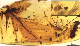
So what does a museum want with a powerful X-ray beam? One of our research fellows, Lauren Sumner-Rooney, is particularly interested in studying the eyes and brains of spiders. So the team, led by Lauren, went to Didcot to create some X-ray images of spiders from the Museum’s collections.

Ready for your X-ray close -up? A spider specimen is mounted in the I13-2 beamline at Diamond Light Source. Image: Lauren Sumner-Rooney

You may not have looked too closely at a spider’s head before, but they usually have eight eyes as well as eight legs. That said, there is actually quite a lot of variation in the number and structure of eyes between different species, and Lauren is interested in documenting this variation across selected spider families to investigate how it affects spiders’ brain structures.

Using the I13-2 beamline at Diamond, and fighting severe sleep deprivation with the aid of strategically-selected songs and snacks, the team was able to visualize details measuring less than one thousandth of a millimetre without damaging the precious specimens. They were assisted by Andrew Bodey, a senior support scientist at Diamond, and Emelie Brodrick, a PhD student at the University of Bristol.

The research team following 72 hours of very little sleep! From left to right: Ricardo Pérez-de la Fuente, Lauren Sumner-Rooney, Imran Rahman, Jack Matthews and Emelie Brodrick. Image: Emelie Brodrick

Over the course of 72 straight hours, the team scanned 116 spiders, creating about 14 terabytes of data. This will form the basis of a variety of exciting scientific research projects at the Museum over the coming years. Watch this space for the results!

*Didcot Town were fourth in the Southern League Division One West table on 8th March. The Museum accepts no responsibility for any change in their position after this date.

Top image of Pholcus moca courtesy of Smithsonian Institution.

Kelp our corals!

Many people know about the importance of conserving coral reefs to protect marine biodiversity, but here at the museum we also need to conserve the corals that are in our collections. These specimens provide a valuable picture of the diversity of life in the ocean, and document changes seen over time, which is more important than ever. So it’s essential that our conservation team make sure these corals are in the best shape possible. Stefani Cavazos, an intern from UCL’s MSc in Conservation for Archaeology and Museums, tells us how they’re going to do it.

As part of the ongoing effort to improve the museum’s collections storage we decided to give our soft corals and sponges a bit of TLC through some repacking and reorganisation.

This collection – a mix of old display material and specimens not formally accessioned to the museum collection – isn’t currently stored as well as it could be and there is a danger of breakages and damage. The specimens are packed in non-conservation grade materials, such as cardboard boxes, which are notorious for creating acidic gases that can damage delicate specimens.

The current housing of our soft coral and sponge collection

So a new project, Kelp our Corals, will focus on two areas of improvement.

First, we’ll remove all old packaging and repack using new bespoke storage boxes made from conservation grade materials. At the same time, specimens will be photographed, catalogued, and given accession numbers.

The goal is not only to rehouse the coral and sponge collection, but to also make it more accessible to the public for use in teaching and for research. We don’t have a lot of documentation for these corals, so hopefully the project will help us fill in some gaps: Where did these specimens come from? What can they tell us about life on a reef?

Large specimens are improperly laid on their sides with no protection from the environment and dust, causing weight stress on the specimen

Would you like to kelp, er, sorry – help? We are looking to recruit volunteers to help us with the work. We’re aiming to start in mid-February and finish by May this year. If you are interested in gaining some museum and conservation experience, or like to work with your hands, please do get in touch at volunteering@museums.ox.ac.uk.

Credit for image at top of post: USFWS/Jim Maragos via Creative Commons

Amour for armour

If you pop in to the Museum at 2.30pm on a Monday-Thursday afternoon, you’ll meet one of our Museum experts with some of their favourite specimens. Here Eileen Westwig, Life Collections Manager, shares one of her recent Spotlight Specimens.

Last month, as part of our regular Spotlight Specimens activity, I chose to highlight armadillo specimens. They got lots of attention, which is not surprising considering how amazing armadillos are. The word armadillo is Spanish meaning ‘little armoured one’. It is true that all armadillos have armour wrapping around their body as protection. Their size, however, varies a lot. The smallest one is the Pink Fairy Armadillo (Chlamyphorus truncatus), which grows up to 18cm (including tail length) and weighs up to a tiny 100g. At the other end of the spectrum, the aptly named Giant Armadillo (Priodontes maximus) is the largest, and can grow up to 150cm (head to tail) and weigh up to 60kg.

Giant Armadillo from the OUMNH collection. Sharp, big claws help to scratch and dig for food, such as tubers and termites, and dig burrows for sleeping.

Armadillos are found in South and Central America. However, the common Nine-banded Armadillo (Dasypus novemcinctus) has spread over the last hundred years, all the way into the southern United States. What makes it so successful is its varied diet of tubers, termites, ant larvae and other insects, as well as snails and bird eggs found on the ground. The expanse of ranching and the absence of natural predators such as cougars have made it easy for this long-nosed armadillo to spread as far as Texas and Florida.

Beside their stiff protective armour, all armadillos are capable of curling up their body to some extent, in order to protect the soft and vulnerable underside. Only one armadillo is the true champion when it comes to rolling up tightly into a perfect sphere. This astonishing achievement can be found in the Southern Three-banded Armadillo (Tolypeutes matacus). In the picture at the top of this page, you can see two armoured triangles in the middle, which are its head (on the left) and tail (on the right).

Common Nine-banded Armadillo showing its body plates, which usually lie underneath a layer of horn.

The armour of armadillos is made out of two layers. There are bony scute plates (visible in white in the picture above) that are overlaid with horny plates. The horny plates are made of keratin, the same material as hair and fingernails.

Nine-banded Armadillo made into a basket as souvenir

Sadly the existence of this amazing creature is threatened by loss of habitat and hunting. Not only are armadillos widely eaten, they are also made into tourist souvenirs, such as this basket.

According to the Centers for Disease Control and Prevention, some armadillos from the southern USA are naturally infected with the bacteria (Mycobacterium leprae), that cause leprosy (Hansen’s disease). Most people (95%) are immune to it, but please use caution if you’re ever in a position to handle an armadillo!

What’s in a name?

By Duncan Murdock, research fellow

Whether it’s the Physeter macrocephalus (Sperm Whale) whose jaw greets our visitors, the Apus apus (European Swift) which spend the summer nesting in the tower, or the Raphus cucullatus (Dodo) on our Museum’s logo, all animals, plants, fungi and microbes, living and extinct, have scientific names – or at least once they have been properly described in a scientific paper they do. Usually found tucked away on specimen labels, scientific names carry much more significance than just a convenient means of reference.

The jaw of the Sperm Whale (Physeter macrocephalus)

The scientific name, also known as a binominal or Latin name, consists of two basic parts, and should be written in italics. The first part is the genus (the plural is genera), which refers to anything from one to thousands of kinds of creature that are more closely related to each other than anything else. Genera are always capitalised, such as Panthera (big cats).

The second part is the specific name, written in lower case. Together these define one species; for example a tiger is Panthera tigris. Sometimes, subspecies or varieties are written after the species name, such Panthera tigris tigris, which is the Bengal Tiger. They can also be abbreviated by replacing the genus with just an initial followed by a full stop, hence the ever-popular T. rex, or Tyrannosaurus rex.

T. rex in the Museum’s centre court

Some binomials are pretty easy to decipher: no prizes for working out Gorilla gorilla*. Others can seem pretty cryptic or even positively confusing – Puffinus puffinus anyone? Yep, that’s right, the Manx Shearwater**. Nevertheless, once translated they are often enlightening as to the appearance, distribution, behaviour, or history of the critter in question.

Here are a few examples. Ailuropoda melanoleuca, meaning ‘black and white cat-foot’, describes the appearance of the Giant Panda pretty well; Megaptera novaeangliae, or ‘giant-wing of New England’, alludes to both the anatomy and chequered history of the humpback whale; and while Pteropus vampyrus, or ‘wing-footed vampire’, is a bit of a misnomer for the flying fox, which is a large fruit-eating bat, it does reflects our changing understanding of the animal.

Gorilla gorilla, the Western Gorilla
Magpie (Pica pica)

Some names are elegantly concise: Pica pica, the magpie. Some are tongue-twisters: Phalacrocorax carbo, the Great Cormorant. And some, such as Synalpheus pinkfloydiare entertaining. But they are all more than just names; they are the most visible aspect of the science of taxonomy.

Carl Linnaeus (1707-1778) first formalised the system we use today, which has allowed us to divide all the many species into not just genera, but a nested hierarchy of ever-more inclusive groups.

With this system we can not only be sure we are using a common language to precisely refer to the right species, but we can also then ask questions about how the staggering diversity of life that we see evolved. And from this we start to build ‘a tree of life’. But this will be the subject of a future article…

* Bonus points for knowing it’s the Western Gorilla, as opposed to Gorilla beringei, which is the Eastern Gorilla.

** Common Puffins, by the way, go by the delightful name Fratercula arctica, the ‘little friar of the north’.

Bound by blood

It may sound like we’ve stumbled into a script-writing session for Jurassic Park, but one of our research fellows, Dr Ricardo Pérez-de la Fuente, along with an international team, has discovered a parasite trapped in amber, clutching the feather of a dinosaur. This small fossilised tick, along with a few other specimens, is the first direct evidence that ticks sucked the blood of feathered dinosaurs 100 million years ago. Ricardo tells us all about it…

The paper that my colleagues and I have just published provides evidence that ticks fed from feathered dinosaurs about 100 million years ago, during the mid-Cretaceous period. It is based on evidence from amber fossils, including that of a hard tick grasping a dinosaur feather preserved in 99 million-year-old Burmese amber.

Fluorescence detail of the studied hard tick grasping a dinosaur feather. Extracted from the publication.

The probability of the tick and feather becoming so tightly associated and co-preserved in resin by chance is virtually zero, which means the discovery is the first direct evidence of a parasite-host relationship between ticks and feathered dinosaurs.

Fossils of parasitic, blood-feeding creatures directly associated with remains of their host are exceedingly scarce, and this new specimen is the oldest known to date. The tick is an immature specimen of Cornupalpatum burmanicum; look closely under the microscope and you can see tiny teeth in the mouthparts that are used to create a hole and fix to the host’s skin to suck its blood.

The structure of the feather inside the amber is similar to modern-day bird feathers, but it could not belong to a modern bird because, according to current evidence at least, they did not appear until 26 million years later than the age of the amber.

Feathers with the same characteristics were already present in multiple forms of theropod dinosaurs –  the lineage of dinosaurs leading to modern birds – from ground-runners without flying ability, to bird-like forms capable of powered flight. Unfortunately, this means it is not possible to determine exactly which kind of feathered dinosaur the amber feather belonged to.

But there is more evidence of the dinosaur-tick relationship in the scientific paper. We also describe a new group of extinct ticks, created from a species we have named Deinocroton draculi, or “Dracula’s terrible tick”. These novel ticks, in the family Deinocrotonidae, are distinguished from other ticks by the structure of their body surface, palps and legs, and the position of their head, among other characteristics.

Blood-engorged Deinocroton draculi tick (female). Extracted from the publication.

This new species was also found sealed inside Burmese amber, with one specimen remarkably engorged with blood, increasing its volume approximately eight times over non-engorged forms. Despite this, it has not been possible to directly determine its host animal:

Assessing the composition of the blood meal inside the bloated tick is not feasible because, unfortunately, the tick did not become fully immersed in resin and so its contents were altered by mineral deposition.
Dr Xavier Delclòs, an author of the study from the University of Barcelona and IRBio.

But there was indirect evidence of the likely host for these novel ticks in the form of hair-like structures called setae from the larvae of skin beetles, or dermestids, found attached to two Deinocroton ticks preserved together. Today, skin beetles feed in nests, consuming feathers, skin and hair from the nest’s occupants. But as no mammal hairs have yet been found in Cretaceous amber, the presence of skin beetle setae on the two Deinocroton draculi ticks suggests that their host was in fact a feathered dinosaur.

The hair-like structures, or setae, from skin beetles (dermestids) found attached to two Deinocroton ticks fossilised inside amber, in comparison with extant ones. Modified from the publication.

Together, these findings tell us a fascinating story about ancient tick behaviour. They reveal some of the ecological interactions taking place among early ticks and birds, showing that their parasite-host relationship has lasted for at least 99 million years: an enduring connection, bound by blood.

The paper “Ticks parasitised feathered dinosaurs as revealed by Cretaceous amber assemblages” is published as open access in Nature Communications. Direct link: http://dx.doi.org/10.1038/s41467-017-01550-z

Clean as a new pin

The spiky customer above has enjoyed a serious spruce-up from Stefani Cavazos, our current intern from UCL’s MSc in Conservation for Archaeology and Museums. Stefani tells us how she got this Spot-fin Porcupinefish looking shipshape, without receiving any serious injuries.

So far at the Museum I have been working on a range of specimens, from taxidermy and wet specimens to cleaning the whales, but my favourite project so far has been the conservation of this Spot-fin Porcupinefish from the displays. It is part of what is known as the Christ Church Collection, which came to the Museum in 1860. This makes the Porcupinefish at least 150 years old.

The specimen itself was covered in dust and all five of its fins were backed with deteriorating cardboard pieces. These were most likely attached to give some support during a previous restoration attempt. Unfortunately, cardboard is not a conservation grade material because over time it becomes acidic. Temperature and humidity changes in the Museum have caused it to bend forward, pulling the fins out of shape, so we felt it should be removed to prevent further damage.

The Spot-fin Porcupinefish (Diodon hystrix) before conservation treatment (left images), and close ups of the cardboard backings on the left pectoral fin (right images), where staining and bending are visible. The red arrow indicates where the paper has separated from the fin, and how thin the fins are.

The first step in the treatment was to clean the surface of the Porcupinefish using warm water and a cotton swab. This allowed me to get into the nooks and crannies of the body whilst (mostly) avoiding being poked by its spines. Next, the cardboard backings were softened with water vapor, causing them to break apart so they could be removed easily using tweezers and a scalpel blade.

Conservation can feel like detective work since we often uncover interesting information about specimens as we work on them. In this case, as we removed the cardboard pieces, we found writing on the underside. It appears to be from a shoe box! Though unexpected, it wasn’t entirely surprising. Preparators in the past used whatever materials were available to them at the time.

(Left) The cardboard was carefully removed from the caudal (tail) fin. (Right) The cardboard once removed from the fin, it appears to be from a shoe label.

After detaching the cardboard from all the fins, the remaining ink and adhesive residues were removed using a 50/50 alcohol and water mixture applied with a cotton swab. The edges of the fins were then coated with two thin layers of an acrylic adhesive to prevent any further breakage and to offer some support to the weakest areas. Cleaned, and free of damaging materials, this Porcupinefish is now ready to go back on display!

Photos of the Spot-fin Porcupinefish after treatment was completed. Without the cardboard backings, the fins are somewhat translucent.
Stefani takes her finished work out to one of our regular Spotlight Specimen sessions, giving visitors the chance to get a closer look and ask questions.