Odd egg out

This is a great time of year to hear the distinctive call of the Cuckoo (Cuculus canorus) as it spends the summer in the UK. Collections Manager Eileen Westwig recently shared Cuckoo specimens with the public in one of our Spotlight Specimens sessions. You missed it?! No problem, here she is with the fascinating story of this threatened bird…

Cuckoos could be described as absent mothers, laying their eggs into the nest of a ‘host bird’, such as Dunnocks, Meadow Pipits, Garden Warblers, Whitethroats or Flycatchers. When she finds a suitable nest, the female Cuckoo will remove one of the host’s eggs and lay hers in its place. She lays between 12 and 22 eggs in a season, all in different nests. No worries befall her about building a nest, brooding out any eggs or raising her young as she leaves it all to strangers. One challenge for the Cuckoo is to make sure her trickery is not discovered.

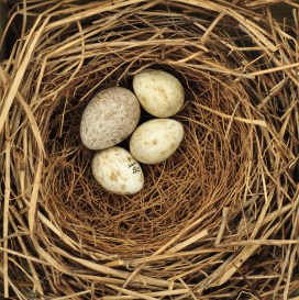
When the female host returns to her nest, she will inspect it for any changes and if she discovers the intruder’s egg, she will simply toss it out. So the female Cuckoo has to be pretty good at forgery and mimic the host bird’s egg ‘signature’, copying the colour, pattern and shape of the original eggs. This is the only way to get away with her ‘brood parasitism’. Around 20% of Cuckoo eggs never make it. In the top picture, you can see the nest of a Garden Warbler with three Warbler eggs and one larger Cuckoo egg, on the top left.

An adult Garden Warbler (Sylvia borin borin) can reach a weight of 16-22g with a wingspan of 20-24.5cm

After twelve days, the Cuckoo hatches and pushes the other nestlings out. As the single remaining occupant of the nest, it has the full attention of the host parents, which try to feed a nestling soon outweighing. An adult Cuckoo is more than 6 times the weight of an adult Garden Warbler. The Cuckoo young will leave the nest after 19 days, but gets fed by the parents for a further two weeks. That is one busy summer.

OUMNH.ZC.11868_Cuculus_canorus_canorus_Eileen_Westwig
An adult Cuckoo (Cuculus canorus) can reach a weight of 105-130g with a wingspan of 55-65cm.

According to the RSPB, there are about 15,000 breeding pairs in the UK and Cuckoos are now included on the Red List, giving them the highest conservation priority. Ten years ago, numbers of this migrant bird fell by 21% and more than half of the population has disappeared in the past 25 years. Threats include damage to the bird’s winter habitats and a decline in large insect species that are its major food source.

Cuckoos migrate to West Africa over the winter months and can be seen in the UK from late March or April through July or August. Young birds leave a month or so later to give them time to grow and prepare for the long journey ahead. Wintering grounds are not exactly known but include Cameroon, Gabon and other African nations.

Who shot the Dodo?

By Scott Billings, Digital Engagement Officer

If ever the Oxford Dodo were to have squawked, its final squawk may have been the saddest and loudest. For the first time, the manner of death of the museum’s iconic specimen has been revealed: a shot to the back of the head.

This unexpected twist in the long tale of the Oxford Dodo has come to light thanks to a collaboration between the Museum and the University of Warwick. WMG, a cutting-edge manufacturing and technology research unit at Warwick, employed its forensic scanning techniques and expertise to discover that the Dodo was shot in the neck and back of the head with a 17th-century shotgun.

Mysterious particles were found in the specimen during scans carried out to analyse its anatomy. Further investigation of the material and size of these particles revealed them to be lead shot pellets of a type used to hunt wildfowl during the 1600s.

The Oxford Dodo specimen, as it has come to be known, originally came to the University of Oxford as part of the Tradescant Collection of specimens and artefacts compiled by father and son John Tradescant in London in the 17th century. It was thought to have been the remains of a bird recorded as being kept alive in a 17th-century London townhouse, but the discovery of the shotgun pellets cast doubt on this idea, leaving the bird’s origins more mysterious than ever.

Dodos were endemic to the island of Mauritius in the Indian Ocean. The first European accounts of the bird were made by Dutch explorers in 1601, after they rediscovered the island in 1598. The last living bird was sighted in 1662.

The story of the Oxford Dodo is especially significant because it represents the most complete remains of a dodo collected as a living bird – the head and a foot – and the only surviving soft tissue anywhere in the world.

This discovery reveals important new information about the history of the Oxford Dodo, which is an important specimen for biology, and through its connections with Lewis Carroll and Alice’s Adventures in Wonderland of great cultural significance too.
– Professor Paul Smith, Museum director

The Oxford Dodo represents the only soft tissue remains of dodo in the world. This iconic specimen was taken from the Museum to WMG at the University of Warwick for CT scanning.

WMG’s CT scans show that this famous symbol of human-caused extinction was shot in the back of the head and the neck, and that the shot did not penetrate its skull – which is now revealed to be very thick.

The discovery of such a brutal demise was quite a surprise as the scans were actually focused on discovering more about the Dodo’s anatomy, as well as how it lived and died. This work will continue, but we now have a new mystery to solve: Who shot the Dodo?

What’s the next step? It is possible that the isotope of lead in the shot could be analysed and traced to a particular ore field. This might tell us what country it was mined in, and perhaps what country is was made in, and ultimately reveal who shot the Dodo.

Amour for armour

If you pop in to the Museum at 2.30pm on a Monday-Thursday afternoon, you’ll meet one of our Museum experts with some of their favourite specimens. Here Eileen Westwig, Life Collections Manager, shares one of her recent Spotlight Specimens.

Last month, as part of our regular Spotlight Specimens activity, I chose to highlight armadillo specimens. They got lots of attention, which is not surprising considering how amazing armadillos are. The word armadillo is Spanish meaning ‘little armoured one’. It is true that all armadillos have armour wrapping around their body as protection. Their size, however, varies a lot. The smallest one is the Pink Fairy Armadillo (Chlamyphorus truncatus), which grows up to 18cm (including tail length) and weighs up to a tiny 100g. At the other end of the spectrum, the aptly named Giant Armadillo (Priodontes maximus) is the largest, and can grow up to 150cm (head to tail) and weigh up to 60kg.

Giant Armadillo from the OUMNH collection. Sharp, big claws help to scratch and dig for food, such as tubers and termites, and dig burrows for sleeping.

Armadillos are found in South and Central America. However, the common Nine-banded Armadillo (Dasypus novemcinctus) has spread over the last hundred years, all the way into the southern United States. What makes it so successful is its varied diet of tubers, termites, ant larvae and other insects, as well as snails and bird eggs found on the ground. The expanse of ranching and the absence of natural predators such as cougars have made it easy for this long-nosed armadillo to spread as far as Texas and Florida.

Beside their stiff protective armour, all armadillos are capable of curling up their body to some extent, in order to protect the soft and vulnerable underside. Only one armadillo is the true champion when it comes to rolling up tightly into a perfect sphere. This astonishing achievement can be found in the Southern Three-banded Armadillo (Tolypeutes matacus). In the picture at the top of this page, you can see two armoured triangles in the middle, which are its head (on the left) and tail (on the right).

Common Nine-banded Armadillo showing its body plates, which usually lie underneath a layer of horn.

The armour of armadillos is made out of two layers. There are bony scute plates (visible in white in the picture above) that are overlaid with horny plates. The horny plates are made of keratin, the same material as hair and fingernails.

Nine-banded Armadillo made into a basket as souvenir

Sadly the existence of this amazing creature is threatened by loss of habitat and hunting. Not only are armadillos widely eaten, they are also made into tourist souvenirs, such as this basket.

According to the Centers for Disease Control and Prevention, some armadillos from the southern USA are naturally infected with the bacteria (Mycobacterium leprae), that cause leprosy (Hansen’s disease). Most people (95%) are immune to it, but please use caution if you’re ever in a position to handle an armadillo!

Steven’s Christmas spiders

by Steven Williams, Oxford Brookes University research student

Described by A.G. Butler in 1873 as ‘the most gorgeously coloured spider in this genus’, Gasteracantha scintillans, with its metallic green iridescent abdomen, is the first of my Christmas Spiders.

img_0532
Spiky and sparkly: Gasteracantha scintillans

The beautiful colour of the abdomen certainly has a very festive feel and it would not be out of place next to a bauble on a Christmas tree; at least not in my house. This species and the other closely-related metallic Thorn Spiders are currently only found on the Solomon Islands.

Christmas Island, in the Indian Ocean, is one of the locations where my second Christmas spider, Austracantha minax, can be found. Although not as striking as the metallic green of Gasteracantha scintillans, the layout of the abdominal spines on this spider almost give it the appearance of a star – perfect for the top of a Christmas tree, no?

Austracantha minax: starry, and found, amongst other places, on Christmas Island

The common name of ‘Christmas Spider’ is attributed to this species because in areas of Western Australia it is associated with the arrival of Christmas as the males reach maturity in mid-December and females in January.

Did you know that there is also an Eastern European folk tale of how tinsel came to be included in Christmas tree decorations? The legend tells of how spiders spun cobwebs on a poor family’s undecorated Christmas tree overnight. In the morning the webs turned to gold and silver and the family never lived in poverty again. So when you put the tinsel on the tree this year you could imagine you are a spider spinning a web!

With that spidery festive thought, have a very Merry Christmas from everyone at the Museum!

Nature’s medals

By Sarah Joomun, Documentation officer

In the 1820s a young geologist named Charles Lyell travelled around France studying the landscape and rock formations to try and work out the processes that created them.

In between these field-trips, he met the people who had been studying the geology of France and from these discussions and his observations he created The Principles of Geology, one of the first significant popular science books on the subject and a foundation for the methods of modern geology.

pal_cl01718_01

Lyell collected many samples from the rocks he studied, amassing thousands of fossils during his lifetime. The Museum has a collection of some 16,000 of them, around 90 per cent of which are shells, mostly gastropods (snails) and bivalves (clams), many collected during his travels in France.

The reason for this prodigious collection of fossil shells, or testacea as they were then known, was that Lyell believed them to be the most useful clue to understanding the Earth’s history.

The testacea are by far the most important of all classes of organic beings which have left their spoils in the subaqueous deposits : they are the medals which nature has chiefly selected to record the history of the former changes of the globe.

– Lyell’s Principles of Geology, Vol III, 1833.

Fossil shells can show how the animal that lived inside the shell behaved, and whether it lived on the land, in freshwater or in the sea. Species of shelled animals have a wide geographical range and individual species survive for a long time, so they can be compared across time and space.

pal_cl01742_02

This allowed Lyell and his colleagues to determine the relative ages of the rock layers that the fossil shells came from. He looked at the proportion of shells that belonged to living species and determined that the rock layers with the lowest proportion of living species were likely to be older than rocks with higher proportions of living species.

And so three main groups of rock layers were found: the Eocene, containing fewer than 4% living species; the Miocene, with fewer than 18% living species; and the Pliocene, with more than a third of living species.

Although what is now known as the Eocene (from 56 to 34 million years ago), Miocene (23 to 5.3 million years ago) and the Pliocene (from 5.3 to 2.6 million years ago) don’t denote exactly the same periods as Lyell described, we still use these terms for some of the youngest geological epochs today.

Art of glass

Spotlight Specimens by Mark Carnall

From the comfort of our own homes, or even on a mobile device, we are accustomed to watching video footage from the most remote environments on Earth, and beyond. It is easy to take for granted this kind of visual access but we don’t have to go too far back in time to reach a point when the uninhabitable parts of the world remained much more mysterious. Then, the only windows into the nature of exotic locations were through drawings, paintings or collected specimens.

In museums, illustrations of nature were – and are – used in teaching to show what certain animals or environments look like. Along with our biological specimens, the Museum’s collections contain representations of animals whose natural appearance is not preserved after death, including a set of beautiful glassworks of British sea anemones.

These delicate models were created by the Blaschkas, a family which specialised in glasswork and ran a business spanning 300 years and nine generations. But it was only from the late 19th century that Leopold Blaschka, later to be joined by his son Rudolf Blaschka, turned his skills to making models of microscopic organisms and soft-bodied invertebrates for museums and universities.

Blaschka anemones still
The only Blaschka models at the Museum today form a series of British anemones, many of which are recognisable as the species and even individual animals illustrated in British Sea-Anemones and Corals published in 1860 by Philip Henry Gosse

Inspired by zoological specimens, scientific papers, and observation of living animals, as well as artworks showing colours and structures that were difficult to preserve or too small to show, the Blaschkas created thousands of glass models before they accepted a contract in 1886 to work exclusively at Harvard University on the Ware Collection of plant models.

historyofbritish00goss_0299
Plate from Philip Henry Gosse, 1860. British Sea-Anemones showing many of the species in the Oxford University Museum of Natural History series. Public domain image from the Biodiversity Heritage Library. http://www.biodiversitylibrary.org

It is somewhat surprising that these incredibly fragile specimens made their way to museums and universities across the globe back in the 19th century and even more surprising that any have survived 150 years later.

Anemones tend to lose their shape and colour when preserved in fluid

Earlier this year the Corning Museum of Glass published an interactive map of marine invertebrate models showing the known locations of collections, or records of collections, of Blaschka glass models.

The models at the Museum, acquired in 1867, are thought to be some of the oldest surviving Blaschka glass models. Even though they are over 150 years old, and in some cases slightly inaccurate representations of species, they still show the vibrant colours and alien shapes of British anemones in a way that can’t be seen outside their living environments.