The latest display in our changing Presenting… series showcases some of the incredible colours seen in many insects. Zoe Simmons, collections manager in our Life Collections, explains how such wonderful hues are created.
Reflected and refracted light creates the many bright and shining colours found in some insects. The dazzling natural display shown in the specimens here is formed through a combination of embedded pigments and sculpted surfaces on each insect’s external skeleton.
Some species can be variable in colour. Here a pair of Lamprima, a genus of Stag Beetles, shows off the range of colours present in the species.
Different pigment chemicals are responsible for different colours. Carotenoids produce yellow, orange and red hues, while bilins may be green, or blue if linked with proteins. They reflect and absorb different wavelengths of light, and the wavelengths that are reflected are the ones that we see as colour. Typically humans can see wavelengths of 390-700 nanometres, with the lower wavelengths perceived as blue, and the higher ones as red.
Many of the Leaf Beetles (Chrysomelidae) exhibit metallic colours.
Many insects also have multiple thin layers over their upper surfaces to help protect them and prevent dehydration. Variations in thickness and chemical composition of these layers can interfere with the transmission of light, refracting and scattering it back.
Some of the most striking metallic colours are found in the genus Chrysina, where species can be rose, silver or gold.
The shape of the surface layer can reflect light in a multitude of directions, with micro-folds, grooves, pits, hairs and scales all helping to produce complex colours and effects.
The formation and purpose of these colours is scientifically interesting, with research having applications in areas such as nanotechnology. But these insects are also simply beautiful examples of the spectacular diversity of the natural world.
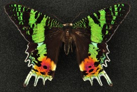
Sunset moths (Uraniidae)are so called because of the dazzling array of colours on their wings. As day-flying moths they are brightly coloured like many butterfly species.
by Chris Stimpson, visiting researcher from Queen’s University Belfast
Visitors to the museum will be familiar with the striking parade of mammal skeletons in the court, where they can get a close look at a polar bear’s jaws and peer up through the rib cages of Indian and African elephants, amongst many other things. But these mounted specimens are just a small sample of the animal skeletons that are looked after by the museum.
The main collection of skeletons is carefully stored in behind-the-scenes spaces such as the museum’s Tradescant Room. For researchers who work on animal bones found in archaeological sites, collections like these are not just important – they are essential.
Comparison of an archaeological pig astragalus (ankle bone, left) with an articulated reference specimen from the museum collection (opposite leg, OUMNH.ZC.19948) of an Indonesian wild boar (Sus scrofa). Radiocarbon dating of charcoal indicates the archaeological specimen is over 17,000 years old.
Differences in size, shape, proportion, and the number and arrangement of bones and teeth are a great aid to identification. Teeth in particular often have features that help identify the animal they came from. Bones also have articulations and facets which can be helpful, though identification can be more challenging than with teeth.
Comparison of an archaeological premolar (top), with the upper right tooth row of a goat-like animal called a serow (Capricornis sumatraensis) from the museum’s collection (OUMNH.ZC.21654). Radiocarbon dating of charcoal from the site indicates the archaeological specimen is over 5,000 years old.
These challenges are part of the work I am doing on the SUNDASIA Project which is undertaking archaeological and palaeoecological investigations in the Tràng An World Heritage Area, in Ninh Binh Province, Northern Vietnam. Working with Vietnamese colleagues, we are investigating climatic and landscape changes that have affected – and may affect – the limestone karst forest over thousands of years. In particular, we’re looking at the responses of human, animal and plant communities to these changes.
The limestone karst landscape of the Trang An World Heritage Area
During our cave excavations we have recovered bones from a variety of birds, mammals, reptiles, fish and amphibians. Radiocarbon dates from charcoal in the cave deposits suggest this material ranges from 30,000 to 5,000 years old. This is great, but what can these bones tell us of animal life and human hunters at different times in the past? What has changed and why? And what could it mean for the future of Tràng An?
Excavations underway in Hang Moi, a cave site in Trang An
Before we can begin to answer juicy research questions like these, we need to identify the bones. This is where collections like those held in the museum really come into play. Only with access to skeletons of known animals – where there is knowledge of family, genus or species classification – can we compare the excavated material and identify what we have found.
And while old bones and skeletons may smack rather of death, with a little patience and a good comparative collection like that in the museum, it is remarkable what a few specimens can tell you of life in different times and places that we otherwise know little about. Museum collections are a key to the past, present, and perhaps even to the future.
This month marks three years since the completion of our ‘Once in a Whale’ project. The initial conservation undertaken in 2013 focused on the cleaning and stabilisation of five whale skeletons, which had hung from the roof of the Museum for over 100 years.
The skeletons were lowered into a special conservation space, where the team were able to work up close with the specimens. As well as the cleaning, they improved incorrect skeletal anatomy, replacing old corroded wiring with new stainless steel. For final display, the specimens were put into size order and rigged using new steel wiring, with the larger specimens being lifted higher into the roof space to make them a more prominent display than previously. You can read all about the project on our blog, Once in a Whale.
Three years on, our conservation team felt it was a good time to check on the specimens to see how they’re coping, post-treatment, in the fluctuating museum environment.
Conservation intern Stefani Cavazos works on high to clean the Beaked Whale
It’s been wonderful to see the whales on display and their new position looks very impressive. However, when the time came for making this recent conservation assessment, the new height was greater than any of our ladders could reach. Specialist scaffolding was brought in to allow the conservators to access the specimens. Starting at the highest level, with our Beaked Whale, cleaning was completed using a vacuum and soft brush for delicate areas. This removed a thick layer of dust and particulate debris: especially satisfying work!
Dust gathered on the Beaked Whale fin
With cleaning complete, visual assessments could then be undertaken. These showed that while the specimens were still very stable, a few areas of bone have continued to deteriorate, visible in cracking and flaking of the surface. In other areas, the fatty secretions which we previously removed using ammonia had once again started to emerge. We had expected to see this though, because, in life these whales’ bodies contained a lot of fat, deep within the bones and this is notoriously impossible to completely remove.
Lubricant stain seen on a vertebra
It was also observed that the lubrication used on the new rigging bolts had melted and dripped down the wires. You can see in the photo above how this has become drawn into the vertebrae of the Orca and Common Dolphin, staining them yellow. While no conservation treatment was undertaken due to time restrictions, thorough photography was performed to document these changes and once time permits this can be carried out.
This shows how conservation work, especially with natural history specimens, is a gradual, ongoing process. With frequent check-ups and specialist attention, these whales will be able to continue their life as our beautiful display specimens.
One of the most common questions asked about our specimens, from visitors of all ages, is ‘Is it real?’. This seemingly simple question is actually many questions in one and hides a complexity of answers.
In this FAQ mini-series we’ll unpack the ‘Is it real?’ conundrum by looking at different types of natural history specimens in turn. We’ll ask ‘Is it a real animal?’, ‘Is it real biological remains?’, ‘Is it a model?’ and many more reality-check questions. Here’s your final installment…
There’s nothing like standing under a huge T.rex skeleton, staring up at its ferocious jaws, to get the blood pumping. Visitors often ask “Is it real?” and look rather deflated when they find out it’s a cast. So why do we include casts, models or replicas in our displays, if they don’t have the same impact as the real deal? The truth is that they’re valuable additions to museum displays, allowing the public to engage with specimens that would otherwise be hidden behind the scenes.
Please touch! A cast of the famous Oxford Dodo helps visitors explore this fragile specimen.
On any visit to the Museum, you’ll come across labels that tell you the object you’re looking at is a cast. It could be a dinosaur skeleton, a brightly coloured fish, an amphibian specimen or even the head of the Oxford Dodo. But what is a cast? Casts are made by taking a mould of bones, or sometimes whole animals, then filling that mould with resin, plaster or fibre glass to make a copy. They can be incredibly accurate or lifelike.
It’s extremely rare to find whole dinosaur skeletons, and very difficult to mount heavy fossils (weighing tonnes) onto large armatures. Our Tyrannosaurus rex is a cast of the famous Stan, found in South Dakota, USA, and one of the best preserved skeletons of its kind in the world. But the “real” Stan is kept at the Black Hills Institute of Geological Research, so the only way we can offer the breath-taking experience of standing beneath a T. rex here in Oxford is by using a cast.
The Dodo Roadshow in 2015 would have been a lot less fun without our life-size dodo model
Even Stan has some bones missing, so sometimes casts are made up of several individual skeletons. Copies can also be made to give the impression of a more complete skeleton. For example, if a left bone is missing, a mirror of the right hand bone can be created. We call these specimens “composites”.
Animals such as fish and frogs aren’t easy to taxidermy; their skins shrivel, dry out, lose their colour and crack. Painted casts are a good way to show what these animals look like.
A model allows us to show the intricate scales of this Blue Morpho butterfly up close.
Models, such as the giant insects on the upper gallery and the Archaeopteryx in the Evolution of Flight display (at the top of this post), are very clearly not real. These are made by model makers to show something that can’t be seen or shown with real specimens. The giant insects are a way of showing the detail of very small creatures. The palaeontological models show what we think extinct animals might have looked like in life. They’re hypothetical models based on the latest scientific research, which can change very quickly, and always have an element of artistic assumption or speculation in the details.
In this series we’ve talked about taxidermy, skeletons, fossils and more, but these are just a few of the kinds of specimens we have on display. There are also nests, plastinated models, microscope slides and dioramas, which all have a mix of real and non-real elements. When you are looking around the Museum try to think about which specimens are real and which aren’t… and how does that make you think about the specimen?
You’re reading this, so I’m guessing you like museums. But have you ever heard of living fossils? Animals such as sharks and crocodiles are often referred to as ‘living fossils’ because they appear pretty unchanged from their ancient fossilized relatives. Of course, by definition, you can’t be both alive and a fossil. But fossils allow us to become primary eyewitnesses to ancient life; we can literally see what life used to look like, how cool is that? They can also dole out some pretty valuable advice, if we just choose to listen.
This summer during a visit to England, I spent some time at the Museum studying another so-called living fossil, the horseshoe ‘crab’. The horseshoe crab is not actually a crab, but is instead more closely related to spiders, scorpions and ticks. In fact, they are the closest living relatives of the extinct trilobites. But unlike their famous trilobite cousins, horseshoe crabs have survived all five of Earth’s major mass extinction events. Today, as a direct result of their ability to survive, the four remaining species of horseshoe crab play a vital role in global medical safety.
The Museum’s fossil specimen of Mesolimulus walchi, from the Upper Jurassic (163-145 million years ago), Solnhofen Germany, shows how little the form of the horseshoe crab has changed since
Not only do living horseshoe crabs look very similar to their early relations, they are also able to survive surprisingly severe injuries that often leave them missing body parts. Being able to see, through fossil evidence, how little their form has changed over time has helped to uncover the answer to this secret superpower. It lies in a very special life-saving trick that the crabs have kept for millions of years: a coagulating blood protein.
Horseshoe crabs on display in the Museum may provide food for thought for visitors
The blood of the horseshoe crab is able to clot quickly if bacteria are introduced, preventing infection, and saving the crab’s life. Since this discovery in the 1970s, this life-saving protein has been extracted from horseshoe crab blood and used in human medicine to test the safety of vaccines, medical laboratories, intravenous drugs, implants, and much, much more. The chances are that you owe a great deal of gratitude to the horseshoe crab.
But after all that surviving, horseshoe crabs, like many species, are now struggling for survival. They are losing their spawning grounds because of coastal development, industry, housing, marinas and coastal defense structures; they are collected and killed by the millions for bait, and bloodlet in their hundreds of thousands for medical use every year. It is likely that horseshoe crabs will not survive much longer.
But don’t despair. Museums are critical because they hold collections that can unlock knowledge about environmental change, and we can use that knowledge to protect life. Of course, horseshoe crabs are not alone in telling their stories through the fossils they leave – natural history museums are full of stories in stone, bones, pollen, and other traces. If you want to learn about and protect biodiversity, visit your local museum, or support organisations like Oxford’s Environmental Change Institute.
And to help the ancient horseshoe crab itself, join in with the efforts of the Ecological Research and Development Group – the crabs have saved us, so let’s return the favour.
In the process of researching or conserving old pinned insects, it’s common to find a green deposit clustered around the pin. This is known as verdigris and is a natural patina created when the metal oxidizes over time. Katherine Child is Image Technician in the Museum’s Life collections and takes photos of insects for researchers, students, artists and publications. She is also an artist in her own right, so when she witnessed verdigris being removed during a conservation project, she came up with an inspired idea.
A clearwing moth before conservation, showing verdigris spreading where the metal and the insect fats, or lipids, react.
A few years ago I read a book called Colour: Travels Through the Paintbox, by Victoria Finlay, and was interested to learn that verdigris was once used as a pigment. Verdigris, which I now know translates from French as ‘Green of Greece’, is a word that’s been in my vocabulary since I was small. I loved its rich bright blue-green colour, which is often seen on old copper piping or copper statues.
Verdigris forms when copper or a copper alloy reacts with water, oxygen, carbon dioxide or sulphur.
L: Three years’ worth of verdigris, ground and ready to make into paint. R: A second attempt at mixing the paint, this time using linseed oil.
As early as 5thcentury AD, it was used in paint-making, and until the late 19th century it was the most vibrant green pigment available. But it was unstable – Leonardo da Vinci warned that it ‘vanishes into thin air if not varnished quickly.’ These days synthetic pigments provide a more constant alternative.
Despite its past uses, verdigris is a big problem in pinned insect collections. Nowadays stainless steel pins are used, but pins containing copper still remain in old collections and these react with air and insect fats. The more fatty the insect, the more verdigris tends to form and, if left, it can damage a specimen irreparably.
Comprising around five million or so insects, the Hope Entomological Collections here in the Museum take quite a bit of looking after. A few years ago a project to catalogue and conserve many of its butterfly and moth specimens was undertaken and the removal of verdigris and repining of insects was part of this.
With paint-making in mind, I asked that the beautiful, but problematic, substance be saved. About three years on I finally got around to using the pigment, which I had also been adding to while photographing the collections.
I chose a variety of differently shaped moths to paint (most of the verdigris came from moths, so moths seemed the most apt subject). To narrow my options further I went for green moths. Some of the specimens I chose had verdigris on their pin, so I was able to take pigment and use it to paint the very specimens from which it came!
Katherine tested out the newly made verdigris paint in her sketchbook.
After a first failed attempt to make watercolour paint (during which pigment and water remained stubbornly separate due to the greasy insect fats still present), I tried again, this time using linseed oil to make oil paint – and it worked! Traditionally a flat bottomed tool called a muller was used to press pigment into the water or oil. Not having one of these, I used the flat end of a pestle and a mortar which did the trick.
A Miscellany of Moths, the finished verdigris painting.
The paint went surprisingly far and, following on from the 14 green moths, I plan to use up the remainder to paint beetles.
Katherine’s Miscellany of Moths painting can be seen on display in the Museum’s Community Case until 18th October.