Bursting into life

By Ricardo Pérez-de la Fuente, Museum Research Fellow

One of the earliest and toughest trials that all organisms face is birth. In egg-laying animals, the egg shell that has protected the embryo during its early development ultimately becomes a hard barrier between the animal and its life out in the world. The bursting of the egg is literally a threshold moment, and there are many ways to crack an egg…

Some animals break the egg membranes using dissolving chemicals; others physically, mechanically tear their way through the shells. Among the latter, a great diversity of animals use specialised devices called egg bursters. These vary greatly among the many arthropods and vertebrates that use them, but perhaps the most famous example is the ‘egg tooth’ that is present on the beak of newborn chicks.

Four complete Tragychrysa ovoruptora newborns preserved together with egg shell remains and one egg burster. Modified from the open access Palaeontology paper.

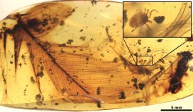
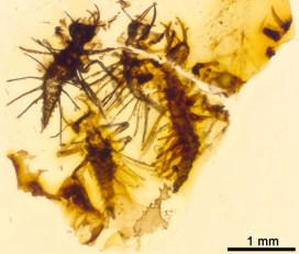
My colleagues and I have found an exceptional fossil in 130 million-year-old Lebanese amber. Inside, trapped together are newborn larvae from Green Lacewings, the split egg shells from where they hatched, and the minute egg bursters that the hatchlings used to crack the egg. This is a first: no definitive evidence of these specialised egg-bursting structures had been reported from the fossil record of any egg-laying animals, until now.

The finding has been recently published as open access in the journal Palaeontology. Because multiple newborns were ensnared and entombed in the resin simultaneously, the fossil larvae have been described as the new species Tragichrysa ovoruptora, meaning ‘tragic green lacewing’ and ‘egg breaking’. A sad event, indeed, taking place in an ordinary day 130 million years ago in the Cretaceous forests of Lebanon, yet a happy circumstance now that we can take a privileged glimpse into the adaptations and behaviours of these fascinating tiny creatures.

The hatchlings from modern Green Lacewings open a slit on the egg with a ‘mask’ bearing a saw-like blade. Once used, this ‘mask’ is shed together with the embryonic cuticle and is left attached to the empty egg shell.

With the help of Amoret Spooner, Collections Manager at the Museum, egg clutches from modern green lacewings were found in the Museum collections. These eggs happened to have the intact egg bursters still attached to them, and proved to be crucial to understand that we had the same structures preserved in the amber together with the newborn larvae.

Two Tragychrysa ovoruptora newborns preserved together with egg shell remains and two visible egg bursters (right inset). Modified from the open access Palaeontology paper.

Green Lacewing larvae are small predators that often carry debris as camouflage, using their sickle-shaped jaws to pierce and suck the fluids of their prey. Whereas the larvae trapped in amber differ significantly from modern-day relatives, in that they possess long tubes instead of clubs or bumps for holding debris, the studied egg shells and egg bursters are remarkably similar to those of today’s green lacewings.

The larvae were almost certainly trapped by resin while clutching the eggs from which they had freshly emerged. Such behaviour is common among modern relatives while their body hardens and their predatory jaws become functional. Indeed, the two mouthparts forming the jaws are not assembled in most of the fossil larvae, which indicates, together with the large relative size of the head and legs, that they were recently born.

Detail of a head with the jaws still dislodged, indicating that the larva was recently hatched when it was ensnared by amber and the jaws had not yet had time to fully assemble.

It may seem reasonable to assume that traits controlling a life event as decisive as hatching would have remained largely unchanged during evolution. In fact, we see in very closely related insect groups different means of hatching that can entail the loss of the egg bursters. So the persistence of a hatching mechanism in a given animal lineage through deep time can’t be determined without direct proof from the fossil record.

Reconstruction of two Tragichrysa ovoruptora newborns clutching the eggs from where they recently hatched, moments before they were trapped by resin. Larvae colour and egg stalks are conjectural. Extracted from the open access Palaeontology paper.

This new discovery shows that the mechanism green lacewings use to crack the egg was already established 130 million years ago. Overall, it represents the first direct evidence of how insects hatched in deep time, egg-bursting their way through into life.

*

The hatching mechanism of 130-million-year-old insects: an association of neonates, egg shells and egg bursters in Lebanese amber by Ricardo Pérez-de la Fuente, Michael S. Engel, Dany Azar and Enrique Peñalver is published as open access in Palaeontology this month.

Life’s big bang?

by Harriet Drage and Scott Billings

You may have heard of the Cambrian Explosion, an ‘event’, starting roughly 540 million years ago, when all the major animal groups suddenly appear in the fossil record, an apparent explosion of life and evolution.

But was there really an evolutionary explosion of all these animal groups, or is the lack of evidence from earlier periods due to some peculiarity of the fossilisation process? The debate has rumbled on for a number of years.

Now, a new study from our research team, the University of Oxford’s Department of Zoology, and the University of Lausanne, claims that the early Cambrian saw the origins and evolution of the largest and most important animal group on Earth – the euarthropods – in a paper which challenges two major pictures of animal evolution.

Euarthropoda contains the insects, crustaceans, spiders, trilobites, and a huge diversity of other forms alive and extinct. They comprise over 80 percent of all animal species on the planet and are key components of all of Earth’s ecosystems, making them the most important group since the dawn of animals over 500 million years ago.

Exceptionally preserved soft-bodied fossils of the Cambrian predator and stem-lineage euarthropod Anomalocaris canadensis from the Burgess Shale, Canada. Top left: Frontal appendage showing segmentation similar to modern-day euarthropods. Bottom right: Full body specimen showing one pair of frontal appendages (white arrows) and mouthparts consisting of plates with teeth (black arrow) on the head. Images: A. Daley.

A team based at the museum, and now at Lausanne, conducted the most comprehensive fossil analysis ever undertaken on early euarthropods, to try and establish whether these animals really did emerge in the early Cambrian period, or whether fossilisation just didn’t occur in any earlier periods.

In an article published today in the Proceedings of the National Academy of Sciences they show that, taken together, the total fossil record does show a gradual radiation of euarthropods during the early Cambrian, 540-500 million years ago, challenging other ideas that suggest either a rapid explosion of forms, or a much slower evolution that has not been preserved in the fossil record.

Each of the major types of fossil evidence has its limitation and they are incomplete in different ways, but when taken together they are mutually illuminating
Professor Allison Daley

Reconstruction of the Cambrian predator and stem-lineage euarthropod Anomalocaris canadensis, based on fossils from the Burgess Shale, Canada. Reconstruction by Natalia Patkiewicz.

By looking at a huge range of fossil material the researchers ruled out the possibility that Pre-Cambrian rocks older than around 541 million years would not have preserved early euarthropods. The only plausible explanation left is that the origins of this huge animal group didn’t evolve until about 540 million years ago, an estimate which also matches the most recent molecular dating.

The timing of the origin of Euarthropoda is very important as it affects how we view and interpret the evolution of the group and its effects on the planet. By working out which groups developed first we can trace the evolution of physical characteristics, such as limbs.

Exploring all the evidence like this allows us to make an informed estimate about the origins of key animal groups, leading to a better understanding of the evolution of early life on Earth.

Model of the Cambrian stem lineage euarthropod Peytoia, based on fossils from the Burgess Shale. Top left: Closeup of the mouth parts and frontal appendages. Bottom right: Overall view of the body. Model and image: E. Horn.

Who shot the Dodo?

By Scott Billings, Digital Engagement Officer

If ever the Oxford Dodo were to have squawked, its final squawk may have been the saddest and loudest. For the first time, the manner of death of the museum’s iconic specimen has been revealed: a shot to the back of the head.

This unexpected twist in the long tale of the Oxford Dodo has come to light thanks to a collaboration between the Museum and the University of Warwick. WMG, a cutting-edge manufacturing and technology research unit at Warwick, employed its forensic scanning techniques and expertise to discover that the Dodo was shot in the neck and back of the head with a 17th-century shotgun.

Mysterious particles were found in the specimen during scans carried out to analyse its anatomy. Further investigation of the material and size of these particles revealed them to be lead shot pellets of a type used to hunt wildfowl during the 1600s.

The Oxford Dodo specimen, as it has come to be known, originally came to the University of Oxford as part of the Tradescant Collection of specimens and artefacts compiled by father and son John Tradescant in London in the 17th century. It was thought to have been the remains of a bird recorded as being kept alive in a 17th-century London townhouse, but the discovery of the shotgun pellets cast doubt on this idea, leaving the bird’s origins more mysterious than ever.

Dodos were endemic to the island of Mauritius in the Indian Ocean. The first European accounts of the bird were made by Dutch explorers in 1601, after they rediscovered the island in 1598. The last living bird was sighted in 1662.

The story of the Oxford Dodo is especially significant because it represents the most complete remains of a dodo collected as a living bird – the head and a foot – and the only surviving soft tissue anywhere in the world.

This discovery reveals important new information about the history of the Oxford Dodo, which is an important specimen for biology, and through its connections with Lewis Carroll and Alice’s Adventures in Wonderland of great cultural significance too.
– Professor Paul Smith, Museum director

The Oxford Dodo represents the only soft tissue remains of dodo in the world. This iconic specimen was taken from the Museum to WMG at the University of Warwick for CT scanning.

WMG’s CT scans show that this famous symbol of human-caused extinction was shot in the back of the head and the neck, and that the shot did not penetrate its skull – which is now revealed to be very thick.

The discovery of such a brutal demise was quite a surprise as the scans were actually focused on discovering more about the Dodo’s anatomy, as well as how it lived and died. This work will continue, but we now have a new mystery to solve: Who shot the Dodo?

What’s the next step? It is possible that the isotope of lead in the shot could be analysed and traced to a particular ore field. This might tell us what country it was mined in, and perhaps what country is was made in, and ultimately reveal who shot the Dodo.

Bound by blood

It may sound like we’ve stumbled into a script-writing session for Jurassic Park, but one of our research fellows, Dr Ricardo Pérez-de la Fuente, along with an international team, has discovered a parasite trapped in amber, clutching the feather of a dinosaur. This small fossilised tick, along with a few other specimens, is the first direct evidence that ticks sucked the blood of feathered dinosaurs 100 million years ago. Ricardo tells us all about it…

The paper that my colleagues and I have just published provides evidence that ticks fed from feathered dinosaurs about 100 million years ago, during the mid-Cretaceous period. It is based on evidence from amber fossils, including that of a hard tick grasping a dinosaur feather preserved in 99 million-year-old Burmese amber.

Fluorescence detail of the studied hard tick grasping a dinosaur feather. Extracted from the publication.

The probability of the tick and feather becoming so tightly associated and co-preserved in resin by chance is virtually zero, which means the discovery is the first direct evidence of a parasite-host relationship between ticks and feathered dinosaurs.

Fossils of parasitic, blood-feeding creatures directly associated with remains of their host are exceedingly scarce, and this new specimen is the oldest known to date. The tick is an immature specimen of Cornupalpatum burmanicum; look closely under the microscope and you can see tiny teeth in the mouthparts that are used to create a hole and fix to the host’s skin to suck its blood.

The structure of the feather inside the amber is similar to modern-day bird feathers, but it could not belong to a modern bird because, according to current evidence at least, they did not appear until 26 million years later than the age of the amber.

Feathers with the same characteristics were already present in multiple forms of theropod dinosaurs –  the lineage of dinosaurs leading to modern birds – from ground-runners without flying ability, to bird-like forms capable of powered flight. Unfortunately, this means it is not possible to determine exactly which kind of feathered dinosaur the amber feather belonged to.

But there is more evidence of the dinosaur-tick relationship in the scientific paper. We also describe a new group of extinct ticks, created from a species we have named Deinocroton draculi, or “Dracula’s terrible tick”. These novel ticks, in the family Deinocrotonidae, are distinguished from other ticks by the structure of their body surface, palps and legs, and the position of their head, among other characteristics.

Blood-engorged Deinocroton draculi tick (female). Extracted from the publication.

This new species was also found sealed inside Burmese amber, with one specimen remarkably engorged with blood, increasing its volume approximately eight times over non-engorged forms. Despite this, it has not been possible to directly determine its host animal:

Assessing the composition of the blood meal inside the bloated tick is not feasible because, unfortunately, the tick did not become fully immersed in resin and so its contents were altered by mineral deposition.
Dr Xavier Delclòs, an author of the study from the University of Barcelona and IRBio.

But there was indirect evidence of the likely host for these novel ticks in the form of hair-like structures called setae from the larvae of skin beetles, or dermestids, found attached to two Deinocroton ticks preserved together. Today, skin beetles feed in nests, consuming feathers, skin and hair from the nest’s occupants. But as no mammal hairs have yet been found in Cretaceous amber, the presence of skin beetle setae on the two Deinocroton draculi ticks suggests that their host was in fact a feathered dinosaur.

The hair-like structures, or setae, from skin beetles (dermestids) found attached to two Deinocroton ticks fossilised inside amber, in comparison with extant ones. Modified from the publication.

Together, these findings tell us a fascinating story about ancient tick behaviour. They reveal some of the ecological interactions taking place among early ticks and birds, showing that their parasite-host relationship has lasted for at least 99 million years: an enduring connection, bound by blood.

The paper “Ticks parasitised feathered dinosaurs as revealed by Cretaceous amber assemblages” is published as open access in Nature Communications. Direct link: http://dx.doi.org/10.1038/s41467-017-01550-z

Rehoming a dinosaur

Last summer we ran an unusual competition: finding a new residence for our four metre-long model of Utahraptor ostrommaysorum. It had been hibernating in one of our off-site stores for a while, but following a reorganisation of collections we needed to find a new place for it to live. The competition to rehome the dinosaur was fierce, with 200 venues across the world vying to become the Utahraptor‘s new keeper…

It’s taken some time, thanks to logistics and admin, but one year later we are really delighted to reveal that the Utahraptor has now been installed at the Children’s Hospital at the John Radcliffe Hospital in Oxford.

The bid to take in the Cretaceous creature came from Sarah Fletcher, who now works at the Churchill Hospital in Oxford. Sarah nominated the Children’s Hospital so that the dinosaur could amaze and inspire the young patients.

The idea of having a model Utahraptor in the hospital seemed like a lot of fun. Having been through the Children’s Hospital with my family, I knew that it would make such a difference to everyone who walks through those doors. But I never thought in a million years that we would win it – I am thrilled!
– Sarah Fletcher

The Children’s Hospital team celebrate the arrival of their new ‘pet’

The model has been installed in the main entrance of the hospital, complete with new shadow-casting lighting, thanks to support from Oxford Radcliffe Hospitals Charitable Funds.

The team are now looking to develop new arts projects for young patients, themed around the dinosaur, including an all-important naming competition. We all hope it will bring pleasure to patients, provide a welcome distraction, and make their hospital visit a little more fun.

Patients, staff and visitors can peer at the dino on the way to the wards

Crayfish of the world united

by Sammy De Grave, head of research

How many species of crayfish can you name? Not many, or perhaps none? Well today, for the first time, a list of all the species of crayfish in the world has been published, thanks to a collaborative effort between Professor Keith Crandall at George Washington University and Dr Sammy De Grave, head of research here at the Museum.

The new list draws together much recent work and gives biologists access to a single, comprehensive summary of all the recognised species of crayfish for the first time. The new classifications group crayfish into 669 species, 38 genera, and five families, with two superfamilies corresponding to the Northern and Southern hemispheres.

Fallicambarus devastator. Image: Chris Lukhaup

On the occasion of this taxonomic triumph it seems like a good opportunity to take a look at some interesting crayfish from around the world.

Outside biological taxonomy, crayfish are much better known as a source of food. They are eaten worldwide, but especially in the southern US, Australia, and Europe, where the Red Swamp Crayfish (Procambarus clarkii) is most commonly on the menu. As a result, the Red Swamp Crayfish has been introduced into several countries and has out-competed the local species.

Several other species are also known as invaders. The Signal Crayfish (Pacifastacus leniusculus), native to North America, is now very abundant in Europe, and is out-competing the native Noble Crayfish (Astacus astacus).

The Noble Crayfish (Astacus astacus), above, is native to Europe, but is being out-competed by the introduced Signal Crayfish (Pacifastacus leniusculus). Image: Chris Lukhaup

Another remarkable crayfish is the Marmorkrebs, a species which still has no official taxonomic name. It was first noticed in the aquarium trade in Germany in the 1990s, but no natural populations are known. But the really interesting thing about this species is that all known individuals are female: it is parthenogenetic, which means the females reproduce from eggs without fertilisation – no males involved!

The Marmorkrebs crayfish has no official taxonomic name and is parthenogenetic – all individuals are female, genetically identical and reproduce without males. Image: Chris Lukhaup

Unfortunately, Marmorkrebs has escaped from aquaria in several countries, and is outcompeting local species due to its fast reproduction. Of most concern is its occurrence in Madagascar, where it competes for food and space with the endemic Astacoides crayfish, a much larger but slower-growing species.

Astacopsis madagascariensi, above, is being out-competed in Madagascar by the Marmorkrebs, which has escaped from several aquaria. Image: Chris Lukhaup

The Tasmanian Giant Crayfish (Astacopsis gouldi) is considered to be the largest freshwater invertebrate on the globe. Although its size has declined in recent years due to over fishing, historical specimens weighed up to 6kg and could reach 80-90 cm in length.

The completion of the new world crayfish list allows for further refinements to the conservation status of the animals too. Current Red List assessments show that 32 per cent of crayfish are already thought to be threatened with extinction, a similar number to freshwater shrimps and crabs.

It is really exciting to finally have a single source for the world’s freshwater crayfish taxonomy. Such a resource will impact a wide variety of fields that rely on crayfishes as study organisms. We hope it will also advance conservation efforts of these keystone species of highly endangered freshwater ecosystems.
– Professor Keith Crandall, George Washington University

The paper, An updated classification of the freshwater crayfishes (Decapoda: Astacidea) of the world, with a complete species list, is published today in the Journal of Crustacean Biology.