Skeletons in the closet

Once found in almost every museum and university in the western world, human anatomy collections have slowly disappeared over time, as medical practices have changed. These collections are historically important and medically relevant, providing direct evidence of patients’ lives and illustrating a pivotal period in the history of medicine.

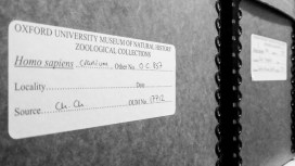
Over the past seven months, Dr Kathryn Krakowka has been going through the Museum’s human remains specimens to create a new detailed database of the contents. Here, Kathryn talks about the fascinating material she is working on…

The Museum holds many specimens that are not on public display, and perhaps one of the largest collections hidden from view is the human remains. This collection was started in the first half of the 18th century by Dr Matthew Lee, and eventually passed to Dr Henry Acland, founder of the Museum. Acland added a lot to the collection, acquiring his own samples as well as purchasing specimens and collections from other physicians.

When Acland gathered material for the new museum building in 1860, he brought this anatomical and physiological material with him, including the human remains. The collection continued to develop throughout the 19th century and grew to contain over 1,400 specimens. It is now one of the few collections from this period still in existence in Britain.

As a specialist in human osteology, I am trained to assess human skeletonised remains for such characteristics as sex, age-at-death, and any pathologies or trauma that the individual was affected by in life.

The Museum’s collection revealed specimens that tell a tale of health and medicine at Oxford in the 18th and 19th centuries. Many of the specimens were used to teach medicine, so many different skeletal pathologies are represented, including bacterial infections, genetic deformities, nutritional deficiencies, and traumatic injuries.

A skull fragment showing a large growth. The four round marks are thought to have been made by a tool during investigations after death.

One example is the frontal bone of a possible child, pictured above. This presents a large porous growth affecting both the outer and inner table of the bone. The characteristics of the lesion suggest osteosarcoma (bone cancer). There are then what appear to be four tool markings on the outer table of bone that indicate some sort of drill may have been used to take samples of the bone after death, possibly in an attempt to determine what was ailing the individual during life.

As this growth is quite large and just above the forehead, it would have been very noticeable while the individual was alive. This specimen provides an interesting example of how medical practices were often developed through exploratory postmortem analysis.

Now that the collection has been fully catalogued and digitised, it is hoped that further research into specimens like this one can help shed light on historical pathologies and the development of medicine as a discipline.

The database and digital images of the specimens will soon be available on the Museum’s website, thanks to funding by the John Fell Fund.
Fossil-finding

By Jack J Matthews, research fellow

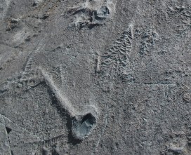
On the southern shores of Newfoundland in Canada lie rocks containing the oldest known evidence of large, architecturally-complex life. Deposited within the Ediacaran Period, some 565 million years ago, these deep marine deposits have been the focus of palaeontological research since the first discovery of fossils there in 1967, and the locality – Mistaken Point Ecological Reserve – now sits in the UNESCO World Heritage list.

As part of my research on these rocks, alongside colleagues from Memorial University of Newfoundland, and the University of Cambridge, I created a new geological map of the area, covering 35 km of coastline in and around the Reserve. As well as providing new insights into the rocks themselves, and what environments they were deposited in, this mapping had an unexpected outcome – the discovery of some totally new fossil sites.

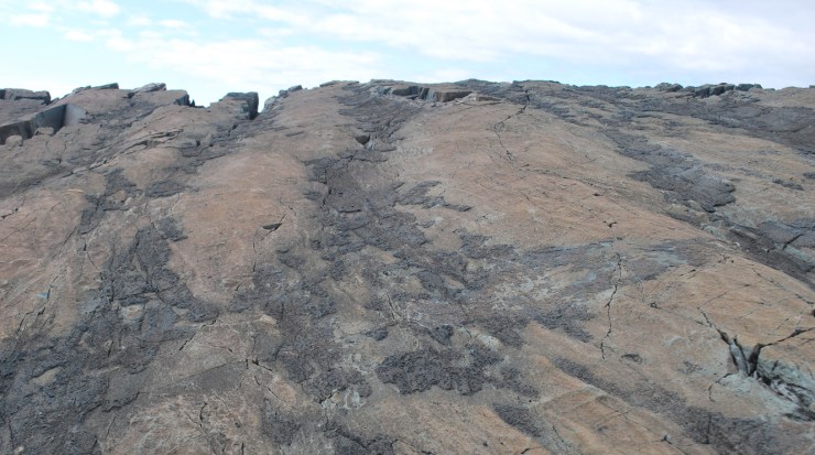
Overview of the Mistaken Point outcrop of the famous ‘E’ Surface

One site in particular, dubbed the ‘E’ surface, is the focus for Ediacaran fossils in Newfoundland. It is an area about the size of three Olympic boxing rings, containing more than 3,000 fossil organisms. Through the mapping we found a number of other outcrops of this same surface, but each shows slightly different types of fossils.

This is a mystery: if all the outcrops are from the same geological surface, why do they show different fossil assemblages?

The clue to the answer came while photographing the fossils and overlying volcanic ash at Mistaken Point, when I heard a loud, deep boom: a freak wave had struck the bottom of the cliff below the outcrop, sending a large splash of salty spray over much of the surface.

This got me thinking – how are processes such as weathering and erosion affecting the fossil surfaces now? Closer observation revealed those outcrops of ‘E’ with pristine beautiful fossils tended to be further from the sea, have a shallower dip, and the overlying ash tended to fall away in little flakes revealing beautiful, crisp, fossils. Other outcrops with scruffy fossils were usually close to the sea, battered by waves and rocks, steeply dipping, and the overlying ash, and often the fossils below it, would gradually abrade away as they are attacked by the sea.

Looking along the ‘E’ surface showing areas still covered in ash (black) and revealed fossil surface (red and grey)

Palaeontologists often discuss how changes during the fossil preservation of an organism can affect what we discover today, but they rarely discuss how processes occurring after preservation – metamorphism, exhumation, weathering, erosion, and even the time, manner, and conditions in which the fossil is recorded – might all affect how we analyse and interpret the original community of life which became fossilised.

Our new paper, published by the Geological Society of London, talks about these Post-Fossilization Processes, and recommends that when researchers are collecting fossil data they consider how their measurements might have been biased by such factors.

For 50 years now, the coastline of Newfoundland has yielded some of the most important finds in understanding the rise of the early life of the Ediacara, and through that the first evidence of animal life. Discoveries over the past few years show there is still much more to be found, and we’ll just have to hope that the post-fossilization processes fall in our favour to allow for many more significant discoveries.

Which one’s Pink?

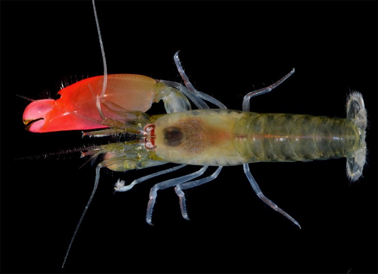
Image: Arthur Anker
In 1975, on Have a Cigar, Pink Floyd wryly sang “The band is just fantastic / That is really what I think / Oh, by the way, which one’s Pink?”

Well, in the rather different world of snapping shrimps there really is no question which one’s pink; and, unlikely as it seems, these two worlds have now overlapped…

The strikingly bright pink-clawed species of pistol shrimp pictured above, and discovered on the Pacific coast of Panama, has been given the ultimate rock and roll name in recognition of the discoverers’ favourite rock band – Pink Floyd. In a paper published today, and co-authored by our head of research Sammy De Grave, it has been named as Synalpheus pinkfloydi.

Just like all good rock bands, pistol shrimps, or snapping shrimps, have an ability to generate substantial amounts of sonic energy. By closing its enlarged claw at rapid speed the shrimp creates a high-pressure cavitation bubble, the implosion of which results in one of the loudest sounds in the ocean – strong enough to stun or even kill a small fish.

Combined with its distinct, almost glowing-pink snapping claw, Synalpheus pinkfloydi is aptly named by the report’s authors: lead author Arthur Anker of the Universidade Federal de Goiás in Brazil, Kristin Hultgren of Seattle University in the USA, and Sammy De Grave here at the Museum.

If Synalpheus pinkfloydi had adorned the cover of Pink Floyd’s 1977 album Animals, rather than the famous dirigible pig. Image: Chris Jarvis
Sammy has been a lifelong Pink Floyd fan and has been waiting for the opportunity to name the right new species after the band.

I have been listening to Floyd since The Wall was released in 1979, when I was 14 years old. I’ve seen them play live several times since, including the Hyde Park reunion gig for Live8 in 2005. The description of this new species of pistol shrimp was the perfect opportunity to finally give a nod to my favourite band.

Synalpheus pinkfloydi is not the only pistol shrimp with such a lurid claw. Its closely-related and similar-looking sister species, Synalpheus antillensis, scientifically described in 1909, is found in the western Atlantic, including the Caribbean side of Panama. But the authors of the new paper found that the two species show considerable genetic divergence, granting S. pinkfloydi a new species status and its very own rock and roll name.

Arthur Anker, the report’s lead author, says:

I often play Pink Floyd as background music while I’m working, but now the band and my work have been happily combined in the scientific literature.

Another Shrimp in the Wall featuring Synalpheus pinkfloydi, the Oxford University Museum of Natural History building, and other Pink Floyd references. Artwork by Kate Pocklington.
Animals feature frequently in the Floyd back-catalogue. Indeed, the 1977 album Animals includes tracks titled Dogs, Sheep, and a suite of music dedicated to pigs. Then there’s Several Species of Small Furry Animals Gathered Together in a Cave and Grooving with a Pict from 1969’s Ummagumma. In fact, other biologists have already named a damselfly after that album: Umma gumma, in the family Calopterygidae.

However, until today there have been no crustacean names known to honour the band.

The full paper, Synalpheus pinkfloydi sp. nov., a new pistol shrimp from the tropical eastern Pacific (Decapoda: Alpheidae), by Arthur Anker, Kristin M. Hultgren, and Sammy De Grave is published by Zootaxa.

Ask a neuroscientist

Can your brain rewire itself? How is the brain built and what can go wrong? And should the secondary school day start later to compensate for teenage sleep patterns?

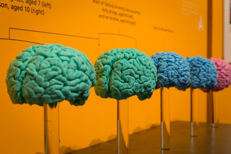
Neuroscientists from the University of Oxford address all these questions in our new exhibition – Brain Diaries – Modern Neuroscience in Action. Created in partnership with Oxford Neuroscience, the exhibition and event programme kicked off last week to coincide with international Brain Awareness Week, which runs from 13-19 March.

Here we present a selection of videos from the exhibition. The full set is available on our YouTube channel now. And if you’re not able to visit the exhibition itself, we’ve built a special Brain Diaries website which contains all that neuroscience goodness.

Is brain-building a tricky business?
Professor Zoltán Molnár of the University of Oxford specialises in the development of the brain. In this video he talks about the complex processes at play during the brain’s early development, including how things can sometimes go wrong.

Can my brain rewire itself?
Associate Professor Holly Bridge works in the Nuffield Department of Clinical Neurosciences in the John Radcliffe Hospital in Oxford. Her research focuses on using MRI scans of the human brain to understand the organisation of the visual system in people with normal vision and in those with abnormal visual function. Here she talks about how the brain can rewire itself to compensate for damage to certain sensory areas.

School’s out – should the school day start later?
Dr Christopher-James Harvey works at the Sleep and Circadian Neuroscience Institute at the University of Oxford. As part of the Teensleep research project, he is investigating how changes in the natural rhythm of sleep in adolescents, and the effects of sleep education, might impact on academic, health and sleep outcomes. Here he talks about initiatives to trial a later starting time for the secondary school day.

To read more about Brain Diaries and see the full programme of public events see braindiaries.org.

Birth of a species

img_4402
Under the sea, Curaçao, Caribbean. Image: Sancia van der Meij

By Sancia van der Meij, Research Fellow

Biologists often refer to the word “species” when they are talking about the animals or plants that they study, but just what exactly is a species? Defining ‘species’ is actually quite tricky…

A basic definition is based on the work of a German biologist called Ernst Mayr, whose simplified description is “a group of interbreeding populations that are reproductively isolated from other groups”. This is a great starting point, but it is difficult to use when studying animals in the field. Biologists therefore use breeding experiments in laboratories and, increasingly, genetics to help determine what a species is.

How and under which circumstances new species evolve remains an important topic in biology. Quite a lot is known about geographical barriers causing the formation of new and distinct species through evolution – a process known as speciation. Mountains, rivers and ocean currents, for example, can divide populations of single species and in the long run – thousands or millions of years – this isolation can cause different populations to evolve in separate, new species.

g-van-moorsel-ecosub
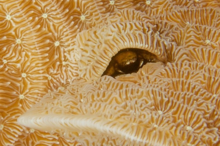
Gall Crab inhabiting a small tunnel in an Agaricia coral. Image: G van Moorsel

But a more difficult concept in speciation is how species can evolve in the same geographical area. Together with a colleague, I studied the genetic composition of Opecarcinus hypostegus, a tiny crab species, around 5 mm in size, that only occurs in the Atlantic Ocean. These Gall Crabs are adapted to living in stony corals and often show a clear preference for inhabiting closely related coral species.

img_4234
Overhang in a Agaricia coral where a gall crab dwells. Image: Sancia van der Meij

We studied over 200 specimens from five different coral species, all collected from the Caribbean island of Curaçao. The results showed that O. hypostegus should be considered a single, valid species. But to our surprise, when we zoomed into the details of the genetic composition of the crab,  we noticed small differences in the DNA of the crabs inhabiting the various coral species. With statistical tests we could prove that the variation in DNA was significantly different between the crabs inhabiting these five different Agaricia corals.

img_3808
Gall crab dwelling in Agaricia coral. Image: Sancia van der Meij

Despite the fact that all the crabs live around the same small Caribbean island, it does appear that we see the very first signs of future speciation in the crab’s DNA. Unfortunately we will not be around to witness the new species as it will likely take several hundreds of thousands of years before the making of these new crab species has neared completion. But how exciting to witness its new beginnings?


Mysteries of the deep

by Sammy De Grave, Head of Research

It is often said that the final frontier of our exploration of the Earth lies deep in the oceans. Covering 70 per cent of the planet’s surface, the oceans nevertheless remain 95 per cent unexplored, according to the National Oceanic and Atmospheric Administration in the United States. And they know, for they are doing the exploring…

Since 2008, the Okeanos Explorer at the NOAA has been investigating deep water ecosystems and has live-streamed many of its remotely operated vehicle (ROV) dives for scientists and the public to observe. Last year, Okeanos was engaged in an exploration of the Mariana Trench Marine National Monument in the Pacific Ocean. During a single dive at an impressive 4,826 metres, on a muddy bottom at a site nicknamed Twin Peaks, a large shrimp was observed, which the participating scientists did not recognise.

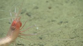
Spotted 4,826m below sea level, in the Mariana Trench Marine National Monument: the shrimp Bathystylodactylus
Spotted 4,826m below sea level, in the Mariana Trench Marine National Monument: the shrimp Bathystylodactylus cf. bathyalis

This was an unexpected find. Although there are about 4,000 species of caridean shrimps in the world’s oceans, very few live below 1,000 metres, and fewer than 20 species are known from depths lower than 3,000 metres. Those that do are usually only found as broken specimens, creatures damaged by the trawls which also bring them to the surface.

Photos of the deep-dwelling shrimp were duly sent to two experts: Dr Mary Wicksten at Texas A&M University and Dr Sammy De Grave, head of research here at Oxford University Museum of Natural History. Both immediately recognised the specimen as belonging to the rare genus Bathystylodactylus, which comprises of only three scientifically-described species, which in turn are only known from six specimens, all damaged.

The bristly legs, called setose legs, are used in passive filter feeding
The bristly legs are used in passive filter feeding

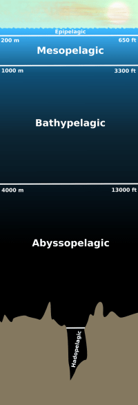
wfm_pelagicThe Okeanos team had made something of a discovery, one which was published in the open access journal Zookeys. The posture of the shrimp clearly show it to be a filter-feeding species, with its long, bristly legs facing into the current. This type of feeding behaviour has not been seen in caridean shrimps before.

Surprisingly, although the shrimp was observed at almost 5,000 metres below the surface, it is not the deepest recorded shrimp. That honour goes to a different species, Glyphocrangon atlantica, living in the western Atlantic, which has been trawled from as deep as 6,373 metres. Blurry photographs from 6,890 metres in the Kermadec Trench might indicate that carideans live even deeper still, maybe even as far down as a related group of shrimps called dendrobranchiates, which are known from depths down to 7,703 metres.

These findings show that life of many kinds continues to be discovered in regions of the planet once thought to be completely inhospitable.