The ancient ‘Kite Runner’

72dpi Press%205

An ancient creature which carried its young like tiny, swirling kites is the latest discovery by researchers at the Museum, working with Yale Peabody Museum of Natural History, Leicester University, and Imperial College London.

Found in a deposit of rocks known as the Herefordshire Lagerstätte, which preserves ancient remains with superb detail, the 430 million year old fossil shows that the marine animal carried its young in kite-like capsules tethered to the parent’s body, earning it the moniker “Kite Runner” after the 2003 novel by Khalid Hosseini.

The small creature has been officially named Aquilonifer spinosus, from “aquila”, meaning eagle or kite, and “fer” which means carry. It grew to just over a centimeter long, not including the tail spines, and there is only one known fossil of the animal.
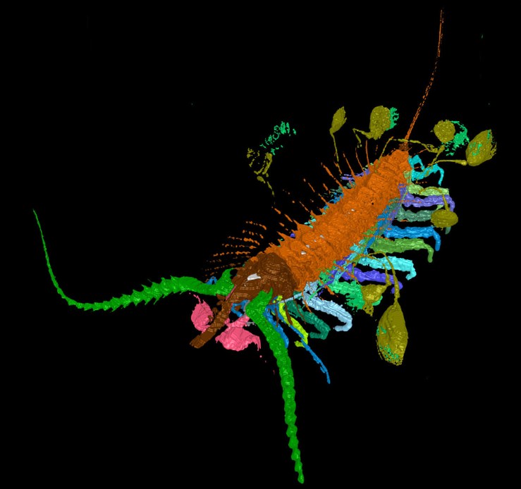
Kite runner. Anterior oblique.The arthropod Aquilonifer spinosus
Reconstruction of Aquilonifer spinosus

Modern crustaceans employ a variety of strategies to protect their eggs and embryos from predators — attaching them to limbs, holding them under a carapace, or enclosing them within a special pouch until they are old enough to be released — but this example is unique. We know of nothing alive today which attaches the young by threads to its upper surface; perhaps this strategy was less successful and became extinct.

Kite runner. Two young in capsules of the arthropod Aquilonifer spinosus
Two capsules of juveniles tethered to the parent’s body

Aquilonifer spinosus lived on the seafloor during the Silurian period, with a variety of other animals including sponges, brachiopods, worms, snails and other mollusks, a sea spider, a horse-shoe crab, various shrimp-like creatures, and a sea-star.

The researchers were able to describe Aquilonifer spinosus in detail thanks to a virtual reconstruction. They reconstructed the animal and the attached juveniles by stacking digital images of fossil surfaces that were revealed by grinding away the fossil in exceptionally thin increments. You can see this animation here:

The Yale Peabody Museum of Natural History, the Natural Environmental Research Council, the John Fell Oxford University Press Fund, and the Leverhulme Trust supported the research.
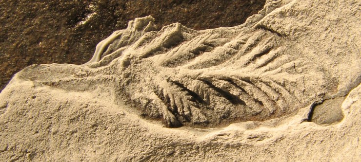


The world’s first fussy eaters?

OUMNH_Blog_Post_Rahman_Figure1

by Dr Imran Rahman, Research Fellow

I have just returned from visiting colleagues at Vanderbilt University in Nashville, Tennessee. Nashville is the home of country music, hot chicken and, most importantly for my work, a brand new CT scanner. Together with Drs Simon Darroch, Marc Laflamme and Rachel Racicot, I used the CT scanner to create 3-D models of 560-million-year-old fossils which will be used to learn more about how such ancient organisms lived and fed.

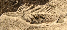
These fossils are some of the strangest ever described. They come from the ‘Ediacara biota’, which is approximately 542 to 600 million years old. These include the first large organisms on Earth, some of which might be early animals, but placing these fossils in their correct place in the tree of life is extremely controversial. In fact, despite extensive study by palaeontologists for many years, we know very little about what these organisms were like when they were alive.

In order to better understand these enigmatic fossils, we used Simon’s CT scanner to study them. The scanner works by using X-rays to create cross-sectional images through the specimen, which can be then used to digitally reconstruct it in 3D. We scanned a range of different fossils and were able to describe their morphology in exceptional detail, as shown in the image below.

OUMNH_Blog_Post_Rahman_Figure2
3D reconstruction of the fossil shown at the top of the article: a 560-million-year-old organism from Spaniard’s Bay, Newfoundland (width of fossil = c. 8 mm). Source: Marc Laflamme.

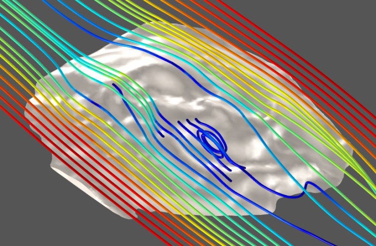
These 3D reconstructions will serve as a basis for computer simulations of water flow around the fossils, which will allow us to evaluate ideas about how these organisms might have fed. Research we carried out last year suggested that some Ediacaran organisms may have fed in a more complex way than previously thought, and we would like to test if this applies to other species from the same time period.

Simon, an Assistant Professor in Vanderbilt’s Department of Earth and Environmental Sciences, brought us together to carry out this research. We will also be working closely with Simon’s Grad Student, Brandt Gibson, who is creating 3D models of fossils using computer graphics software. Ultimately, we hope to gain a better understanding of Ediacaran ecosystems, which will provide important new insights into the early evolution of complex life.

Computer simulation of water flow around a 3-D reconstruction of a 555-million-year-old fossil organism from Flinders Ranges, Australia (width of image = c. 30 mm). Source: Imran Rahman.

The value of collections

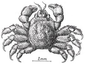
Gall crab illustration
Figure after Kropp and Manning (1987: Fig. 4)

by Sancia van der Meij, Research Fellow

Scientists often use DNA to study the position of a species in the tree of life. By sequencing DNA from different species we can begin to see how closely they are related, but sometimes it can be difficult to obtain specimens to carry out this type of research. This is where museum collections become invaluable.

I have been investigating a tiny coral-dwelling crab, Detocarcinus balssi, which is less than 1cm in size and lives in the Atlantic ocean. In a paper just published in the Journal of Zoological Systematics and Evolutionary Research, my co-writer and I show how we have resolved where this crab is placed, taxonomically-speaking.

To establish this we needed DNA, but unfortunately the crab has only been collected on a few occasions since the discovery of the species in 1956. Luckily, the Smithsonian Institution in Washington D.C. houses several specimens of D. balssi that were collected in the 1980s.

Gall crab females carry many, many eggs, so with permission of the curator at the Smithsonian a special DNA protocol for old and degenerated material was used to extract small fragments of DNA from the eggs of a D. balssi specimen collected in 1984.

Gall crab
Hapalocarcinus marsupialis, a gall crab similar to Detocarcinus ballsi, with big bundle of eggs

The DNA sequence that was obtained offered a surprise: the Atlantic species D. balssi is more closely related to an Indo-Pacific species Utinomiella dimorpha than any other Atlantic species. This may seem odd at first, but gall crabs probably originated before the Panama Isthmus – the landmass connecting North and South America – was fully closed. At the time, species could freely pass between what we now know as the Pacific Ocean and the Atlantic Ocean.

With this little part of the puzzle solved, the relationship between Atlantic and Indo-Pacific gall crab species can be studied in more detail.

Without the scientific collections held in museums this research could not have been completed. Researchers increasingly use museum collections, including the famous Dodo in this Museum, to answer important questions about where to fit a species in the taxonomic scheme of things – on the grand tree of life.

Plesiosaur puzzle

IMG_7075

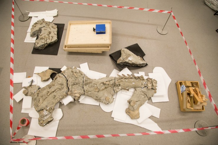
An exciting new resident arrived at the Museum recently, having lain in the Cambridgeshire earth for around 165 million years. Discovered in a quarry near Peterborough, the skeleton of a 5.5 metre plesiosaur has been donated to the Museum and is now awaiting reconstruction and further study in our labs.

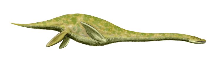
Plesiosaurs were long-necked sea creatures that lived during the time of the dinosaurs, but died out 66 million years ago.

Muraenosaurus
Artist’s impression of Muraenosaurus leedsii, a similar plesiosaur from the Middle Jurassic of Europe. Image by Nobumichi Tamura.

The fossilized remains of the marine reptile were discovered at a site owned by building product manufacturer Forterra, which has kindly allowed the material to be added to the Museum’s collections.

The creature was first spotted by Oxford Clay Working Group member Carl Harrington who noticed a tiny fragment of bone sticking out of the clay. Over the course of four days, Carl and eight others dug up more than 600 pieces of fossilised bone. Carl then spent over 400 hours cleaning and repairing the specimen.

I’d never seen so much bone in one spot in a quarry. As I was digging amongst the wet clay, the snout of a plesiosaur started to appear in front of me. It was one of those absolute ‘wow’ moments – I was the first human to come face to face with this reptile.

IMG_7059
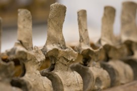
The plesiosaur’s neck vertebrae

The plesiosaur had a 2.5 metre long neck, a barrel-shaped body, four flippers and a short tail. Its skull is still preserved inside a block of clay, and the painstaking task of removing it will now be undertaken here.

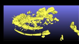
A CT scan of the plesiosaur's skull, which is still inside a clay block
A CT scan of the plesiosaur’s skull, which is still inside a clay block

DrJames Neenan, one of our research fellows, and Professor John Hutchinson from the Royal Veterinary College have already CT-scanned the block to reveal the location of the bones inside. This will help in removing it from the clay.

Next week pupils from a visiting secondary school will get the chance to see the plesiosaur find for themselves and to ask our Earth Collections manager Dr Hilary Ketchum all about it. Hilary says:

We are so excited that the plesiosaur has come to the Museum where it will be used for research, education and display.

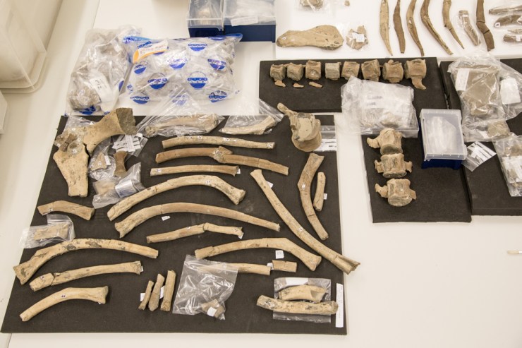
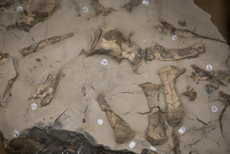
The plesiosaur’s ribs and vertebrae still inside the rock

Hilary will now begin the task of reconstructing the plesiosaur from the remains you can see in the photos here – a combination of individual separate bones and those still contained in clay nodules. Ultimately, we hope to articulate and suspend the specimen for public display.

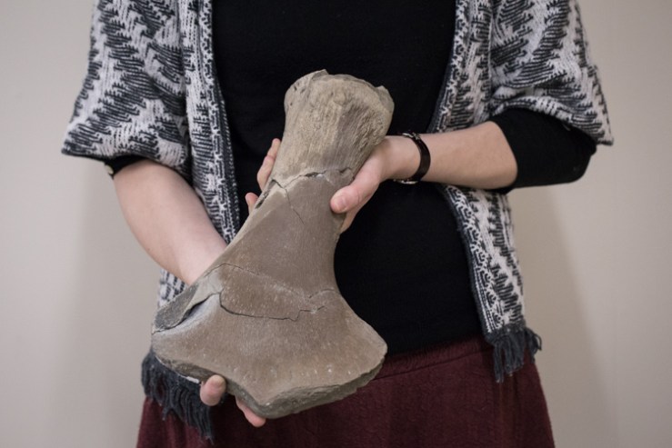
Hilary holds out the plesiosaur’s arm bone (humerus)

Part of the study will be to determine whether this is a known or new species of plesiosaur. Early indications suggest that it might be a species new to science, but more investigation is needed before we will know for sure.

Watch this space…

IMG_7078

Temporarily misplaced

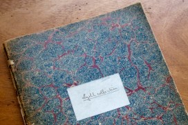
Scouring the archives, and receiving an unexpected package, help our documentation officer Sarah Joomun in her investigations into the Museum’s Lyell collection of fossil material.

Read more on the latest post from Past to Present…

Sarah Joomun's avatarPast to Present

When I started work on the Lyell project in July of this year, I was very keen to know more about the history of the collection, both before and after it arrived at the museum. Collections often arrive at the Museum with associated material such as catalogues, letters or notebooks and after they arrive any activity related to the collection should be documented.

The first step in investigating the history of the collection was to find out what was in the Museum’s records. I began by looking at the donors database; this recorded the date that the Lyell collection arrived (1903) and the donor, Sir Leonard Lyell, Charles Lyell’s nephew. There was a little more information in the donors card index, which mentioned the fact that the collection came in two parts, the bulk of the collection in 1903 and then additional Italian specimens in 1907. The Collections Manager, Eliza…

View original post 674 more words

So long, 2015…

IMG_6901-Edit

As you can tell from the adornment of our Red Deer, Christmas is upon us, so it’s nearly time to bid farewell to another year. It’s been another remarkable twelve months here at the Museum so here’s a little round up a few highlights from 2015…

As winter gave forth to spring
News emerged of a heartwarming thing
The Art Fund whispered in our ear
We were nominees for Museum of the Year!

Although eventual winners we were not
It mattered really not one jot
For in celebration we embarked
On the Dodo Roadshow – a tremendous lark

Back in April we’re pleased to say
Another award came our way
Goes to Town gave creatures free reign
And grabbed a gong for Marketing Campaign

But we weren’t always on the road
In our exhibitions many stories were told
Of evolution, geology and sensory powers
Science and research passed the visitors’ hours

Our doors were open without interruption
While out on the lawn was a volcanic eruption
University scientists had plenty to say
On a really Super Science Saturday

So to our schools, and families, and adults and more
Thank you, cheers, and thank you some more

Here’s our programme for January to April. See you in 2016…