Close up of OUMNH-ZC-7483 Section of blue whale intestine with mysterious acanthocephalan parasites

Worms of Discovery

By Mark Carnall, Life Collections manager

The Museum’s zoology collections contain a dizzying diversity of animal specimens. It is a collection that would take multiple lifetimes to become familiar with, let alone expert in. So we benefit hugely from the expertise of visiting researchers – scientists, artists, geographers, historians – to name just a few of the types of people who can add valuable context and expand our knowledge about the specimens in our care.

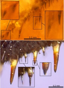
Earlier this year, Dr Andrew McCarthy of Canterbury College (East Kent College Group) got in touch to ask about our material of Acanthocephala, an under-studied group of parasitic animals sometimes called the spiny-headed worms.

Although there are around 1,400 species of acanthocephalans, they are typically under-represented in museum collections. Dr McCarthy combed through the fluid-preserved and microscope slide collections here, examining acanthocephalan specimens for undescribed species, rare representatives and unknown parasitic associations.

Close up of OUMNH-ZC-7483 Section of blue whale intestine with mysterious acanthocephalan parasites

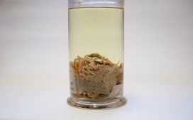
One such specimen, catchily referenced OUMNH.ZC.7483, was of particular interest. It is a section of blue whale intestine packed with acanthocephalan adults, labelled ‘Echinorhynchus sp. “Discovery Investigations”’, and dated 13 March 1927. Drawing on his expert knowledge, Dr McCarthy spotted an unusual association here because the genus Echinorhynchus was not known to infect Blue Whales, meaning the specimen could represent a species to new science.

However, identifying different species of acanthocephalans cannot be done by eye alone, so Dr McCarthy requested to remove one of the mystery worms from the intestine and mount it on a slide to examine its detailed anatomy. When we receive a destructive sampling request like this it triggers an investigation of the specimens in question: we need to weigh up their condition, history, and significance against the proposed outcome of the research before we decide whether the permanent alteration of the specimen justifies the outcome.

Image of Oxford University Museum of Natural History zoology collections accession register entry for this specimen showing the donation of the specimen and collector information.

This particular investigation began to yield a much richer story than the Museum’s label suggested. It turned out that the specimen was collected by Sir Alister C. Hardy who was serving as zoologist on RRS Discovery’s scientific voyage to the Antarctic. Fortunately, Discovery’s scientific findings were meticulously documented and published by many libraries of the world, including the fantastic Biodiversity Heritage Library where it was easy to find the report mentioning acanthocephalans collected during the voyage.

Alongside descriptions of acanthocephalans from seals, dolphins and icefish there is no mention of Echinorhynchus sp. from Blue Whales, though there are a few references to another genus, Bolbosoma, collected from Blue Whales on seven occasions: a single individual of Bolbosoma hamiltoni, so obviously not this specimen, and six occurrences of Bolbosoma brevicolle from the intestines of Blue Whales from South Africa and South Georgia.

These specimens and others reported in the Discovery reports. Image from Biodiversity Heritage Library

Piecing together the evidence, the association with Hardy, the dates, and the descriptions of RRS Discovery’s acanthocephalans, it seems likely that our specimen is one of the six samples of Bolbosoma brevicolle and not Echinorhynchus at all. So in this instance we decided not to grant destructive sampling as the likelihood of identifying a new species seemed much lower when all the information was brought together.

Although sampling wasn’t granted, Dr McCarthy was delighted that his initial research request had prompted the discovery of some important historical connections to the humble specimen, and the new identification seemed to fit.

We still weren’t sure when or why this specimen was mislabelled some time between the Discovery reports and its donation to the Museum in 1949, so Dr McCarthy conducted some further investigations. He found out that Echinorhynchus was the original name combination for Bolbosoma brevicolle, and that H. A. Baylis, a parasitologist and author of Discovery reports, had links with the University of Oxford.

This story is just one example of how visiting researchers enrich knowledge and information about our collections, and it illustrates nicely why our work with broader research communities is so important.

Sight without eyes

By Lauren Sumner-Rooney, Research Fellow

Vision is among the most important innovations in animal evolution. The ability to see predators, prey, mates, and the environment transformed the way animals interact with each other and the world around them. Eyes can take many different forms, but this month saw the description of a visual system unlike almost any other known to science, found in a brittle star called Ophiocoma wendtii.

Brittle stars are marine invertebrates related to starfish. They have long, slender arms connected to a central disk, but no head, no brain, and – so we thought – no eyes. But recent experiments have shown that some brittle stars are able to see the world around them.

Ophiocoma wendtii is a common species found throughout the Caribbean Sea and the Gulf of Mexico. If you rummage around in coral rubble in shallow water, you’ll probably find Ophiocoma hiding underneath rocks and other debris, sheltering from their fishy predators. It has beautiful bright red tube feet (small, water-filled tentacles) and a neat party trick: it changes colour. During the day, the animals are a deep reddish-brown colour, but after dark they become beige with dark stripes.

The red brittle star, Ophiocoma wendtii

For more than thirty years, O. wendtii has been something of a mystery to scientists like myself who are interested in animal vision. It’s covered in light-sensing cells – thousands of them – and it hates being exposed to bright light, quickly dashing for cover if possible. However, it’s possible to head for dark, shadowy places without vision; you only need to be able to tell that one direction is brighter than the other. So, with a team of colleagues from Germany, Sweden and the USA, we set about giving the brittle stars an eye-test.

Lauren Sumner-Rooney, collecting specimens of Ophiocoma wendtii. Image: Jane Weinstock

We know that when they’re exposed to sunlight, O. wendtii try to hide underneath nearby rocks or other objects, so we designed a circular arena with a stimulus printed on one side – the idea is that the stimulus might resemble an object under which the animals can shelter, and the animal will move towards it.

We ran three experiments, changing the stimulus and background of the arena in each to test whether the brittle star can just see relative light or dark areas, or whether it can resolve finer points of contrast. To my surprise, O. wendtii moved towards the stimuli in all three experiments significantly more frequently than expected by random chance, as you can see in the video below. This was super exciting, as it represents not only the very first evidence of vision in these animals, but the second known example of any animal that can ‘see’ without having eyes (the first is a close relative, a sea urchin).

While O. wendtii is known to shelter during the day, we were also curious to test its behaviour at night. Running the same experiments again in natural darkness, we found that animals no longer moved towards any of the stimuli. There could be a whole number of reasons behind this, so we devised tests that eliminated several possibilities, and were left with a remaining explanation that the animal’s colour-change between night and day was somehow responsible.

Close-up of the arm plates of Ophiocoma wendtii

Colour-changing in the brittle star is controlled by the expansion and contraction of cells, called chromatophores, that are filled with pigment granules. These sit inside pores in the skeleton, alongside the light-sensing cells. During the day, the chromatophores expand, pushing up through the pores and spreading over the body surface. The pigment is spread over the outside of the animal, which looks dark brown as a result. During the night, the chromatophores contract, bringing all the pigment granules back inside the skeleton and giving a paler appearance.

The red brittle star, Ophiocoma wendtii. Image: Heather Stewart

We thought that during the day the pigment granules surrounding the light-sensing cells might block light reaching them from most directions. To test this, we constructed digital models of the visual system, creating 3D models of the light-sensing cells, the skeleton, and the pigment granules.

We found that in light-adapted systems, those with pigment, light could only reach the sensory cells from an angle of around 60° out of 360° which, though probably very coarse, could support vision. By removing the pigment from the models, vision was made impossible, as light could reach the sensory cells from too many different directions. It looked as though it was the chromatophores that made all the difference.

This is the first proposed example of whole-body colour change enabling and disabling vision in any animal, and raises many new questions about image formation and information processing. There are exciting parallels with the only other example of ‘extraocular’ (=without eyes) vision, the sea urchin we mentioned earlier: these sea urchins can also change colour in response to light levels, using similar chromatophores. Have they independently evolved a similar trick?

Top image: Heather Stewart

On the trail of the Piltdown hoax

The latest display in our single-case Presenting… series takes a look at the famous Piltdown Man hoax, and Life Collections manager Mark Carnall tells us how the display came about…

Visiting researchers to the zoology collections at the Museum often give us an excuse to dig deeper into our own material, and one such recent enquiry led me into the intriguing story of the Piltdown Man hoax.

Professor Andrew Shortland from Cranfield University contacted us to enquire about the Piltdown Man material in our collections, as part of research for a book on hoaxes and forgeries in anthropology that he is writing with Professor Patrick Degryse of KU Leuven.

I knew we had some Piltdown material here thanks to this page written by Malgosia Nowak-Kemp, but I hadn’t had an excuse to investigate any further. The enquiry was also timely as we’d just transferred a collection of palaeoanthropology casts, models and reconstructions from our Earth collections to bring our human collections into one place. I knew from our move project team that there was some Piltdown material awaiting processing – perfect.

For those who don’t know the Piltdown Man story, a short history is in order. In the early 20th century, amateur fossil hunter Charles Dawson brought a collection of human remains excavated from gravel pits in Sussex to the attention of Arthur Smith Woodward, then Keeper of Geology at the British Museum (Natural History). Woodward and Dawson collected further material and presented the remains as those of Eoanthropus dawsoni (‘Dawson’s dawn man’), an important fossil human from Britain.

Group portrait of the Piltdown skull being examined. Back row (from left): F. O. Barlow, G. Elliot Smith, Charles Dawson, Arthur Smith Woodward. Front row: A. S. Underwood, Arthur Keith, W. P. Pycraft, and E. Ray Lankester. Charles Darwin looks on from a portrait on the wall. Image via Wikipedia.
R.F. Damon-produced endocast and associated label recording the presentation of this specimen to the Museum by Arthur Smith Woodward

The discovery looked set to put Britain on the map when it came to evidence of human evolution, but suspicions were quickly raised about the authenticity of the material. Such was the skill of the forgery – meticulous breaking, abrading and staining of various archaeological and historic specimens – that it wasn’t until dating techniques, chemical analyses and some experimental palaeoanthropology in 1953 that the hoax was conclusively put to bed.

In turned out that the Piltdown ‘remains’ were a mix of medieval bone, an orangutan jaw, and chimpanzee teeth maltreated to look like an evolutionary intermediate between humans and other apes.

For 40 years or so the hoax refused to go away and numerous casts, models and reconstructions of Piltdown Man were made, sold, exchanged and gifted to museums and universities. These included casts of the original material as well as reconstructions of the skull and even reconstructions of the endocast – a cast of the inside of the skull.

The Museum has a selection of this material, but as Professor Shortland examined the collections, two specimens stood out.

The first is an R. F. Damon-produced endocast presented to the Museum by Arthur Smith Woodward himself. Smith Woodward was known as an expert on fossil fish but published widely on zoological topics. As a scientist of some repute there’s been long-standing speculation about his role in the hoax. Was he wholly duped by Dawson, or was he in on the hoax from the beginning? If it’s the former, then the presentation of this endocast shows Smith Woodward disseminating research he presumably took some pride in. If it’s the latter, perhaps it was a way of cementing the hoax as legitimate by spreading specimens far and wide.

Joseph Weiner’s experimental fake created by modifying an orangutan jaw, alongside a cast of the Piltdown jaw

The second significant specimen is a worked orangutan jaw produced by Joseph Weiner, one of the three authors who debunked the hoax in a 1953 Nature paper titled The Solution of The Piltdown Problem. Weiner modified the orangutan jaw to replicate the original hoax specimen. Thanks to Professor Shortland’s knowledge of the hoax, he sent through a copy of Weiner’s book on the Piltdown Man where this exact specimen is pictured.

The Piltdown Man hoax wasn’t the first and certainly won’t be the last hoax, fake or forgery in the history of science, but it remains one of the most well-known and stands as a warning of the dangers of hubris in the discovery and description of the natural world.

The Weiner jaw and Damon endocast will be on display alongside other Piltdown Man material in our Presenting… case from 9 January to 8 March 2020.

Image credit - Flickr/milo bostock, licensed under CC BY 2.0

Changing climate is narrowing options for migrating birds

This article is taken from European research magazine Horizon as part of our partnership to share natural environment science stories with readers of More than a Dodo.

Across an entire desert or ocean, migratory birds make some of the most extreme journeys found in nature, but there are still huge gaps in our understanding of how they manage to travel these vast distances and what a changing climate means for their migration patterns.

‘Some species of migrants might be affected by a changing climate,’ said Professor Stuart Bearhop, an animal ecology expert from the University of Exeter. ‘There is evidence from a number of populations that climate change probably is going to have some impact on the demography (population levels).’

Bearhop ran the STATEMIG project, which studied the migration of Brent Geese along their journey from Ireland to the Arctic where they breed. He found that the volatility of today’s seasons was affecting the geese’s population levels because the weather was playing havoc with their breeding patterns.

‘Wet years are predicted to increase with climate change as temperature rises, but, of course, because they travel so far north, it doesn’t mean rain, it means snow,’ he said. Brent Geese are more likely to breed when the weather is cold and clear, but when there is more snow there are fewer places to safely raise their young and feed.

The team observed that in the colder years the birds were breeding later in the year, causing ripple effects for their populations. The geese did not have enough time to raise their offspring to independence before winter, or there was not enough food for them to survive.

Bearhop says the snowy years saw more offspring die or be abandoned by adults. That means if snowy years persist then it could pose a long-term risk to the population of these birds.

Brent Geese

Bearhop chose Brent Geese because they follow a routine migration and their young stay with their parents for at least a year. These reliable patterns reveal useful insights into population levels and what could be affecting their migration.

To gather their data, STATEMIG researchers observed the geese in Ireland and Iceland before the birds flew to the Arctic to breed around July. In Ireland and Iceland they attached identity tags to the birds and took some physical measurements to use as reference points over several years.

When the geese returned to Ireland and Iceland around late August, with their chicks, the researchers could compare the population levels and get an idea of how environmental factors had shaped their journeys.

‘There are multiple factors that have likely driven the evolution of migration, these likely differ among species and the debate is about which ones are most important,’ said Bearhop.

Debate

Bearhop says the two key reasons birds migrate is because of a competition of territory and to take advantage of seasonal ‘pulses’ of vegetation growth or gluts of insects to ensure they have enough food to raise their young.

STATEMIG’s research emphasises the importance of the latter and Bearhop hopes it could lead to further research that explores how changes to feeding grounds will affect populations of migratory birds.

According to Dr Sissel Sjöberg, a bird migration researcher from the University of Copenhagen, Denmark, scientists understand some parts of why birds migrate, like knowing where they eat and breed, but they do not have the tools to accurately understand them during the entire migration.

For instance, there are high resolution tags that can be put on some big birds to track their location, but these do not fit on smaller birds which make up most of the ones migrating.

 Tiny backpacks worn by noctural small birds contain a pressure sensor which provides an update every five minutes of the birds’ behaviour during migration. Image credit - Dr Sissel Sjöberg
Tiny backpacks worn by noctural small birds contain a pressure sensor which provides an update every five minutes of the birds’ behaviour during migration. Image credit – Dr Sissel Sjöberg

These tags also do not provide insights into other aspects, like altitude or how they traverse over huge, inhospitable areas where they may not be able to land, like the Sahara desert or the Pacific Ocean.

Dr Sjöberg is the principal researcher of the BIRDBARRIER project which is putting tiny backpacks on nocturnal small birds migrating long distances, such as red-backed shrikes and great reed warblers. These backpacks contain an activity log with a pressure sensor to determine heights and provide updates every five minutes of their behaviour during the journey, which can be correlated with weather forecasts or detailed landscape maps.

‘It is clear they go higher in their flights then we thought before,’ said Dr Sjöberg, adding that experts previously thought their size limited them to flying at 2,000-3,000 metres above sea-level, but she has observed them fly at almost 6,000 metres.

Dr Sjöberg says they could be doing this to find stronger winds that carry them longer distances, which require less energy to fly in and increase their chances of survival.

She says the biggest risk for these birds is to stop in the hostile terrains they cross because it could be difficult to take off again or find the same heights. Safe places to land are crucial to these birds on their intercontinental journeys because they have favourable conditions, including sources of food, but in some places they are getting smaller, for instance, in the Sahara where the desert is expanding.

‘Those (safe) areas are getting smaller and smaller so there is more competition,’ said Dr Sjöberg, who will continue to collect data from the backpacks for several more months before analysing it for some new insights.

She hopes that her research will help identify the most important areas for birds, which could help inform authorities on how to better protect these safe havens.

The research in this article was funded by the EU. If you liked this article, please consider sharing it on social media.

This post Changing climate is narrowing options for migrating birds was originally published on Horizon: the EU Research & Innovation magazine | European Commission.

First Impressions: exploring early life through printmaking

Dickinsonia by Claire Drinkwater

by Rachel Parle, public enagement manager

In each of our special exhibitions, we complement contemporary scientific research with contemporary art. In recent years this has included Elin Thomas’s crocheted petri dishes, Ian Kirkpatrick’s migration and genetics-themed installation, and who could forget the enormous E. coli sculpture by Luke Jerram?!

First Animals exhibition is on show until 24 February 2020

For our current exhibition, First Animals, we’ve taken this collaboration to a new level by commissioning original works from a total of 22 artists, all part of Oxford Printmakers Co-operative (OPC) – a group of over a hundred printmakers which has been running for more than 40 years.

First Animals looks at the very earliest evidence of life on Earth, dating back half a billion years. Some of the fossils on display are shallow impressions in the rock – the only direct evidence we have that life existed at that time.

Amplectobelua symbrachiata – one of the incredible Cambrian fossils from the Chengjiang site in China

To kick-start the project we ran a series of workshops for OPC artists to meet the Museum researchers working on the exhibition, and to see the fossils first hand. There were also opportunities to draw directly from these unique fossils, many of which have never been displayed in the UK before.

Discussions between researchers and artists revealed fascinating similarities between these ancient fossils and the process of printmaking. Sally Levell, of Oxford Printmakers Co-operative, explains:

I was completely fascinated by the fossil collection in the Museum, especially the fine specimens from Chengjiang and Newfoundland. They are preserved as mere impressions in the rock, so they are, in essence, nature’s prints.

Each printmaker partnered with a researcher who could answer questions, provide extra info and help the artist decide which specimen or subject to depict in their final print. It’s clear from talking to the printmakers that this direct contact with the experts was invaluable and made the work really meaningful.

Xianguangia by Charlie Davies

We couldn’t have worked without the patient explanations and “show and tell” sessions with the three main researchers – Dr Jack Matthews, Dr Imran Rahman and Dr Duncan Murdock. They were just excellent and their dedication to their work was an inspiration to all of us printmakers.

Sally Levell

Over a period of around seven months, ideas blossomed and printing presses were put into action, with the printmakers exploring the forms, textures and evolution of the fascinating first animals. The final result is First Impressions, an enticing art trail of twenty-five prints dotted around the Museum, both within the First Animals exhibition gallery and nestled within the permanent displays.

Ottoia by Jackie Conway

Such a large group of artists brings a huge variety of techniques and styles, all under the umbrella of printmaking; from a bright, bold screen print in the style of Andy Warhol, to a delicate collagraph created from decayed cabbage leaves! To take part in the art trail yourself, simply grab a trail map when you’re next in the Museum.

Workshop printers inking up their plates

But our foray into fossils and printmaking didn’t stop there. OPC member Rahima Kenner ran a one-day workshop at the Museum where participants made their own intaglio prints inspired by the First Animals fossils. The group of eight people featured artists and scientists alike, all keen to capture the unique fossils through print techniques.

Designs were scratched onto acrylic plates and inked up, before a professional printing press created striking pieces to take home. Participants also explored techniques such as Chine-Collé, the addition of small pieces of paper to create texture and colour underneath the print.

It was a delight to be able to share with the group our enthusiasm for these discoveries in the medium of making the drypoint prints and to share their enjoyment of learning and using the new techniques. Some lovely work was produced in a single day.

Rahima Kenner

A plate about to go into the press
A finished print, using intaglio and chine-colle

The First Impressions project has been transformative for the Museum team and for the Oxford Printmakers Co-operative. Catriona Brodribb describes its impact on the printmakers :

It’s been a great opportunity to challenge one’s own artistic boundaries in terms of stretching the imagination, and for our members to throw themselves into something new, and enjoy responding to such ancient material in a contemporary way.

The First Animals and First Impressions exhibitions are open until 24 February 2020 and are free to visit.

A grasp of the past

by Ricardo Perez-De-La Fuente, research fellow

Few creatures look weirder – or are cooler, in my opinion – than mantidflies. There are around 400 species of these small predatory insects known worldwide – a scarce diversity by insect standards.

Like praying mantises, mantidflies have long ‘necks’ and forelegs armed with powerful spines and other structures used to hunt their prey with a sudden lethal grasp. The unfortunate victims become immobilised until they are meticulously eaten alive – not the best way to spend your last minutes on Earth!

Mantidflies belong to the Neuroptera order of insects and so aren’t actually related to praying mantises, but to insects such as lacewings and antlions.

A new paper that a colleague and I have published presents a new fossil mantidfly from Spanish amber that is important in understanding the evolution of their gripping – or raptorial – forelegs. The finding is presented in the open access journal Scientific Reports today.

Although the discovery has just been published, we excavated the new fossil during the scorching summer of 2010 in Teruel, northeastern Spain.

Amber excavations are very romantic – while they take place we carefully store the amber, piece by piece, into muddy plastic bags, remaining oblivious of what creatures are being unearthed because the amber surfaces have become opaque during fossilisation. Later, in the laboratory, the surfaces of the amber pieces are polished and screened for inclusions. Then a first glimpse is gained into what has remained frozen in time for millions of years.

It is only when the amber inclusions are carefully examined and studied that the implications of the specimens that were dug up years earlier start to be revealed. In this case, a specimen that was preserved in fragments, nothing spectacular at first look, ended up being truly exceptional.

Foreleg of Aragomantispa lacerata, showing powerful spines and other structures adapted to strike and hold prey.

Extinct true mantidflies, particularly those preserved in amber, are extremely rare. Our new fossil, pictured above at the top of the article, is 105 million years old, from the Cretaceous period. It currently stands as the oldest true mantidfly known in amber. The new extinct species, named Aragomantispa lacerata, has allowed us to compare the structures of the raptorial forelegs between extinct and extant mantidflies with an unprecedented detail.

Comparison between the foreleg spine-like structures of the new fossil mantidfly (up), with those from a close modern species (bottom).

Present-day mantidflies have forelegs with spines that bear minute cones at their tip. These cones are sensory organs that elicit the striking reflex and feel the prey’s movements once captured and restrained by the mantidfly’s tight embrace.

The forelegs of Aragomantispa lack these cones at the spines’ tip, instead having larger, icicle-shaped tips. We do not know how sensitive the mantidfly forelegs were in the Cretaceous, but the spines of at least some of these insects seem to be not as specialised as those from their present-day relatives.

Some mantidflies have smaller, reclined hair-like structures forming an edge on the leg’s surface opposing the spines. These reinforced edges create a scissor effect that stuns prey when the forelegs strike. Although Aragomatispa has these structures on the forelegs, they are also different in shape to those found on extant mantidflies.

Reconstruction of Aragomantispa lacerata striking at a hypothetical prey on a fern in the Cretaceous Spanish forest.

The fossil record offers the only direct means to assess when and how the traits characteristic of a given animal group originated in time. However, this kind of fossil evidence appears very occasionally. Our discovery shows that the foreleg spine-like structures of recent mantidflies were not fully developed in at least some of their Cretaceous ancestors.

The most exciting part is to think that this story and literally thousands more lie waiting to be discovered – or otherwise forgotten forever – buried underground.