In a series of short videos we look at some of the interesting and sometimes unexpected ways that people use the Museum’s collections.
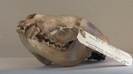
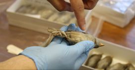
In this video we meet Dr Chris Stimpson, who uses our collections to identify small bones he has collected from ancient Vietnamese caves. His work helps us to understand the impact humans have had on various species of animal over thousands of years.
This article is taken from European research magazine Horizon as part of our partnership to share natural environment science stories with readers of More than a Dodo.
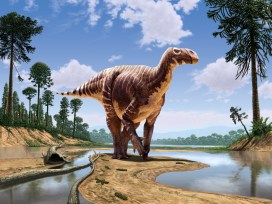
What colour were the dinosaurs? If you have a picture in your head, fresh studies suggest you may need to revise it. New fossil research also suggests that pigment-producing structures go beyond how the dinosaurs looked and may have played a fundamental role inside their bodies too.
The latest findings have also paved the way for a more accurate reconstruction of the internal anatomy of extinct animals, and insight into the origins of features such as feathers and flight.
Much of this stems from investigations into melanin, a pigment found in structures called melanosomes inside cells that gives external features including hair, feather, skin and eyes their colour – and which, it now turns out, is abundant inside animals’ bodies too.
‘We’ve found it in places where we didn’t think it existed,’ said Dr Maria McNamara, a palaeobiologist at University College Cork in Ireland. ‘We’ve found melanosomes in lungs, the heart, liver, spleen, connective tissues, kidneys… They’re pretty much everywhere.’
The discoveries in her team’s newest research, published in mid-August, were made using advanced microscopy and synchrotron X-ray techniques, which harness the energy of fast-moving electrons to help examine fossils in minute detail.
Using these, the researchers found that melanin was widespread in the internal organs of both modern and fossil amphibians, reptiles, birds and mammals – following up a finding they made last year that melanosomes in the body of existing and fossil frogs in fact vastly outnumbered those found externally.
What’s more, they were surprised to discover that the chemical make-up and shape of the melanosomes varied between organ types – thus opening up exciting opportunities to use them to map the soft tissues of ancient animals.
Secondary
These studies also have further implications. For one, the finding that melanosomes are so common inside animals’ bodies may overhaul our very understanding of melanin’s function, says Dr McNamara. ‘There’s the potential that melanin didn’t evolve for colour at all,’ she said. ‘That role may actually be secondary to much more important physiological functions.’
Her research indicates that it may have an important role in homeostasis, or regulation of the internal chemical and physical state of the body, and the balance of its metallic elements.
‘A big question now is does this apply to the first, most primitive vertebrates?’ said Dr McNamara. ‘Can we find fossil evidence of this? Which function of melanin is evolutionarily primitive – production of colour or homeostasis?’
Choosing colours for dinosaur reconstructions is a combination of evidence, modern references, and artistic guesswork. Image copyright: Julius Csotonyi
At the same time, the findings imply that we may need to review our understanding of the colours of ancient animals. That’s because fossil melanosomes previously assumed to represent external hues may in fact be from internal tissues, especially if the fossil has been disturbed over time.
Dr McNamara says her research has also shown that melanosomes can change shape and shrink over the course of millions of years, potentially affecting colour reconstructions.
Further complicating the picture is that animals contain additional non-melanin pigments such as carotenoids and what is known as structural colour, which was only recently identified in fossils. In 2016, a study by Dr McNamara’s team on the skin of a 10-million-year-old snake found that these could be preserved in certain mineralised remains.
‘These have the potential to preserve all aspects of the colour-producing gamut that vertebrates have,’ said Dr McNamara.
She hopes over time that these findings and techniques will together help us to much more accurately interpret the colours of ancient organisms – though in these early days, she doesn’t have examples of animals for which this has already changed.
We’re just at the tip of the iceberg when it comes to fossil colour research.
Dr Maria McNamara, University College Cork, Ireland
Deep time
Many of the significant strides in this area have come out of a project that Dr McNamara leads called ANICOLEVO, which set out to look into the evolution of colour in animals over deep time – or hundreds of millions of years.
The project’s starting point was that previous animal colour studies largely omitted in-depth fossil analysis, leaving a significant gap by basing what we know about colour mainly on modern organisms.
But it has since led to even wider investigation. Dr McNamara says it is providing fresh hints on the kinds of biological structures and processes that are essential for survival in terrestrial and aquatic environments. ‘It looks like we’ll be able to look into much broader, exciting questions about what it means to be an animal,’ she said.
Part of her research on two fossils found in China even showed that flying reptiles known as pterosaurs had feathers, potentially taking the evolution of these structures back a further 80 million years to 250 million years ago. The fossils contained preserved melanosomes with diverse shapes and sizes, one of the tell-tale signs of feathers.
Two fossils found in China showed that flying reptiles known as pterosaurs had feathers, indicating the structures evolved earlier than previously thought. Image credit – Zixiao Yang
‘We were able to show for the first time that not only were dinosaurs feathered, but an entirely different group of animals, the pterosaurs, also had feathers,’ said Dr McNamara.
Another project she worked on, called FOSSIL COLOUR, compared the chemistry of colour patterns between fossil and modern insects. Again, says Dr McNamara, these don’t entirely map onto each other.
‘It’s already clear that the fossilisation process has altered the chemistry somewhat, so we’re doing experiments to try to understand these changes.’
What’s evident is that there’s lots still to find out about colour. ‘We’re just at the tip of the iceberg when it comes to fossil colour research,’ said Dr McNamara.
Thermoregulation
Other researchers agree that there’s more to animal colour than meets the eye. Dr Matthew Shawkey, an evolutionary biologist at Ghent University in Belgium, said that looking into properties and functions beyond colour’s use for visual means like signalling and camouflage will be critical to understanding its true significance.
‘For example, how do colours affect thermoregulation? Flight? Such functions may be complementary to, or even more significant, than purely visual functions,’ he said.
Dr Shawkey is looking into such questions, with one of his recent studies indicating that the wing colour of birds may play an important role in flight efficiency by leading to different rates of heating.
‘What started as a novelty of deciphering dinosaur colours has turned into a very serious field which is studying the origins of key pigment systems, how the evolution of colourful structures may have helped drive major evolutionary transitions like the origin of flight, and how colour is related to ecology and sexual selection,’ said Dr Steve Brusatte, a vertebrate palaeontologist and evolutionary biologist at the University of Edinburgh, UK.
Ultimately, we may be able to find out more about colour than once thought possible. ‘When I was growing up, so many of the dinosaur books I read in school said that we would never know what colour they were,’ said Dr Brusatte. ‘But as is so often the case in science, it was silly to treat this as impossible.’
He said he is excited to see what comes next, with the field just in its infancy: ‘Palaeontologists now have a whole new window into understanding the biology and evolution of long-extinct organisms.’
Top image: Aline Dassel/Pixabay, licensed under Pixabay licence
The research in this article was funded by the EU. If you liked this article, please consider sharing it on social media.
Over the past few months our researchers have been working with University of Plymouth illustration student Abigail Harris, who has delved into the weird and wonderful world of some of the earliest animals. Here, Abigail tells us about the process that led to the creation of her Cambrian artwork, inspired by our First Animals exhibition.
I first visited the Museum in April this year when I was given the opportunity to collaborate with scientists as part of a module in my BA in at the University of Plymouth. Things kicked off with a short talk about the Ediacaran and Cambrian geological periods, when Earth’s first animal life started to appear.
I quickly narrowed my interest down to fossils from the Cambrian period which are more complex life forms, more similar to life today. A collection of small fossils from the Chengjiang fossil site in Yunnan province, China was the inspiration for some initial observational drawings.
A sketchbook page showing initial sketches and observations of OnychodictyonFinal illustration of Cotyledion
After returning to Plymouth University, I began to develop these initial sketches and observations, continuing to research the Chengjiang material and learning more about the characteristics of some of the creatures preserved as fossils.
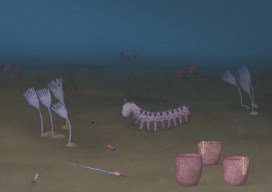
I wanted to create an under-the-sea ecology reconstruction showing a diversity of life forms, focusing on Onychodictyon, Cotyledion, Cricocosmia, Luolishania, and Paradiagoniella.
A five-step process was used for each reconstruction. Initially, I would sketch the fossil as I saw it, then I would research the characteristics and features of that animal, making a list of things to include in my drawing. A second drawing would then include all of these characteristics, not just what was initially visible in the fossil.
These rough sketches were then sent to the scientists for feedback, helping me to redraw and paint the illustrations with watercolour, before scanning and digitally editing each painting. Lastly, I created a background and added my illustrations.
Initial under under the sea ecology reconstruction.
Although the reconstructions were not completely finished by the time of my project deadline, I returned to the Museum in July and was given a tour of the First Animals exhibition by Deputy Head of Research Imran Rahman, as well as the opportunity to discuss how to improve my artworks for accuracy.
Another round of sketching and painting led to the final piece, shown at the start of this article, complete with an added digital background of the seafloor, and darkened to reflect the murky world of a Cambrian ocean, 50 metres below the surface.
The latest exhibition in our Contemporary Science and Society series, First Animals, tells the tale of Earth’s mysterious early animals, which evolved in the sea over half a billion years ago. Here, Dr Imran Rahman, Deputy Head of Research at the Museum, introduces some of the fossils that form a key part of this story.
From sponges to sea slugs and hagfish to humans, all animals alive today trace their roots back to a common ancestor that lived in the ocean more than 600 million years ago. We have no direct evidence of this first animal, but the fossil record reveals some of its earliest descendants. Our First Animals exhibition explores the evidence for Earth’s earliest animal life, attempting to answer the ‘what’, ‘when’, ‘how’ and ‘why’ of the origin of animals.
Yunnanozoon lividum from the Chengjiang fossil site had a long body with several filament-covered arches at the front and a fin-shaped structure towards the back. It cannot be confidently assigned to any known animal group.
First Animals features the oldest animals yet recovered from the fossil record, including specimens from 571-million-year-old rocks in Newfoundland, Canada. These represent the remains of originally entirely soft-bodied organisms, which have proven difficult to classify because they look so different to living species. However, new research on their anatomy and how they grew, including work by Museum researcher Dr Frankie Dunn, suggests they were early animals.
Charnia masoni consisted of alternating branches arranged along a frond. It is thought to be one of the oldest animal fossils yet found.
Microscopic fossils record the first animal skeletons, which first appeared about 550 million years ago. These include the remains of complete animals, as well as fragments such as spines and scales. Work by Museum researcher Dr Duncan Murdock using a particle accelerator to generate X-ray images of these tiny fossils has allowed us to reconstruct how the skeletons changed as they grew. This helps to establish the modern groups to which these ancient animals belonged, and unravels the mystery of why animals evolved hard skeletons when they did.
Virtual cross-sections through small shelly fossils created using X-ray imaging.
The most complete evidence for the early evolution of animals comes from sites of exceptionally-preserved fossils, or Lagerstätten, which retain impressions of soft tissue as well as hard parts, and include rare soft-bodied animals like worms and jellyfish.
First Animals brings together extraordinary specimens from three key fossil sites: Sirius Passet in northern Greenland (518 million years old), Chengjiang in Yunnan province, China (518 million years old) and Burgess Shale in British Columbia, Canada (508 million years old). This includes 55 unique fossils loaned by Yunnan University in China, as well as specimens from the University of Bristol and the Royal Ontario Museum.
The mollusc Halkieria evangelista from the Sirius Passet fossil site had a long body covered in hundreds of overlapping hard plates, with a large shell plate at either end.The arthropod Haikoucaris ercaiensis from the Chengjiang fossil site had a semicircular head shield with a pair of large grasping appendages, a segmented body and a short tail.The worm Ottoia prolifica from the Burgess Shale fossil site had a spiny proboscis and a long trunk that was divided into a series of fine rings.
These exceptionally-preserved fossils reveal the evolutionary diversification of life during the so-called ‘Cambrian explosion’. Through careful study of the fossils, scientists have begun to reconstruct the very first animal ecosystems, which are brought to life in the exhibition through a series of stunning digital reconstructions and the Cambrian Diver interactive installation. This allows visitors to explore a 360-degree oceanic environment in a virtual submersible craft, coming face-to-face with some of the first animals on Earth!
Digital reconstruction of the sea floor 518 million years ago, based on specimens from the Chengjiang fossil site, Yunnan province, China.
Video by Mighty Fossils.
Doctoral researcher Elaine Charwat is exploring the value and meaning of models and casts in the Museum’s collections as part of her PhD. She has recently been studying some fabulous models that help to visualise and understand some of the very, very smallest of specimens…
By Elaine Charwat
The first time I encountered a Radiolarian was in a book – Ernst Haeckel’s (1834-1919) weird and wonderful Kunstformen der Natur (Art Forms in Nature, 1899-1904). It took comparative morphology – comparing the shapes of organisms – to new giddy heights, scientifically, philosophically and artistically. I felt that giddiness when looking at page after page crammed with crustaceans, orchids, hummingbirds, moths and even bat faces, all exquisitely arranged to celebrate their symmetries, the evolution and kinship of their shapes and forms. It also made visible organisms that are normally all but invisible.
Illustration of Cyrtoidea (table 31) from Kunsterformen der Natur (1899 – 1904) by Ernst Haeckel. By permission of the Linnean Society of London.
Foraminifera and Radiolarians are microscopic sea-dwelling organisms. Species may be found as fossils dating from Cambrian times, ca. 500 million years ago, right up to living specimens today.
To Haeckel, they were living proof of Darwin’s theory of evolution, and for his own belief that morphology was the key to understand the actual processes of evolution, catching it in the act. However, these organisms had two big disadvantages – their unwieldy taxonomy, or the way they are classified, and their minute size: they were difficult to examine and display.
Illustrations of Radiolarians, (table 28). from Die Radiolarien (1862) by Ernst Haeckel. By permission of the Linnean Society of London.
Through his illustrations, Haeckel widely popularized them – triggering a Victorian craze for microscopes and microorganisms, as well as influencing art nouveau art and architecture. But there were limits to what an illustration could communicate. Models stepped in, representing these organisms in ways illustrations could not.
Detail from Haeckel’s Kunstformen der Natur (1899 – 1904)
One defining feature of Radiolarians and Foraminifera is their shells – called “tests”. Variations in shapes of the tests not only indicate that they are different species, but also, excitingly, provide clues about space and time. The tests of Neogloboquadrina pachyderma, for instance, record ocean temperature over geological timescales – their shells coil to the left when water temperatures are relatively cold, and to the right when it is warmer. The potential for research into climate change is obvious. Foraminifera are also important “signature fossils”, helping geologists to determine geological strata.
You really need to see them in glorious 3D to appreciate these tests across geological time, to understand their complex, beautiful shapes. And I felt a similar twang of excitement to my first encounter with them through Haeckel when discovering these extraordinary models here in the Museum as part of my PhD research.
Václav Frič (1839-1916) was a natural history dealer based in Prague. He developed a series of 100 plaster of Paris models of Foraminifera (1861), as well as the stunning papier-maché models of Radiolaria (listed in his catalogue of 1878). He worked closely with Ernst Haeckel.
A selection of Frič’s models in the Museum’s stores
The Frič models oscillate between visible and invisible, illustration and model, art and science, philosophy and theory. They bear witness to a key period in the history of science when they were used to give tangible shape and proof to Charles Darwin’s poignant phrase: “[…] from so simple a beginning endless forms most beautiful and most wonderful have been, and are being, evolved.”
Through the models we can “grasp” microorganisms that have been around for over 500 million years; organisms that truly have stood the tests of time.