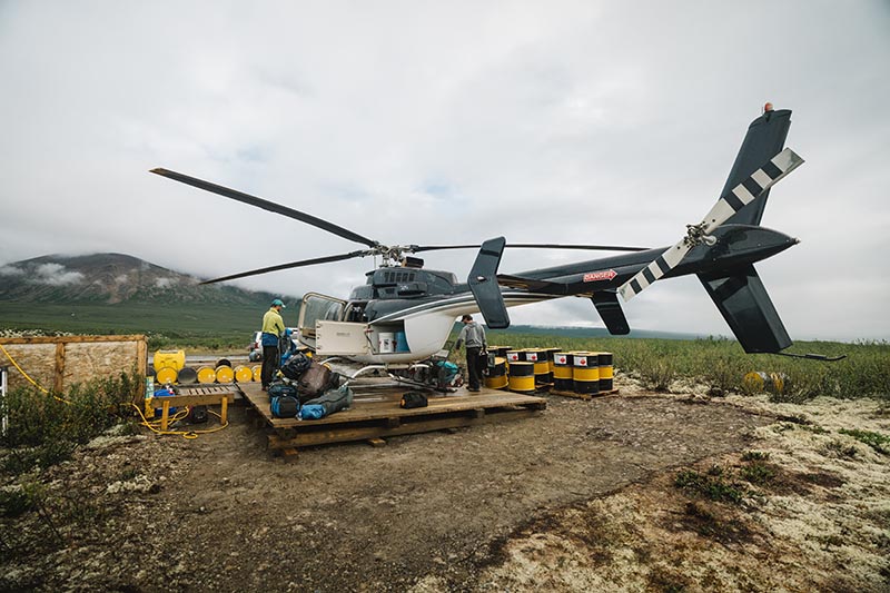
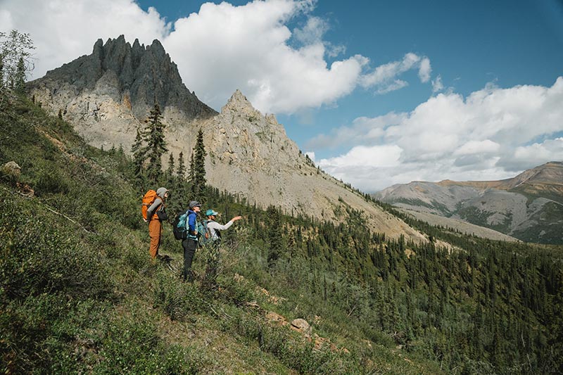
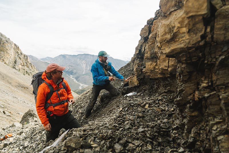
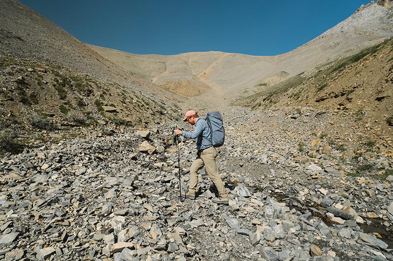
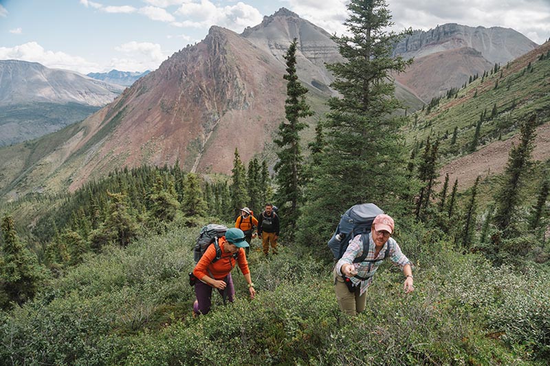
In Summer 2024, a team of palaeontologists and geologists from the University of Oxford, along with colleagues from Dartmouth College, the University of Washington, and Williams College in the USA, undertook an expedition to the Little Dal Group in the Mackenzie Mountains, Northwest Territories, Canada. Our purpose was to uncover some of the oldest fossil ecosystems that record complex life.
Photo: Robert GillPhoto: Robert Gill
Complex life comprises all organisms whose DNA is enclosed in a cell nucleus. This includes animals and plants but excludes bacteria. Today, this complex life accounts for most of the Earth’s biomass, documented biodiversity, and oxygen production. Understanding when and how it first evolved remains one of the central unanswered questions in evolutionary biology.
Photo: Robert GillPhoto: Robert Gill
As palaeontologists, we normally use fossils to reveal the history of life. Fossils tend to preserve larger animals with hard shells or skeletons—creatures such as trilobites, ammonites, dinosaurs, and mammoths. However, the first complex organisms were microscopic and lacked such hard parts. As a result, their soft and fragile cells rarely fossilised. Put simply, we have found it a major challenge to trace the origins of complex life with fossils.
Photo: Robert GillPhoto: Robert Gill
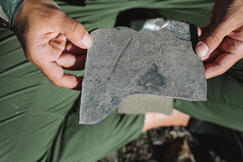
I have argued that finding rocks made up of antibacterial clay minerals holds the key. These minerals can slow the decay of organic cells long enough for them to survive as fossils. The Little Dal Group contains ~900-million-year-old rocks that are rich in just such clays, making it a prime target for new fossils that might help us unravel the origins of biological complexity.
Photo: Robert GillPhoto: Robert Gill
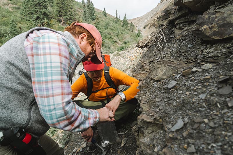
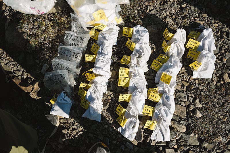
I was joined in Canada by my DPhil student, George Wedlake, from the Department of Earth Sciences. Together we spent two weeks collecting over 100 rock samples. The samples record an ancient tropical sea not unlike the Bahamas today, where early complex life likely flourished.
Back in Oxford, at the Museum of Natural History, George and I are now examining the samples; dissolving the rocks with hydrofluoric acid to extract and study the tiny fossils. We hope these new fossils will transform our understanding of how complex life first took hold on our planet.
Our fieldwork was funded by a Royal Society University Research Fellowship and by the Oxford NERC Environmental Science Doctoral Training Partnership. It was conducted under permit and with the support of the Sahtú Dene people.
Dr Caroline Wood, from the Public Affairs Directorate at Oxford University, takes us behind the scenes to uncover one of the most exciting dinosaur trackways in the world.
The information in a single footprint
The air pulses with seismic activity and under our feet deep vibrations race across the ground. Every so often, a shattering rumble rips out across the surroundings.
Dr Emma Nicholls, a vertebrate palaeontologist at Oxford University Museum of Natural History (OUMNH), has to shout to make her voice heard ‘…they are huge and they can’t see you. Remember, do not leave the designated safety area under any circumstances!’
We all nod diligently, assuring her we have understood. Looking down at the immense footprints a few metres away, I try to imagine how painful it would be to be squashed by the foot of a ten tonne sauropod dinosaur. I’m pretty sure my hard hat wouldn’t be very effective protection… however, it isn’t dinosaurs that are rumbling and thundering all around us today, but thoroughly 21st-century quarry vehicles.
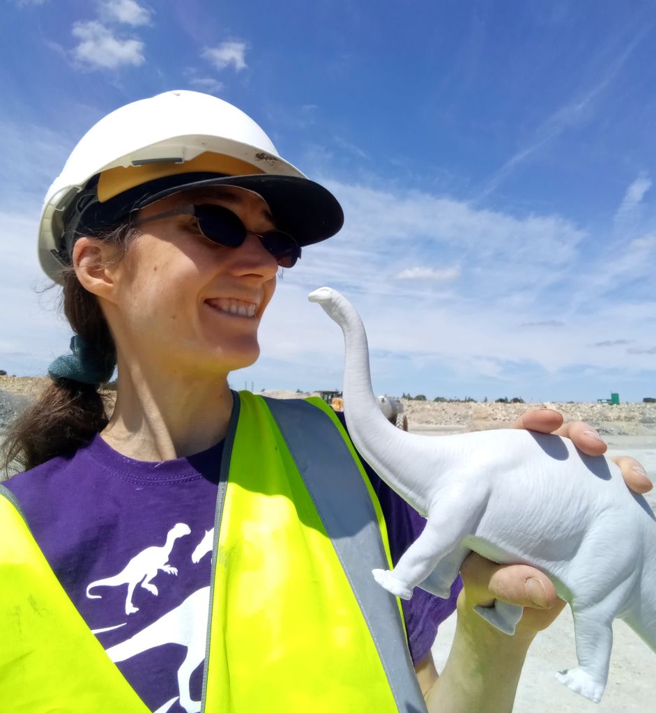
Author Dr Caroline Wood, at the dig site at Dewars Farm Quarry in North Oxfordshire
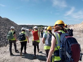
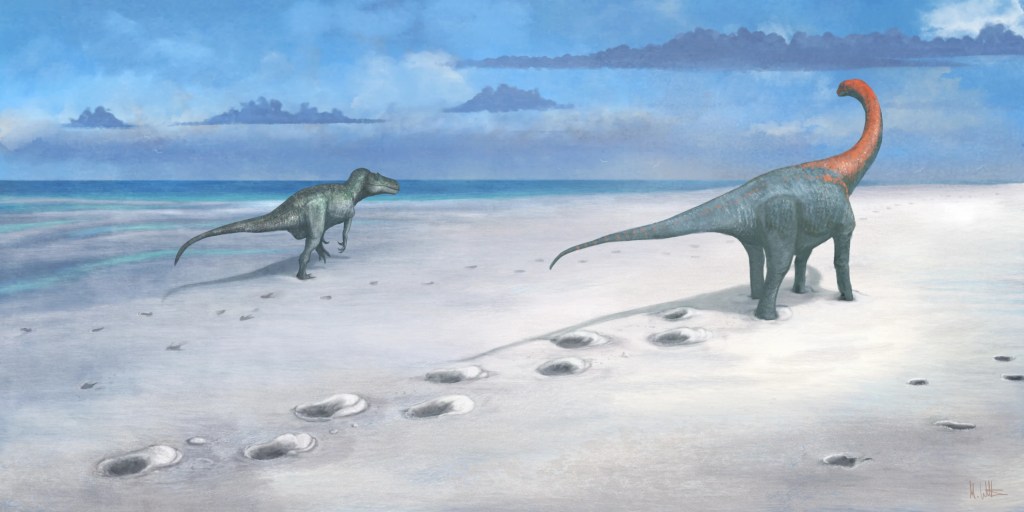
On a scorching hot summer’s day, I’ve come to help uncover a newly-discovered section of one of the longest dinosaur trackways in the world, here in North Oxfordshire. Whilst stripping back clay from the ground with his vehicle, quarry worker Gary Johnson stumbled upon a series of exquisitely preserved dinosaur footprints. The OUMNH team were called, and they soon made a visit to the site with two colleagues from the University of Birmingham. What they found was something really special; tracks from not just one type of dinosaur but at least two: a herbivorous sauropod (thought to be Cetiosaurus) and the terrifyingly-armed carnivore Megalosaurus, both hailing from the Middle Jurassic, approximately 166 million years ago.
This week, Smiths Bletchington have given site access to a team of researchers, students and staff from the Universities of Oxford and Birmingham. Our task today is to uncover the prints as much as possible, capture digital records, then create computer models to enable researchers across the world to study them further. As someone who has been obsessed with dinosaurs practically from birth (my first toy was a Triceratops), it feels like all my Christmases have come at once.
‘It’s amazing how much information you can get from a single footprint’
It is not the first time that footprints from these dinosaurs have been found in the area. In 1997, tracks from the same two types of dinosaur were unearthed at Ardley Quarry and Landfill Site in Oxfordshire, and can now be seen in the ‘Dinosaur garden’ at the Oxfordshire Museum in Woodstock. But the newly-unearthed footprints, which link up with the original, make it by far the largest and most significant dinosaur track site in the UK.
‘Some people may feel they’re not as visually dramatic as fossilised skeletons, but dinosaur footprints are incredibly useful resources for palaeontologists,’ Emma says. ‘They can give us a wealth of information about how these animals moved and travelled. In addition, footprints and other trace fossils can also give direct evidence of the environment within which the organism existed.’
‘It is possible that this huge Jurassic predator was tracking the sauropod to hunt.’
Dr Duncan Murdock
She points to the nearest Cetiosaurus tracks. ‘Each of the sauropod footprints has a distinct, raised ridge at the front. This indicates that the animal was walking in soft, wet sediment, but that it wasn’t so water-logged that the footprint collapsed. When the animal put its foot down, its weight caused the mud to splosh up in front, which has been preserved in situ.’
Dr Duncan Murdock, who is co-leading the excavation with Emma, as well as colleagues Professor Richard Butler and Professor Kirsty Edgar from the University of Birmingham, adds: ‘The climate here in the Middle Jurassic would have been warm and tropical, and the environment essentially a large, muddy lagoon.’ The sediment kicked up at the front of the prints was also the reason that the buried prints came to light in the first place, when quarry worker Gary Johnson felt the huge bumps as he worked to clear the mud with his vehicle.
Dinosaur footprints can also offer valuable clues into how different animals interacted, particularly when their tracks are found together, as they are here. ‘Here, we have trackways from at least four sauropods and one Megalosaurus,’ Duncan says. ‘Interestingly, the sauropods are a mixture of different sizes, so it is possibly a herd with juveniles or perhaps there are more than one type of sauropod represented here.’
At one point, the tracks intersect- which poses an interesting question for the research team – which dinosaur came first?
‘It looks as though the back of the Megalosaurus footprint has squished a section of the bump at the front of the Cetiosaurus print, meaning the carnivore came second,’ says Duncan. ‘Although inconclusive, it is possible that this huge Jurassic predator was tracking the sauropod to hunt.’
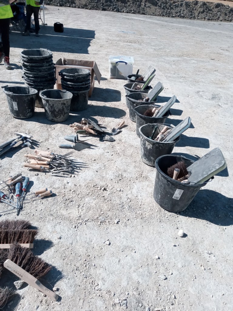
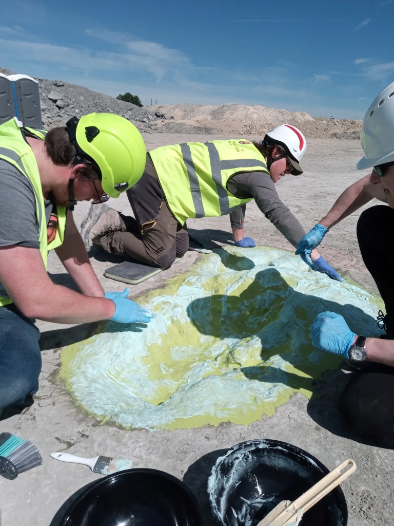
With most of the prints only partially excavated, it’s time I made myself useful. Fortunately, my lack of experience isn’t an issue; instead of high-tech specialist equipment, I am handed a bucket of supplies that could all be sourced from a hardware store. I don gloves and set to work on a sauropod print with a brush, sweeping out dust and loose stones. Besides being a good workout, it is a highly multisensory experience as I look, feel and ‘hear’ my way around the giant print. I am taught how to ‘listen’ for the edge of the print by tapping my shovel gently: the fossilised print gives a sharp, metallic ching whilst the surrounding mud makes a dull thump sound.
Excavation equipment at the dig site. Credit: Caroline Wood.
Slowly, under my hands, the full outline of the 90 cm long print is liberated. It amuses me to think how the enormous creature that stomped this way 166 million years ago would have been oblivious that, one day, a diminutive biped mammal would be sweeping out its footprints with assiduous, almost loving, attention.
I’m not the only one getting goose bumps. Emily Howard, a (second year going into third year) Earth Sciences undergraduate student at Oxford University is working on the footprint next to mine. ‘I feel really lucky to be doing this – there is no analogue for dinosaurs,’ she says. ‘When we have lessons in class, it often feels as though everything has already been found and documented… so to be involved with a new discovery and to play a part in the process of uncovering it is very special.’
‘To me, dinosaur trackways are much more “alive” than fossilised bones, which can only be from dead animals. Similar to when you see human footprints on a path ahead of you, a dinosaur track gives the impression that the creature could be miles away in the direction the tracks march on, but was here only a moment ago.’
Emily Howard
Capturing all the details
Nearby, one of the prints is undergoing more specialised treatment. Juliet Hay, a conservator in palaeontology at OUMNH, is massaging what looks like viscous turquoise toothpaste into the centre of a print. In the intense midday heat (which helps the materials work more quickly than on a cold wet day), the various layers that make the cast will soon bind together and solidify to create a mould that can be peeled off like a beauty mask.
‘Using the mould, we will be able to make 3D casts of the prints from various different materials, both for research and public engagement.’
Juliet Hay
Creating a mould of one of the Megalosaurus footprints. Credit: Caroline Wood.A volunteer takes photographs of one of the Megalosaurus prints. Credit: Caroline Wood.
With so many prints to uncover, staff from all across OUMNH, as well as staff and students from the Universities of Oxford and Birmingham, have come to lend a hand, besides the collections team. ‘All the staff across the museum are excited,’ says Molly Appleby, Visitor Services Assistant at OUMNH. ‘The dinosaurs are such an iconic feature of our exhibits, so it is wonderful that we have all had the opportunity to be involved in this new discovery. This certainly makes a change to my day job!’
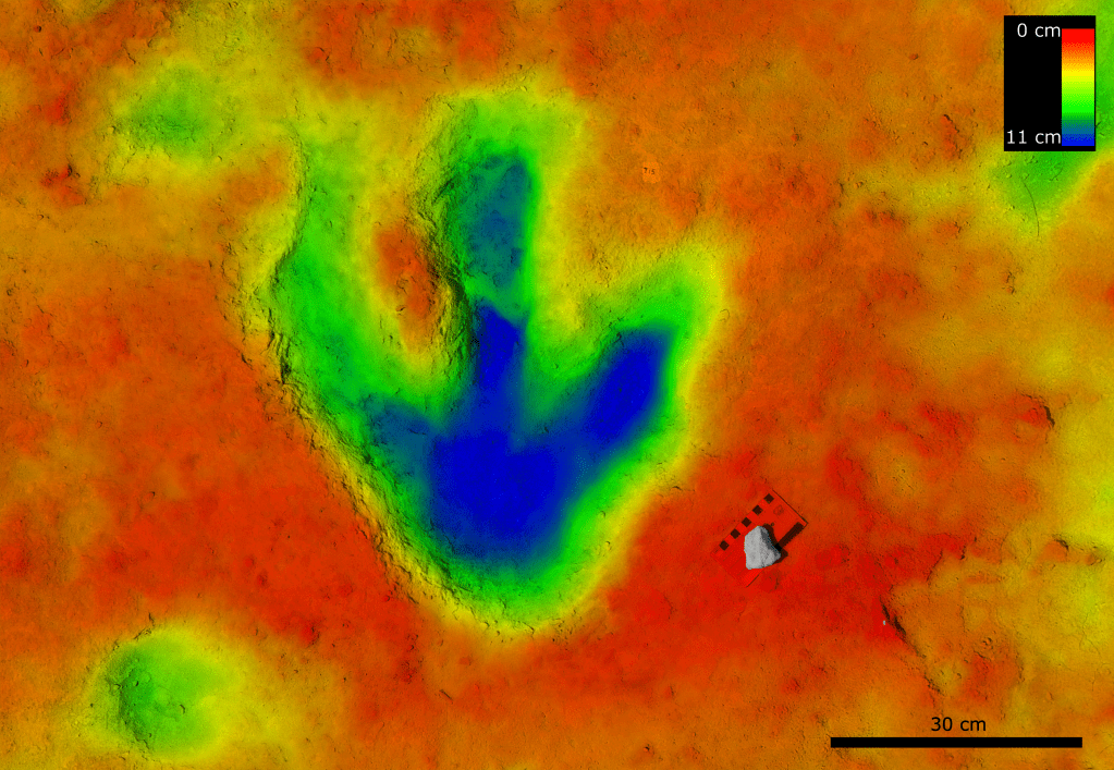
One of the Megalosaurus footprints coloured by depth. Credit: Dr Luke Meade, University of Birmingham.
The team’s aim goes beyond making physical models. A key outcome is to digitally record the prints so that computer software can reconstruct 3D virtual models, that can be used by researchers across the world.
‘Using photogrammetry and computer models, we will be able to work out details such as the height of the animals and their speed,’ Duncan says. ‘On the largest sauropod’s track, one of the prints is slightly out of sequence – almost as though the animal stopped and looked back over its shoulder. Hopefully, the computer models will help solve that mystery.’
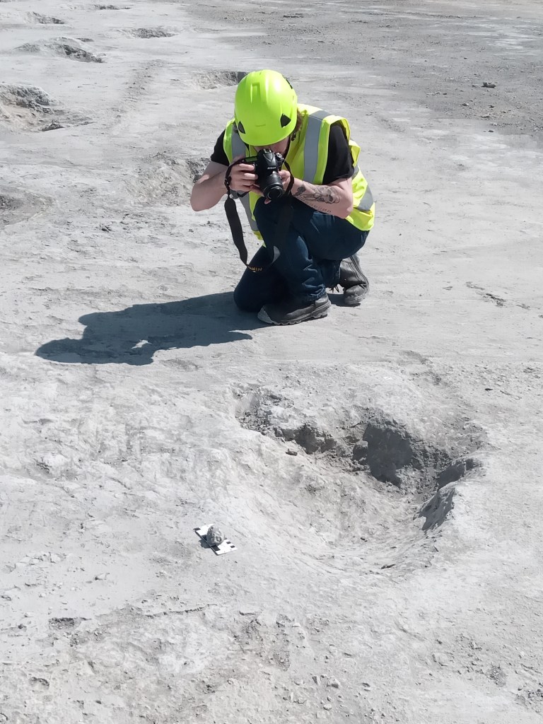
To do this, you need data – and lots of it. I join some of the students who are busy taking close-up photographs of each footprint from as many different angles as possible. Once again, the equipment is straightforward: a standard DSLR camera. In theory, one student tells me, you could even use a mobile phone.
The photographs will be fed into computer software that will identify points of similarity and use trigonometry to reconstruct a 3D model of the print. For each print, between 60 and 100 photos will be taken. I’m told that more photographs are needed for the sauropod prints: being simpler shapes, it’s more taxing for the model to identify reference points.
‘This never ceases to be exciting’
‘Team- breaktime!’ As the sun reaches its noonday zenith, we convene under the OUMNH gazebos to escape into the shade. We refuel and reapply sunscreen, swapping stories of childhood dinosaur addictions and favourite scenes from Jurassic Park. For Emma though, the real-life science of dinosaurs will always trump fictional parodies.
‘Duncan and I have been working with Mark Stanway and the Smiths Bletchington team at the Quarry for nearly two years now, and it never ceases to be exciting,’ she says. ‘Excavating a brand-new Megalosaurus trackway in the 200th anniversary year of the discovery of Megalosaurus – the first dinosaur to be scientifically named and described anywhere in the world – is very special indeed.’
As my eyes are drawn along the length of the largest sauropod trackway, over 150 metres long in total, I realise that the huge footprints disappear under the cliff at the edge of the quarry. There are undoubtedly more tracks to be discovered…who knows what will be found in the future?
The area is still a working quarry with no public access, and will remain so in the medium term. However, Emma, Duncan, Richard and Kirsty are actively working with Smiths Bletchington and Natural England on options for preserving the site for the future.
You can learn more about the discovery and see the original Megalosaurus fossils on display at the Oxford University Museum of Natural History’s Breaking Ground exhibition.
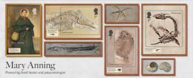
Dr Emma Nicholls and colleagues discuss some of the fascinating stories behind the species and specimens featured on these stamps.
Megalosaurus stamps
Two of the stamps in the Age of Dinosaurs stamp set include artistic reconstructions of Megalosaurus by the palaeo-artist Joshua Dunlop. The animal that nineteenth-century naturalists once understood to be a lumbering long-legged lizard is now depicted as a fearsome Jurassic predator that ran on its hind legs and tore into prey with its large serrated teeth. Dunlop shows Megalosaurus wading through shallow coastal waters, preparing to pounce on Cryptoclidus¸ a plesiosaur that lived alongside Megalosaurus in Jurassic Britain. The artwork also shows Megalosaurus covered in feathers. Although we don’t have any direct evidence that Megalosaurus was a feathered dinosaur, feather-like filaments have been found among the fossils of other dinosaurs such as Sciurumimus, meaning it is highly possible that Megalosaurus had feathers too.
OUMNH’s famous Megalosaurus type specimen, a lower-right jawbone with teethMegalosaurus stamp from the Age of Dinosaurs series
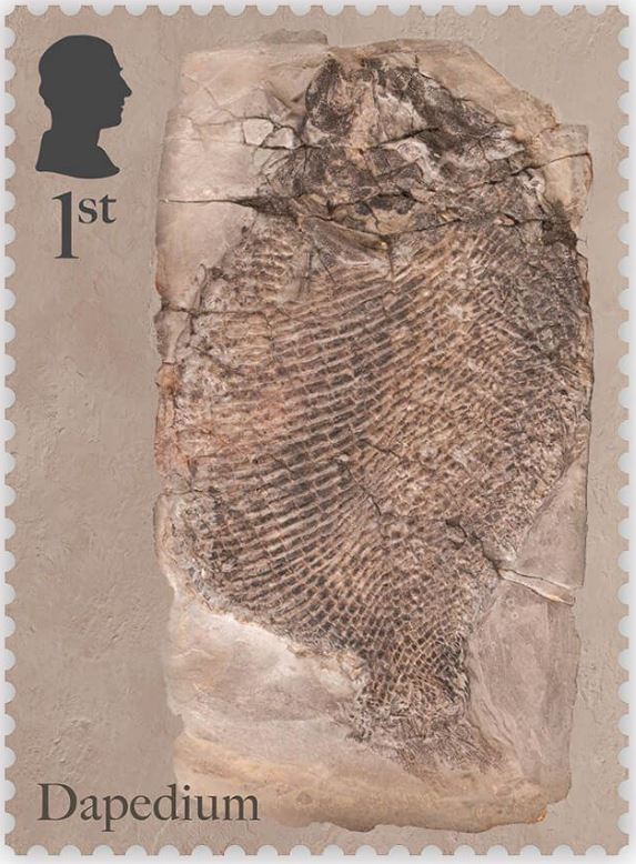
Dapediumstamps
OUMNH collaborated directly with Royal Mail to help produce the Age of Dinosaurs miniature sheet, which showcases fossils collected by Mary Anning. One of the stamps in this collection features a photograph of the fossil of an extinct Jurassic fish, Dapedium, which is housed at OUMNH.
Despite Anning’s illustrious reputation, it wasn’t always known that this Dapedium specimen was connected to her — all that was known about it was that it had probably once belonged to William Buckland and had been collected from Lyme Regis.
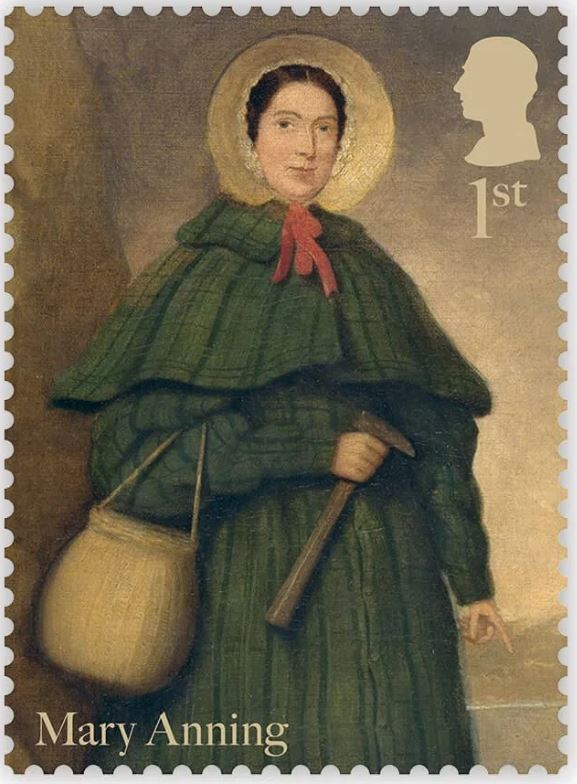
Stamp from the Age of Dinosaurs series featuring a portrait of Mary AnningStamp from the Age of Dinosaurs series featuring a Dapedium fossil from OUMNH
Although Anning is one of the most prolific fossil collectors to have worked in Lyme Regis, naturalists like Buckland often visited Anning to go “fossicking” together, or purchase fossils from her. There are very few archival records of transactions between Anning and other fossil collectors from this time, making it difficult to decipher exactly who extracted fossils such as this, found in nineteenth-century Dorset.
Fortuitously, while Dr Sue Newell was conducting research for her PhD on the Buckland Collection in 2021, she found an exciting letter in OUMNH Archive, dated 3rd September 1829. It was from a former student of Buckland’s, Beriah Botfield, and contained details of two fossils that Buckland had bought from Anning to present to the University of Oxford. Using evidence in the letter, Sue was able to work out that Botfield was referring to a Dapedium fossil which she later recognised tucked away in OUMNH’s fossil store.
Botfield had had the fossil mounted in an expensive (and very heavy!) stone frame, with “Presented by Beriah Botfield Esq. Dapedium politum. Lyme, Dorset” beautifully inscribed on the front surface. At the time, the identity of Anning as the fossil’s original finder, identifier, preparator and vendor, was probably common knowledge and, typically, Botfield did not consider these facts important enough to record on his presentation frame.
The Dapedium fossil is a near-complete example of this Jurassic fish, in which scale patterns and delicate fin structures are preserved in breathtaking detail. Dapedium is the first OUMNH object to grace a Royal Mail stamp – an ideal choice given its scientific and historic importance.
Visit the Museum to see the Dapedium fossil as well as temporary displays about the new stamp collection.
Susan Newell is a doctoral student researching the teaching collections of William Buckland, the first Professor of Geology at Oxford who taught from 1813 to 1849. She reminds us here about Buckland’s role 200 years ago in interpreting the important Pleistocene discoveries being celebrated this year, and the way that Mary Morland, a talented local naturalist, and many others, contributed to making this new knowledge.
This year marks the 200th anniversary of a great advance in our understanding of the geological past… a story which begins in the nineteenth century, with the discovery of a bone-filled cave in Kirkdale, Yorkshire.
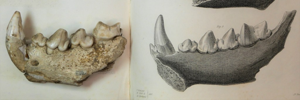
Uncovered by local quarrymen in 1821, the discovery of the Kirkdale cave and its contents of mysterious bone was the source of much intrigue. When news of the discovery reached William Buckland, Professor of Geology at Oxford University, he decided to travel up North to visit the site. However, by the time Buckland arrived at the cave, local collectors had scooped up most of its contents. Nonetheless, he was able to retrieve and examine some of the cave’s remaining material, which led him to an astonishing conclusion — Yorkshire must once have been home to hyaenas, elephants, hippopotamus and rhinoceros, and what was now known as the Kirkdale cave was once a hyaenas’ den.
W. B. Conybeare, lithograph, ‘The Hyaena’s Den at Kirkdale near Kirby Moorside in Yorkshire, discovered A.D. 1821’. Reproduced by kind permission of Christ Church, Oxford. This light-hearted reconstruction of the hyaenas’ den shows Buckland illuminating the scene, in every sense. It is thought to be the first visual reconstruction of the pre-human past.
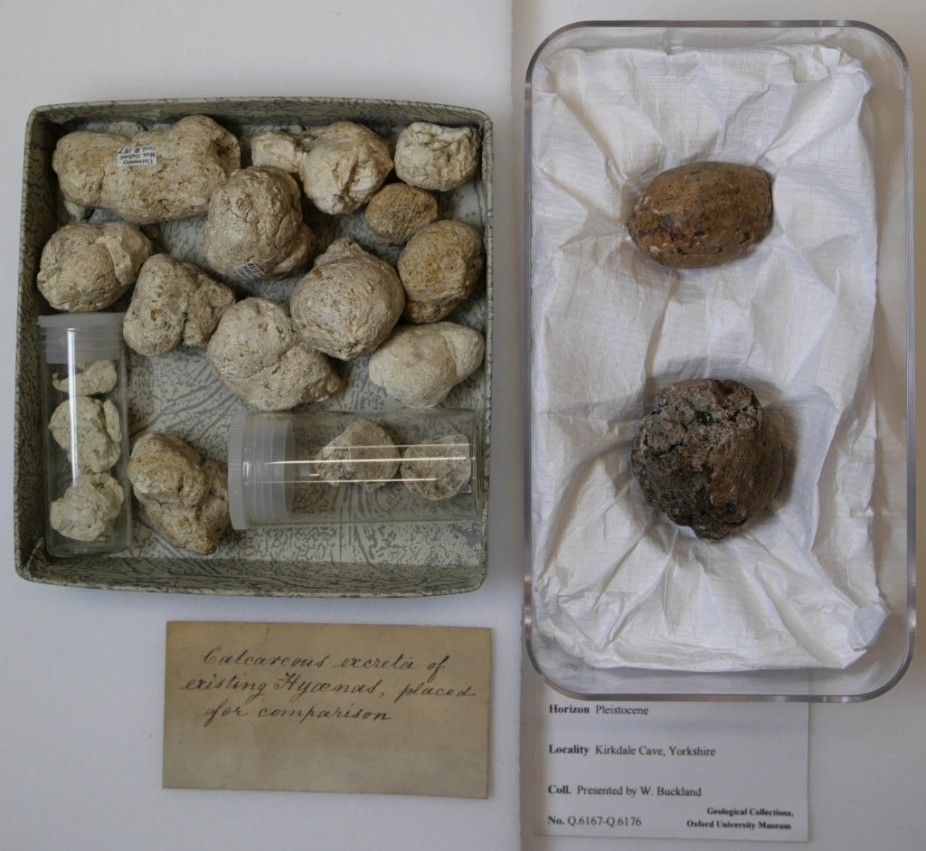
Central to Buckland’s theories were some small white balls that he had found amongst the debris in the cave. Buckland sent these balls to William Wollaston, a celebrated chemist based in London, for analysis. He also asked Wollaston to visit the zoo at Exeter Exchange in London and show the balls to the hyaena’s keeper there. Together with the results from Wollaston’s chemical analyses, the keeper confirmed Buckland’s hypothesis — the balls were droppings from animals very similar to modern hyaenas. Meanwhile, the anatomist William Clift was able to identify the bones from the Kirkdale cave as belonging to other extinct species related to those found living in tropical countries today. Buckland concluded that the cave must have been a den for ancient hyaenas, who would drag parts of the dead animals they had found (or killed) inside and, after feeding on them, leave piles of bones and droppings behind.
In order to strengthen his theory, Buckland discussed the behaviour of hyaenas in the wild with army officers connected to Britain’s colonial expansion in India. These officers also sent Buckland fresh specimens captured by local people. When a travelling menagerie visited Oxford in 1822, Buckland took the opportunity to experiment; feeding bones to a hyaena and noting that the teeth marks matched those on the fossilised bones from the cave.
Left: “Small white balls” of hyaena excrement. The left box probably contains samples collected from the hyaena in the travelling menagerie which visited Oxford, and the right contains fossilized samples collected by Buckland from Kirkdale Cave, now part of the Museum’s Collections. Right: Comparison of two shinbones – one fed to a hyaena by Buckland, and the other collected from Kirkdale cave. The similarity of the gnaw-marks suggest that hyaenas were once present in Kirkdale. On display in the Museum.
Buckland’s findings were something of a shock to his contemporaries. When lecturing, he employed several different methods to try and convince his audiences that his theories were true. This included presenting fossil specimens and bones from living species for comparison, and showing maps, diagrams and drawings. Mary Morland contributed some of these illustrations, including large drawings of living animals, and technical drawings of bones that were later engraved for use in Buckland’s publications. Mary’s Kirkdale drawings seem to have been the first that she produced for William before the couple married in 1825.
Fossil hyaena jaw in the Museum’s collection, possibly the one featured in the engraving alongside it. Engraving is by James Basire after a drawing by Mary Morland. Published in William Buckland’s article in the Royal Society’s journal (1822) on the Kirkdale cave discoveries. [1]
Buckland’s work on the Kirkdale cave was revolutionary, not least because he was the first to make a scientific study of a cache of bones of this type. Although similar bones from ‘tropical’ species had previously been found in Northern Europe, people thought that they had been washed up by a catastrophic flood, believed by many to be the biblical Noah’s Flood. Modern analysis has now allowed us to deduce that the bones date to an Interglacial period when Britain was joined to Europe and had a hot climate, about 120,000 years ago.
Here at the Museum, Buckland’s collections and archives are as much of a treasure trove as the Kirkdale cave. It is through accessing these archives that we can learn about the surprising range of people who contributed to the emergence of new scientific knowledge from the Kirkland cave — quarrymen, collectors, zookeepers, chemists, anatomists, colonial officers in India, workers in India, and artists like Mary Morland. To find out more about the incredible legacy of the Kirkdale Cave, look out for ‘Kirkdale200 – Lost Beasts of the North’, a symposium organised by the Yorkshire Fossil Festival, 12th March 2022.
Mary Morland, watercolour and gouache, lecture illustration of a hippopotamus, signed ‘MM’. Hippopotamus bones were found at Kirkdale cave in Yorkshire, but as there were no living hippos to be seen in Britain at the time, this drawing would have been a valuable teaching aid.
[1] William Buckland, ‘Account of an Assemblage of Fossil Teeth and Bones of Elephant, Rhinoceros, Hippopotamus, Bear, Tiger, and Hyaena, and Sixteen Other Animals; Discovered in a Cave at Kirkdale, Yorkshire, in the Year 1821: With a Comparative View of Five Similar Caverns in Various Parts of England, and others on the Continent’, Phil. Trans., 2 (1815-30), 165-167.
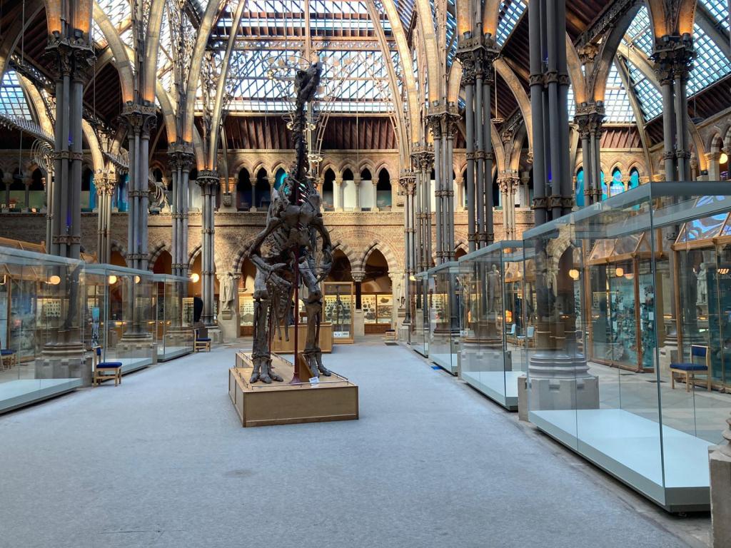
As one of our many invaluable volunteers, Leonie Biggenden has regularly helped to run our Science Saturdays and Family Friendly Sunday activities, both of which take place under the watchful eyes of the large T. rex and Iguanodon skeletons in the Museum’s main court. Having spent so much time beside the Iguanodon, and with a lack of in-person volunteering opportunities in recent months, Leonie decided to find out some of the history of this striking cast. For Volunteers Week this week, she shares what she discovered…
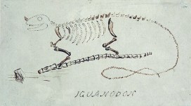
Next year will be the 200th anniversary of the discovery, by a roadside in Sussex, of the first Iguanodon teeth. Found by Mary Mantell in 1822, her husband Gideon saw their similarity with the teeth of modern iguanas and suggested they were from a huge, ancient, herbivorous lizard. He called the animal Iguanodon, and you can see his sketch reconstruction at the top of this post.
However, as an amateur palaeontologist, Gideon Mantell was not initially taken seriously by the scientific establishment. Some claimed the teeth were actually from a rhinoceros, or even a pufferfish! But in 1834, more complete remains were found by workmen who had accidentally blown up a slab of rock in a quarry near Maidstone, Kent. Iguanodon became a rock star of the dinosaur world, being only the second dinosaur – and the first herbivorous one – to be named (the first was the carnivorous Megalosaurus – another famous Museum specimen).
The Iguanodon bernissartensiscast in the centre court of the Museum.
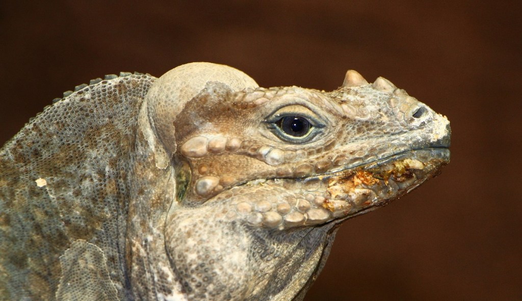
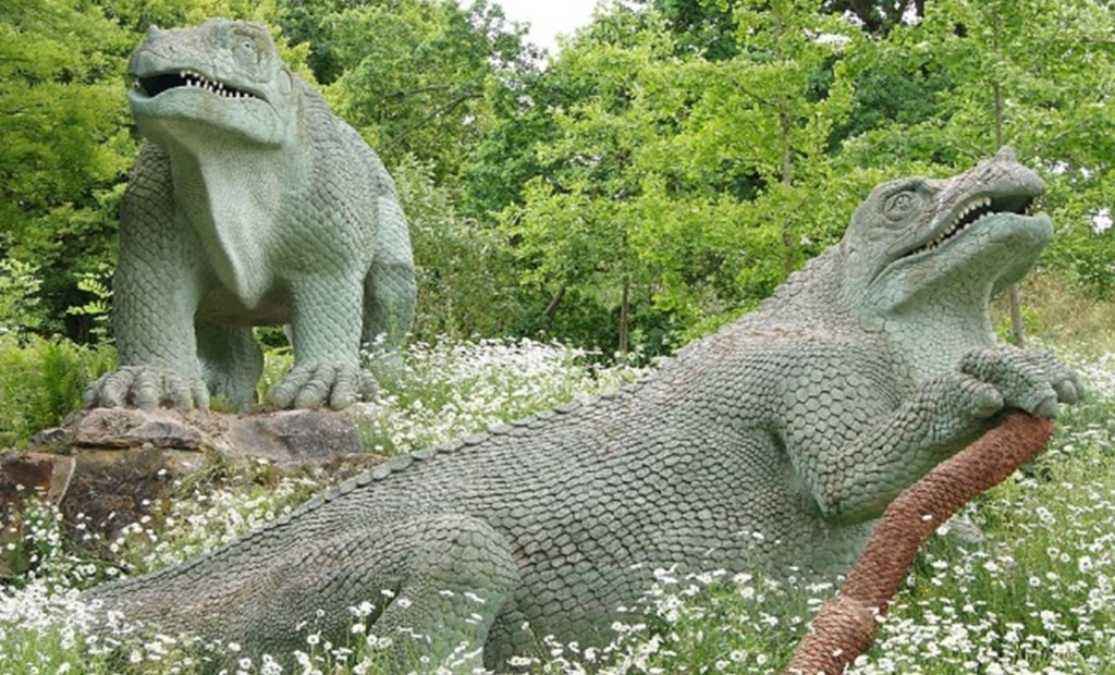
Twenty years later, a model of an Iguanodon was constructed by sculptor Benjamin Waterhouse Hawkins as one of a set of 30 life-sized models of extinct animals for the relocated Crystal Palace Gardens in South London. It was mounted in a rhinoceros-like pose, with what we now know as a thumb spike placed as a nose horn. Scientists always look to the information they have available to them, including observation of living animals, and there is an iguana called Cyclura cornuta – the Rhinoceros Iguana – which does indeed have nose horns, so at the time the nose horn made sense.
Rhinoceros Iguana, showing a nose horn. Image: H. Zell, CC BY-SA 3.0 , via Wikimedia Commons
Another 20 years on and a most significant find was made in southern Belgium. In February 1878, more than 30 fully articulated, adult Iguanodon fossil skeletons were found by miners Jules Créteur and Alphonse Blanchard, 322 m deep in the Sainte Barbe coal mine. Louis de Pauw from the Belgian Royal Museum of Natural History started to excavate the skeletons. It was a risky undertaking. In August an earthquake cut them off for two hours, and in October they were forced to return to the surface as the mine flooded.
The fossils were wrapped in damp paper, covered in protective plaster, and divided into 600 blocks. Each specimen was given a number and each block a letter, to record their exact positions in the mine. The 130 tonnes of specimens, rock, iron reinforcing rods, and plaster were then brought to the surface of the mine by horse drawn trucks and transported to Brussels.
For the first time, scientists, and later the public, could see complete dinosaur skeletons. This was important because scientists learned that the unusual spike found in the scattered fossils in the UK was a thumb spike rather than a nose horn, and they ditched rhino resemblance too, though not in time for the Crystal Palace reconstruction!
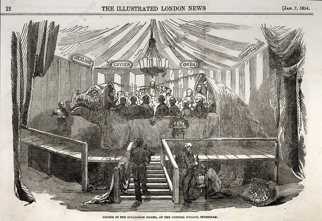
Illustrated London News January 7 1854, page 22. The Iguanodon model at the Crystal Palace in London was large enough for several people to dine inside it.
The models now in Crystal Palace park in South London. In these reconstructions the thumb spike was placed as a nose horn, and the animal is positioned in a rhinoceros-like pose. Image: Chris Sampson, CC BY 2.0 , via Wikimedia Commons
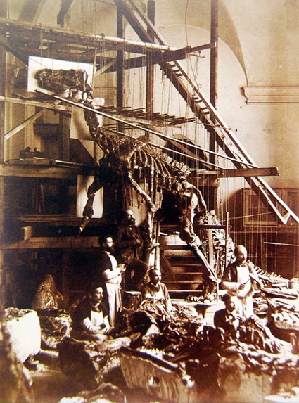
In 1882, de Pauw began assembling at least 38 Iguanodon skeletons under instruction from Louis Dollo, another famous Belgian palaeontologist. The aim was to put them in their most probable living position. A room with a high ceiling was needed because of their size, and a chapel was chosen. Scaffolding was built with hanging ropes being adjusted so the fossilized bones could be moved into their most likely position and then fixed and reinforced with iron rods.
Early reconstructions of Iguanodon showed the dinosaur standing in a kangaroo-like stance. Image: Hutchinson, H. N., Public domain, via Wikimedia Commons
Workmen mounting the first Iguanodon bernissartensis skeleton in the St. George Chapel in Brussels, 1882.
Iguanodon bernissartensis, like the one on display here in the Museum, was a new species, named in 1881. It lived about 125 million years ago. The first assembly was revealed in 1882 and went on public display in Brussels in 1883. Points of reference used for the pose were the skeleton of a cassowary and a kangaroo.
On the Museum’s cast skeleton you can see rod-like structures going across the blade-like, bony processes on the back. These are ossified, or hardened, tendons and would help to stiffen the tail and therefore restrict its movement. They have been broken where the bend in the tail was made to resemble a kangaroo-like stance. The displacement shows that the true position of the tail should be straight.
But having such a straight tail would mean that the Iguanodon would need its head and arms nearer the ground for better balance. The strong hind limbs suggest it would usually walk on two legs with its tail held aloft, as does the fact that fossil Iguanodon footprints are three-toed, and the three-toed limbs are the back ones.
By the end of 1883, six Iguanodons had been mounted this way and positioned in their own glass cage in the courtyard of the Brussels museum. So Iguanodon was one of the very first dinosaurs to be recovered in its entirety and mounted in three dimensions as though a living animal!
—
Leonie is a longstanding Public Engagement volunteer at the Museum. Unable to volunteer in the normal way during the lockdown, she researched the history of this favourite specimen and shared what she learned in a talk for other volunteers as part of an online ‘social’. This article has been adapted from that presentation.
Around 120 years ago, William Sollas, Professor of Geology at the University of Oxford, developed a special technique for grinding down and imaging certain kinds of fossils. Sollas was based at the Museum at the time, and the process he pioneered is still used here today, as our PalaeobiologyTechnician Carolyn Lewis explains to mark the anniversary of Sollas’ birthday on 30 May.
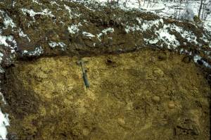
Site of the Herefordshire Lagerstätte, showing the nodules embedded in soft volcanic ash.
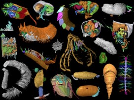
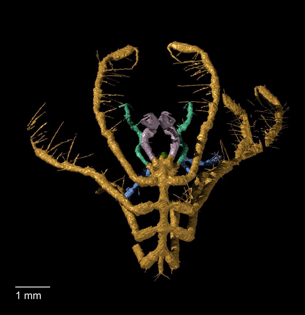
Here at the Museum, I work on a collection of exceptionally well-preserved fossils from the Silurian Herefordshire Lagerstätte. They were deposited on the seabed 430 million years ago when the animals were buried by a volcanic ash flow. The fossils range in size from less than a millimetre up to a few centimetres, and represent a diverse collection of marine invertebrates that includes sponges, echinoderms, brachiopods, worms, molluscs and a wide variety of arthropods.
These Herefordshire Lagerstätte fossils are unusual in that many of them have preserved soft tissues in remarkable detail, including eyes, legs, gill filaments, and even spines and antennae only a few microns in diameter. The key to this extraordinary preservation is that as the fossils developed, calcium carbonate nodules formed around them, protecting and preserving the fossils since the Silurian Period.
Usually, only the hard parts of fossil invertebrates are preserved – the carapace of trilobites or the shells of brachiopods, for example – so the Herefordshire material provides us with a great opportunity to work out the detailed anatomy of these early sea creatures.
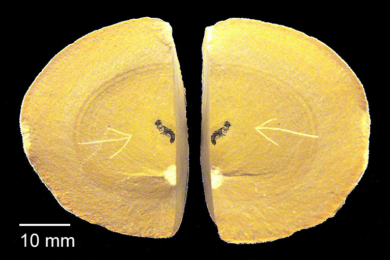
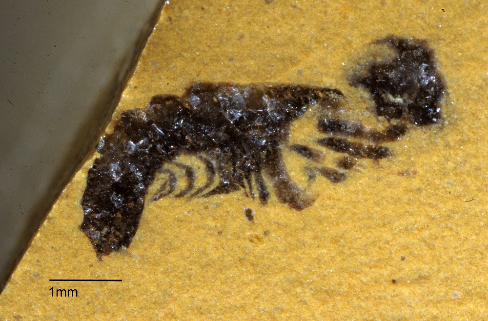
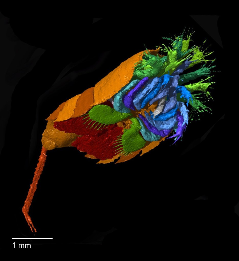
Split rock nodule showing fossil of Offacolus kingi inside.Close-up of the fossil of Offacolus kingi
But the problem we face is how to extract the specimen from the rock nodule without losing the information it contains. The fossils cannot be separated from the surrounding rock by dissolution, because both fossil and nodule are made mainly of calcium carbonate, so would dissolve together. And they are too delicate to be extracted mechanically by cutting and scraping away the surrounding nodule. Even high resolution CT scans cannot, at present, adequately distinguish between the fossils and the surrounding rock material.
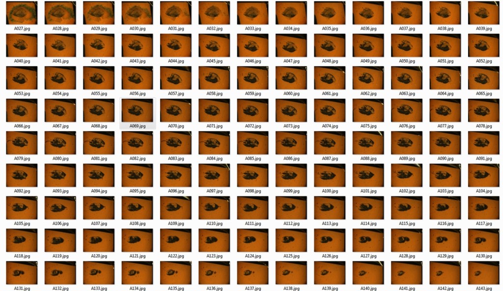
To get round this problem we use a method of serial grinding and photography based on the technique developed by William Sollas in the late 19th century. We grind the fossils in increments of 20 microns then photograph each newly ground surface using a camera mounted on top of a light microscope. This generates hundreds of digital images of cross sections through the specimen.
Then, using specially developed software we convert the stack of two-dimensional images into a 3D digital model that can be viewed and manipulated on screen to reveal the detailed form of the animal. These 3D models are artificially coloured to highlight different anatomical structures and can be rotated through 360o, virtually dissected on screen, and viewed stereoscopically or in anaglyph 3D.
3D virtual reconstruction of Offacolus kingi, a chelicerate arthropod related to horseshoe crabs.
Haliestes dasos (sea spider)
Enalikter aphson (arthropod)
Although our method of serial grinding is still fairly labour intensive, it is far less laborious and time-consuming than the process used by William and his daughter Igerna Sollas. Compared to the photographic methods of the early 20th century, where each photographic plate required long exposure and development times, digital photography is almost instant, enabling us to grind several specimens simultaneously.
Sequential serial grinding images of an ostracod
Computer software also allows us to create 3D virtual models rather than building up physical models from layers of wax. Yet despite our modern adaptations, we are using essentially the same technique that William Sollas developed here at the Museum 120 years ago. And using this technique to study the fossils of the Silurian Herefordshire Lagerstätte has yielded a wealth of new information that opens up a unique window into the evolution and diversification of early life in our oceans.